. 2000 Sep 18;150(6):1507-13.
doi: 10.1083/jcb.150.6.1507.

Tor-mediated induction of autophagy via an Apg1 protein kinase complex

Affiliations

Tor-mediated induction of autophagy via an Apg1 protein kinase complex

Y Kamada et al. J Cell Biol. .

Abstract

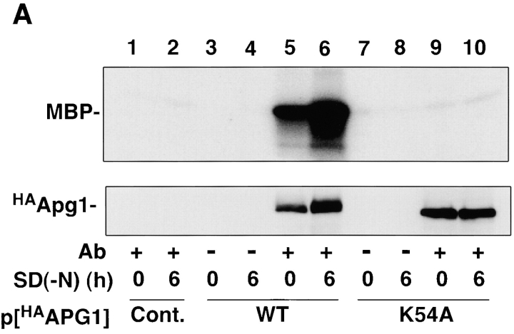
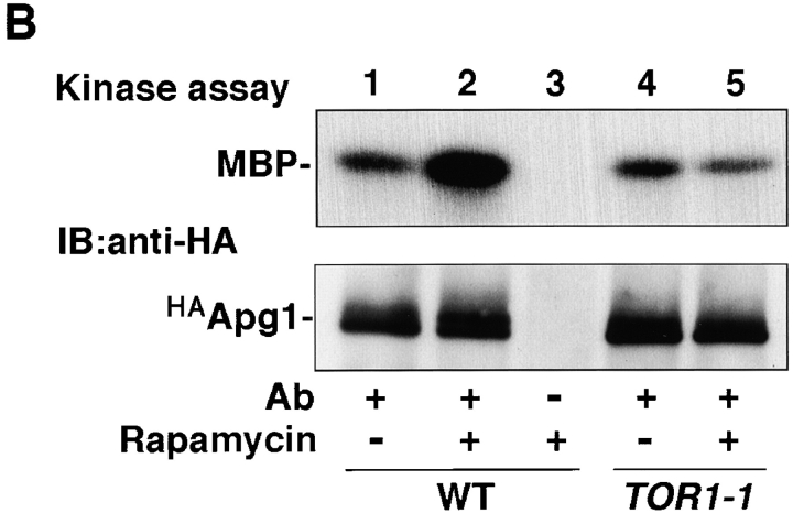
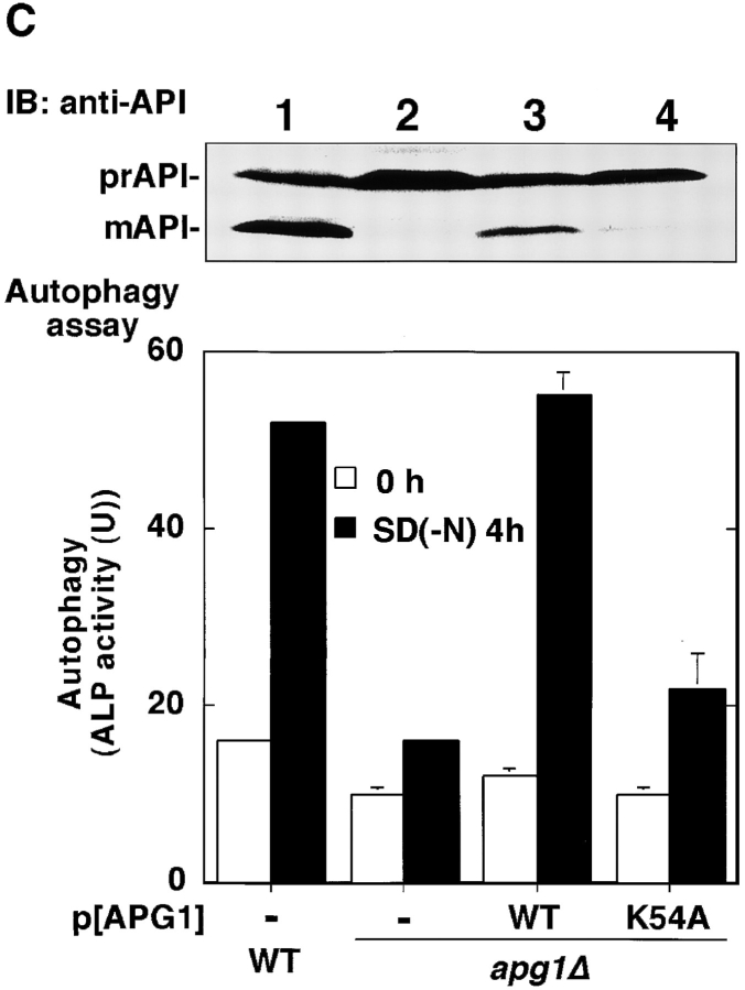
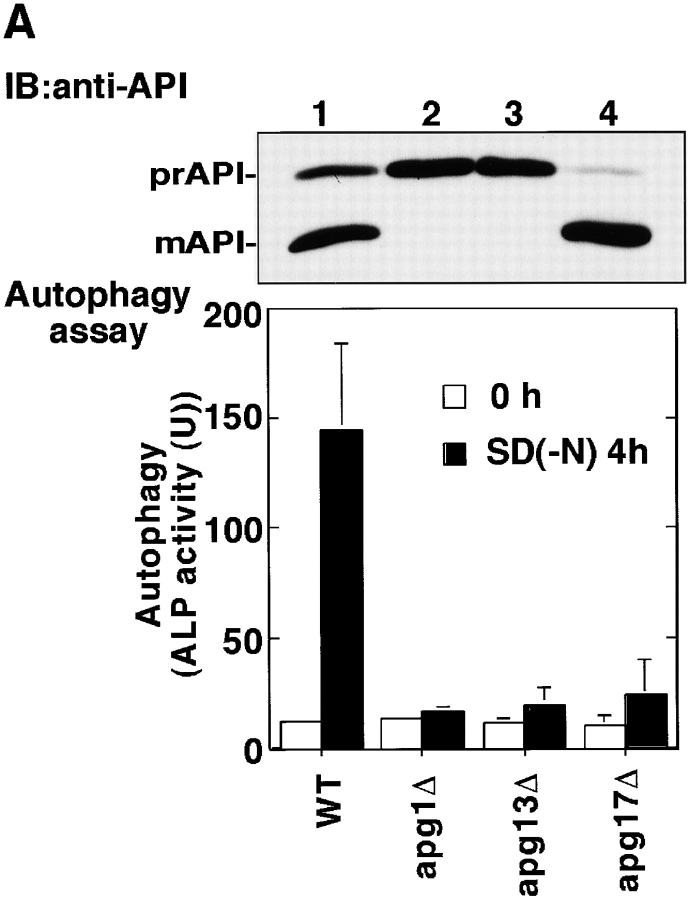
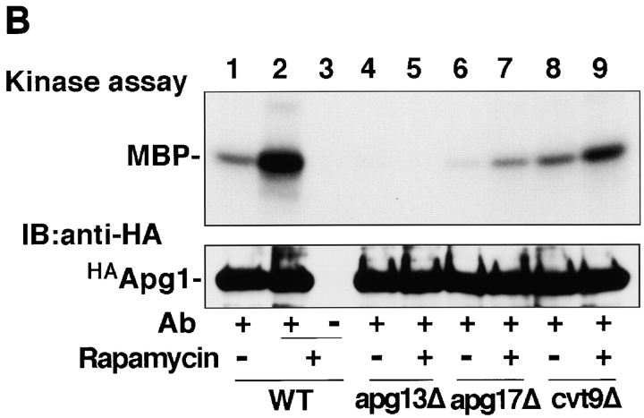
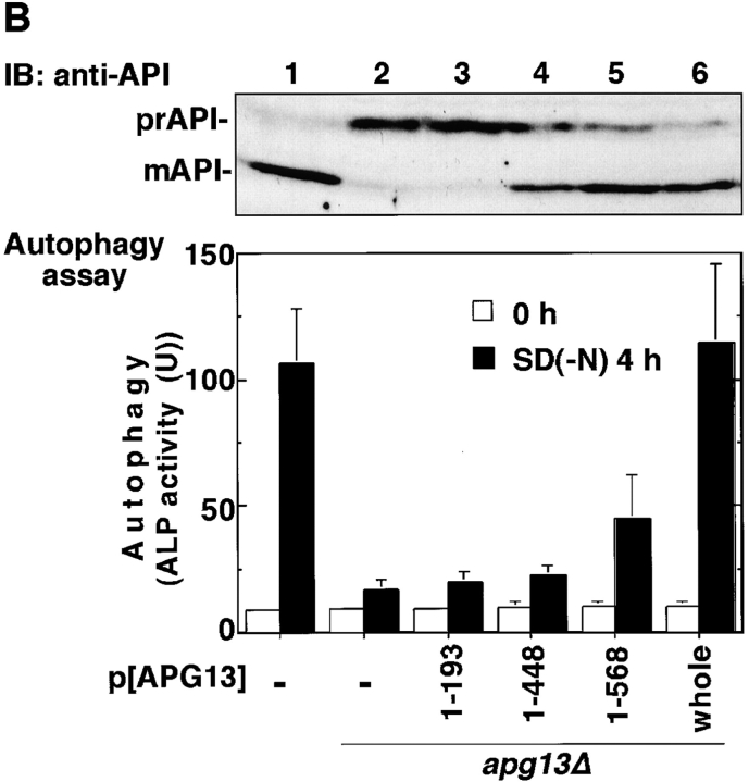
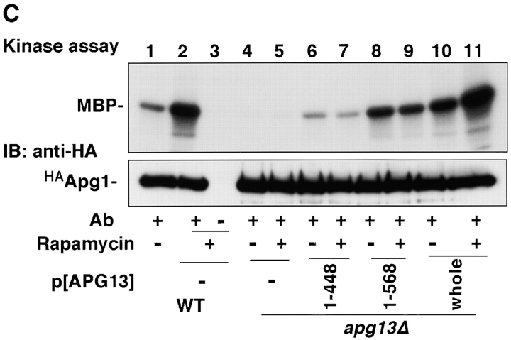
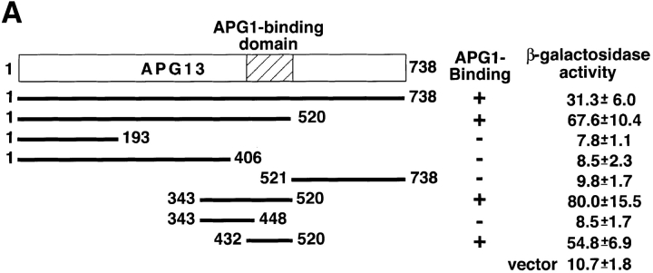
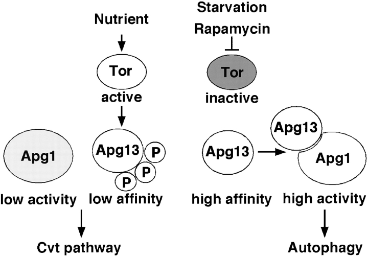
Autophagy is a membrane trafficking to vacuole/lysosome induced by nutrient starvation. In Saccharomyces cerevisiae, Tor protein, a phosphatidylinositol kinase-related kinase, is involved in the repression of autophagy induction by a largely unknown mechanism. Here, we show that the protein kinase activity of Apg1 is enhanced by starvation or rapamycin treatment. In addition, we have also found that Apg13, which binds to and activates Apg1, is hyperphosphorylated in a Tor-dependent manner, reducing its affinity to Apg1. This Apg1-Apg13 association is required for autophagy, but not for the cytoplasm-to-vacuole targeting (Cvt) pathway, another vesicular transport mechanism in which factors essential for autophagy (Apg proteins) are also employed under vegetative growth conditions. Finally, other Apg1-associating proteins, such as Apg17 and Cvt9, are shown to function specifically in autophagy or the Cvt pathway, respectively, suggesting that the Apg1 complex plays an important role in switching between two distinct vesicular transport systems in a nutrient-dependent manner.


Figures

Figure 1
Figure 1
Apg1 is activated by starvation and rapamycin treatment. A, Apg1 is activated by starvation. Cells (KA311A, Cont.) expressing HA-tagged Apg1 (HAApg1, lanes 3–6) or kinase negative HAApg1K54A (lanes 7–10) grown in YEPD at 30°C were incubated in SD(−N) medium for 6 h. Immunoprecipitation of HAApg1 from the cell lysate and protein kinase assay were carried out as described in Materials and Methods (kinase assay, top). Immunoprecipitated HAApg1 was monitored by immunoblot (IB, bottom). B, Apg1 activation is mediated by Tor. Wild-type (WT, JK9-3da, lanes 1–3) or rapamycin-resistant TOR1-1 (JH11-1c, lanes 4 and 5) cells expressing HAApg1 grown in YEPD were treated with or without 0.4 μg/ml of rapamycin for 1.5 h. Apg1 kinase assay and immunodetection were performed. C, Kinase activity of Apg1 is required for both the Cvt pathway and autophagy. Wild-type (TN 125, lane 1) or apg1Δ cells (YYK126, lanes 2–4) transformed with wild-type APG1 (WT, lane 3) or kinase negative apg1 K54A (K54A, lane 4) plasmids were grown in YEPD and subjected to immunoblot (IB) with anti-API antibody (top). The precursor form of API in the cytosol (prAPI) and vacuolar-targeted and processed mature API (mAPI) are indicated in the panel. Progression of autophagy was estimated by monitoring the increase of alkaline phosphatase (ALP) activity (bottom).
Figure 1
Figure 1
Apg1 is activated by starvation and rapamycin treatment. A, Apg1 is activated by starvation. Cells (KA311A, Cont.) expressing HA-tagged Apg1 (HAApg1, lanes 3–6) or kinase negative HAApg1K54A (lanes 7–10) grown in YEPD at 30°C were incubated in SD(−N) medium for 6 h. Immunoprecipitation of HAApg1 from the cell lysate and protein kinase assay were carried out as described in Materials and Methods (kinase assay, top). Immunoprecipitated HAApg1 was monitored by immunoblot (IB, bottom). B, Apg1 activation is mediated by Tor. Wild-type (WT, JK9-3da, lanes 1–3) or rapamycin-resistant TOR1-1 (JH11-1c, lanes 4 and 5) cells expressing HAApg1 grown in YEPD were treated with or without 0.4 μg/ml of rapamycin for 1.5 h. Apg1 kinase assay and immunodetection were performed. C, Kinase activity of Apg1 is required for both the Cvt pathway and autophagy. Wild-type (TN 125, lane 1) or apg1Δ cells (YYK126, lanes 2–4) transformed with wild-type APG1 (WT, lane 3) or kinase negative apg1 K54A (K54A, lane 4) plasmids were grown in YEPD and subjected to immunoblot (IB) with anti-API antibody (top). The precursor form of API in the cytosol (prAPI) and vacuolar-targeted and processed mature API (mAPI) are indicated in the panel. Progression of autophagy was estimated by monitoring the increase of alkaline phosphatase (ALP) activity (bottom).
Figure 2
Figure 2
Apg1-associating proteins are required for Apg1 activation. A, Wild-type (TN125, lane 1), apg1Δ (YYK126, lane 2), apg13Δ (YYK130, lane 3), and apg17Δ (YYK121, lane 4) cells were analyzed with anti-API blot (top) and ALP assay (bottom). B, Wild-type (KA311A), apg13Δ (YYK119), apg17Δ (YYK111), and cvt9Δ (YYK107) cells expressing HAApg1 were treated with rapamycin (0.2 μg/ml, 1 h) and analyzed by kinase assay.
Figure 2
Figure 2
Apg1-associating proteins are required for Apg1 activation. A, Wild-type (TN125, lane 1), apg1Δ (YYK126, lane 2), apg13Δ (YYK130, lane 3), and apg17Δ (YYK121, lane 4) cells were analyzed with anti-API blot (top) and ALP assay (bottom). B, Wild-type (KA311A), apg13Δ (YYK119), apg17Δ (YYK111), and cvt9Δ (YYK107) cells expressing HAApg1 were treated with rapamycin (0.2 μg/ml, 1 h) and analyzed by kinase assay.
Figure 3
Figure 3
Tor-dependent phosphorylation of Apg13 inhibits Apg1–Apg13 association. A, Bandshift of Apg13 in response to starvation and rapamycin. YEPD-grown cells (BJ2168) expressing APG13 with YEp352[APG13] (lanes 1 and 6) were transferred to SD(−N) medium (lanes 2–5) or treated with 0.2 μg/ml rapamycin (lanes 7–10) and incubated for the indicated times. Total protein was analyzed by immunoblot using anti-Apg13 serum. B, Apg13 is hyperphosphorylated. Cells (TFD13-W3) overexpressing APG13 were labeled in vivo with 35S (lanes 1–6) or 32Pi (lanes 7 and 8), and shifted to YEPD (lane 5) or SD(−N) (lanes 6 and 8) for 1 h. Apg13 protein was immunoprecipitated and treated with alkaline phosphatase (PPase; lane 4). Immunoprecipitated protein was subjected to SDS-PAGE, followed by autoradiography. Cont, immunoprecipitation was carried out with preimmune serum. C, Tor-mediated phosphorylation/dephosphorylation of Apg13. Wild-type (WT, JK9-3da) or TOR1-1 (JH11-1c) cells expressing Apg13 grown in YEPD were treated with or without 0.2 μg/ml of rapamycin for 1 h. An immunoblot using anti-Apg13 serum was carried out. D, Phosphorylation/dephosphorylation cycle of Apg13 in response to nutrient conditions. YEPD-grown cells (BJ2168) overexpressing Apg13 (lane 1) were incubated in SD(−N) for 1.5 h (lane 2) with or without 30-min treatment with 10 μg/ml cycloheximide (lanes 4 and 5), 0.2 μg/ml of rapamycin (lanes 6 and 7), or both (lanes 8 and 9). Then, half a sample volume of 2× YEPD was added to the cells, after which they were incubated for 10 min (lanes 3, 5, 7, and 9). Apg13 protein was detected by immunoblot. E, Coimmunoprecipitation of Apg13 with Apg1. YEPD grown cells (BJ2168) overexpressing HAApg1 and/or Apg13 with high-copy plasmids pRS426[HA APG1] (p[HAAPG1]) and YEp351[APG13] (p[APG13]) were lysed, and HAApg1 was immunoprecipitated (IP) from the cell lysate. Apg13 and HAApg1 were detected by immunoblot (lanes 1–6). Apg13 and HAApg1 detected in total cell lysate are also shown (lanes 7–9). The hyperphosphorylated form of Apg13 remaining in the supernatant of the immunoprecipitate was still detected (data not shown), excluding the possibility that Apg1-bound Apg13 was dephosphorylated during the experiment. The asterisk shows a band that anti-HA ascite nonspecifically recognized in total lysate. F, Apg1–Apg13 association is promoted by rapamycin treatment. YEPD-grown cells (BJ2168) expressing HAApg1 and Apg13 with low-copy plasmids pRS316[HA APG1] and pRS315[APG13] were enzymatically converted to spheroplasts and treated with 0.2 μg/ml of rapamycin (at time 0). Spheroplasts were harvested at the indicated times, and coimmunoprecipitation was performed. Apg1 protein kinase assay of the immunoprecipitates was also performed (bottom).
Figure 3
Figure 3
Tor-dependent phosphorylation of Apg13 inhibits Apg1–Apg13 association. A, Bandshift of Apg13 in response to starvation and rapamycin. YEPD-grown cells (BJ2168) expressing APG13 with YEp352[APG13] (lanes 1 and 6) were transferred to SD(−N) medium (lanes 2–5) or treated with 0.2 μg/ml rapamycin (lanes 7–10) and incubated for the indicated times. Total protein was analyzed by immunoblot using anti-Apg13 serum. B, Apg13 is hyperphosphorylated. Cells (TFD13-W3) overexpressing APG13 were labeled in vivo with 35S (lanes 1–6) or 32Pi (lanes 7 and 8), and shifted to YEPD (lane 5) or SD(−N) (lanes 6 and 8) for 1 h. Apg13 protein was immunoprecipitated and treated with alkaline phosphatase (PPase; lane 4). Immunoprecipitated protein was subjected to SDS-PAGE, followed by autoradiography. Cont, immunoprecipitation was carried out with preimmune serum. C, Tor-mediated phosphorylation/dephosphorylation of Apg13. Wild-type (WT, JK9-3da) or TOR1-1 (JH11-1c) cells expressing Apg13 grown in YEPD were treated with or without 0.2 μg/ml of rapamycin for 1 h. An immunoblot using anti-Apg13 serum was carried out. D, Phosphorylation/dephosphorylation cycle of Apg13 in response to nutrient conditions. YEPD-grown cells (BJ2168) overexpressing Apg13 (lane 1) were incubated in SD(−N) for 1.5 h (lane 2) with or without 30-min treatment with 10 μg/ml cycloheximide (lanes 4 and 5), 0.2 μg/ml of rapamycin (lanes 6 and 7), or both (lanes 8 and 9). Then, half a sample volume of 2× YEPD was added to the cells, after which they were incubated for 10 min (lanes 3, 5, 7, and 9). Apg13 protein was detected by immunoblot. E, Coimmunoprecipitation of Apg13 with Apg1. YEPD grown cells (BJ2168) overexpressing HAApg1 and/or Apg13 with high-copy plasmids pRS426[HA APG1] (p[HAAPG1]) and YEp351[APG13] (p[APG13]) were lysed, and HAApg1 was immunoprecipitated (IP) from the cell lysate. Apg13 and HAApg1 were detected by immunoblot (lanes 1–6). Apg13 and HAApg1 detected in total cell lysate are also shown (lanes 7–9). The hyperphosphorylated form of Apg13 remaining in the supernatant of the immunoprecipitate was still detected (data not shown), excluding the possibility that Apg1-bound Apg13 was dephosphorylated during the experiment. The asterisk shows a band that anti-HA ascite nonspecifically recognized in total lysate. F, Apg1–Apg13 association is promoted by rapamycin treatment. YEPD-grown cells (BJ2168) expressing HAApg1 and Apg13 with low-copy plasmids pRS316[HA APG1] and pRS315[APG13] were enzymatically converted to spheroplasts and treated with 0.2 μg/ml of rapamycin (at time 0). Spheroplasts were harvested at the indicated times, and coimmunoprecipitation was performed. Apg1 protein kinase assay of the immunoprecipitates was also performed (bottom).
Figure 4
Figure 4
Apg1–Apg13 association is required for autophagy, but not for the Cvt pathway. A, Apg1-binding site of Apg13. Interaction of the indicated Apg13 fragments (prey) with Apg1 (bait) in the yeast two-hybrid system. Binding activity was estimated by β-galactosidase activity (Kaiser et al. 1994, shown as means and errors calculated from three independent experiments). B, Apg1–Apg13 association is required specifically for autophagy. Wild-type cells (TN125) or apg13Δ cells (YYK130) harboring the indicated APG13 constructs grown in YEPD were analyzed by anti-API immunoblot (top). An ALP assay was performed after 4 h SD(−N) incubation (bottom). C, Apg1 kinase assay was performed using the above APG13 transformants before or after rapamycin (0.2 μg/ml, 1 h) treatment.
Figure 4
Figure 4
Apg1–Apg13 association is required for autophagy, but not for the Cvt pathway. A, Apg1-binding site of Apg13. Interaction of the indicated Apg13 fragments (prey) with Apg1 (bait) in the yeast two-hybrid system. Binding activity was estimated by β-galactosidase activity (Kaiser et al. 1994, shown as means and errors calculated from three independent experiments). B, Apg1–Apg13 association is required specifically for autophagy. Wild-type cells (TN125) or apg13Δ cells (YYK130) harboring the indicated APG13 constructs grown in YEPD were analyzed by anti-API immunoblot (top). An ALP assay was performed after 4 h SD(−N) incubation (bottom). C, Apg1 kinase assay was performed using the above APG13 transformants before or after rapamycin (0.2 μg/ml, 1 h) treatment.
Figure 5
Figure 5
Tap42 does not mediate the induction of autophagy. Autophagy is normal in tap42 ts cells. Wild-type (CY5754) and tap42-11 ts (CY5755) cells were incubated for 4 h at the indicated temperature with or without rapamycin (0.2 μg/ml) in the presence of 1 mM PMSF to visualize autophagic bodies. For incubation at the nonpermissive temperature, 1 h preincubation was carried out. DIC images were shown. Bar, 2 μm.
Figure 6
Figure 6
Model of Tor-mediated regulation between the Cvt pathway and autophagy. See text for details.

Similar articles

Cited by

References

    1. Abeliovich H., Darsow T., Emr S.D. Cytoplasm to vacuole trafficking of aminopeptidase I requires a t-SNARE–Sec1p complex composed of Tlg2p and Vps45p. EMBO (Eur. Mol. Biol. Organ.) J. 1999;18:6005–6016. - PMC - PubMed
    1. Baba M., Ohsumi M., Scott S.V., Klionsky D.J., Ohsumi Y. Two distinct pathways for targeting proteins from the cytoplasm to the vacuole/lysosome. J. Cell Biol. 1997;139:1687–1695. - PMC - PubMed
    1. Beck T., Hall M.N. The TOR signalling pathway controls nuclear localization of nutrient-regulated transcription factors. Nature. 1999;402:689–692. - PubMed
    1. Beck T., Schmidt A., Hall M.N. Starvation induces vacuolar targeting and degradation of the tryptophan permease in yeast. J. Cell Biol. 1999;146:1227–1238. - PMC - PubMed
    1. Cardenas M.E., Cutler N.S., Lorenz M.C., Di Como C.J., Heitman J. The TOR signaling cascade regulates gene expression in response to nutrients. Genes Dev. 1999;13:3271–3279. - PMC - PubMed

Publication types

MeSH terms