. 2000 Sep 26;97(20):10832-7.
doi: 10.1073/pnas.170276797.

Modulation of Akt kinase activity by binding to Hsp90

Affiliations

Modulation of Akt kinase activity by binding to Hsp90

S Sato et al. Proc Natl Acad Sci U S A. .

Abstract

Serine/threonine kinase Akt/PKB is a downstream effector molecule of phosphoinositide 3-kinase and is thought to mediate many biological actions toward anti-apoptotic responses. We found that Akt formed a complex with a 90-kDa heat-shock protein (Hsp90) in vivo. By constructing deletion mutants, we identified that amino acid residues 229-309 of Akt were involved in the binding to Hsp90 and amino acid residues 327-340 of Hsp90beta were involved in the binding to Akt. Inhibition of Akt-Hsp90 binding led to the dephosphorylation and inactivation of Akt, which increased sensitivity of the cells to apoptosis-inducing stimulus. The dephosphorylation of Akt was caused by an increase in protein phosphatase 2A (PP2A)-mediated dephosphorylation and not by a decrease in 3-phosphoinositide-dependent protein kinase-1-mediated phosphorylation. These results indicate that Hsp90 plays an important role in maintaining Akt kinase activity by preventing PP2A-mediated dephosphorylation.


Figures

Figure 1
Figure 1
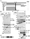
Binding of Akt to Hsp90 in vivo. (A) BALB/3T3 and 293T cell lysates were incubated with protein G agarose conjugated with control sheep IgG or sheep anti-Akt pAb. (B) 293T cells were transfected with mock or the FLAG-tagged WT-akt. The cell lysates were incubated with an anti–FLAG M2 agarose. (C) 293T cells were cotransfected with the V5-tagged WT-hsp90β and the indicated FLAG-tagged plasmids. The cell lysates were incubated with an anti–FLAG M2 agarose. The immunoprecipitated proteins were immunoblotted with the indicated antibodies. Molecular size markers are indicated (in kDa).
Figure 2
Figure 2
Binding properties of wild-type Akt and Akt deletion mutants to Hsp90β. (A) Structural domains of Akt and Akt deletion mutants used in these experiments are represented as black bars. The predicted sites that are involved in the strong and weak Hsp90β binding to Akt are shown schematically (hatched and dotted, respectively). (B–-D) Mock or the V5-tagged WT-hsp90β was transiently transfected into 293T cells together with mock or the indicated FLAG-tagged akt mutants. The FLAG-tagged proteins were immunoprecipitated and immunoblotted with the indicated antibodies. Molecular size markers are indicated (in kDa).
Figure 3
Figure 3
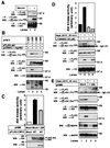
Identification of the Hsp90β domain responsible for binding to Akt. (A) Structural domains of Hsp90β and Hsp90β deletion mutants used in these experiments are represented as black bars. The predicted Akt binding site in Hsp90β is shown schematically (hatched). (B and C) The (His)6-tagged WT-murine akt was transiently transfected into 293T cells together with mock or the indicated FLAG-tagged hsp90β mutants. The (His)6-tagged proteins were immunoprecipitated and immunoblotted with the indicated antibodies. The cell lysates were also immunoblotted with an anti–FLAG M2 mAb. Molecular size markers are indicated (in kDa). (D) Alignment of the amino acid sequence of the human Hsp90β with the equivalent region of human Hsp90α. Identical residues are denoted by white letters on black background. The GenBank accession nos. of human Hsp90β and human Hsp90α were M16660 and X15183, respectively. (E) The V5-tagged WT-hsp90α or WT-hsp90β was transiently transfected into 293T cells together with mock or the indicated FLAG-tagged akt mutants. The FLAG-tagged proteins were immunoprecipitated and immunoblotted with the indicated antibodies. The cell lysates were also immunoblotted with an anti-V5 mAb. Molecular size markers are indicated (in kDa).
Figure 4
Figure 4
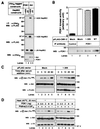
Dephosphorylation and inactivation of Akt after detachment from Hsp90. (A) 293T cells were transfected with the FLAG-tagged WT-akt. After transfection for 24 h, cells were cultured in the medium containing no serum (−) or 10% FBS (+) for 24 h. Then, the FLAG-tagged Akt was immunoprecipitated and immunoblotted with the indicated antibodies. (B) The HA-tagged akt mutants, the V5-tagged WT-hsp90β, and/or the FLAG-tagged WT-akt were cotransfected into 293T cells. The cells were serum-starved for 24 h, and then FLAG-tagged WT-Akt was immunoprecipitated and immunoblotted with the indicated antibodies. The cell lysates were also immunoblotted with the indicated antibodies. (C) 293T cells were transfected with mock or the FLAG-tagged WT-akt together with mock or the HA-tagged 1–309 akt. The cells were serum-starved for 24 h and then harvested. The cell lysates were incubated with protein G agarose conjugated with a control IgG (lane 1) or an anti-Akt pAb (lanes 2 and 3). The Akt kinase activity of the immunoprecipitated Akt was estimated, as described in Materials and Methods. The vertical bars represent SD value of triplicate determinations. The immunoprecipitated proteins and the cell lysates were immunoblotted with the indicated antibodies. (D) HT1080 cells were transfected with the FLAG-tagged WT-akt together with V5-tagged WT-hsp90β. The cells were serum-starved for 24 h and then incubated at 37°C or 45°C for 20 min. In some experiments, cells were treated with 50 μM LY294002 for 10 min before heat shock. The Akt kinase activity of the immunoprecipitated FLAG-tagged Akt was estimated, as described in Materials and Methods. The vertical bars represent SD value of triplicate determinations. The cell lysates were also immunoblotted with the indicated antibodies. (E) HT1080 cells were transfected with the FLAG-tagged WT-akt, and the V5-tagged WT-hsp90β together with mock or the HA-tagged 1–309 akt. The cells were serum-starved for 24 h and then incubated at 37°C (−) or 45°C (+) for 20 min. The immunoprecipitated FLAG-tagged WT-Akt and the cell lysates were immunoblotted with the indicated antibodies. The expression of the HA-tagged 1–309 Akt was confirmed by immunoblot analysis with an anti-HA mAb (data not shown). Molecular size markers are indicated (in kDa).
Figure 5
Figure 5
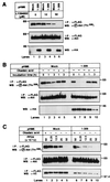
Induction of apoptosis by inhibiting Akt-Hsp90 binding. (A) 293T cells were transfected with mock or the FLAG-tagged 1–309 akt. After transfection for 24 h, the transfected cells were cultured for 24 h in serum-free medium or medium containing 10% FBS. Then the nuclei of the cells were stained with DAPI, as described in Materials and Methods. (B) 293T cells were treated as described in A. The increase in the activity of caspase-3-like proteases (DEVDase activity) was determined, as described in Materials and Methods. The vertical bars represent SD value of triplicate determinations. (C) 293T cells were transfected with mock or the FLAG-tagged 1–309 akt. After transfection for 24 h, the medium was replaced with serum-free medium containing vehicle (lanes 1 and 4), 30 μg/ml of VP-16 (lanes 2 and 5), or 30 μg/ml of VP-16 plus 100 μg/ml of Z-Asp-CH2-DCB (lanes 3 and 6). Cells were further incubated for 24 h and then harvested. The cell lysates were immunoblotted with the indicated antibodies. The expression of the FLAG-tagged 1–309 Akt was confirmed by immunoblot analysis with an anti–FLAG M2 mAb (data not shown). (D) 293T cells were transfected with mock or the indicated FLAG-tagged plasmids. After transfection for 24 h, the medium was replaced with serum-free medium containing 30 μg/ml of VP-16. Cells were further incubated for 24 h. The cell lysates were immunoblotted with the indicated antibodies. Molecular size markers are indicated (in kDa).
Figure 6
Figure 6
Akt binding to Hsp90β does not promote the PDK1-mediated Akt phosphorylation. (A) The (His)6-tagged human PDK1 or WT-murine akt was transfected into 293T cells together with the FLAG-tagged 1–635 hsp90β () or 534–724 hsp90β (ΔN). The immunoprecipitated (His)6-tagged proteins and the cell lysates were immunoblotted with the indicated antibodies. (B) 293T cells were transfected with mock or the FLAG-tagged WT- or 1–309 akt. The cells were serum-starved for 24 h, and then the cell lysates were incubated with a protein G agarose conjugated with a control IgG (Control) or an anti-PDK1 pAb (PDK1). The PDK1 kinase activity was estimated, as described in Materials and Methods. The vertical bars represent SD value of triplicate determinations. The amount of the immunoprecipitated PDK1 proteins was estimated by performing Western blot analysis with an anti-PDK1 mAb. The expression of transfected FLAG-tagged WT- or 1–309 Akt was confirmed by immunoblot analysis with an anti-FLAG M2 mAb (data not shown). (C) Mock or the FLAG-tagged 1–309 akt was transfected into 293T cells together with the HA-tagged WT-akt. The cells were serum-starved for 24 h, and then the medium was replaced with medium containing 10% FBS and 500 nM Okadaic acid. At the indicated time points, cells were harvested and the cell lysates were immunoblotted with the indicated antibodies. (D) The FLAG-tagged WT-akt and the V5-tagged WT-hsp90β were cotransfected into 293T cells. The cells were serum-starved for 24 h and then incubated at 37°C or 45°C for 20 min. The FLAG-tagged WT-Akt was immunoprecipitated and incubated in vitro with the indicated amount of active human PDK1 in the presence (+) or absence (−) of lipid vesicles containing PtdIns(3,4,5)P3 at 30°C for 30 min. The reactions were electrophoresed and immunoblotted with the indicated antibodies. The expression of transfected WT-Akt and Hsp90β was confirmed by immunoblot of the cell lysates with an anti-FLAG M2 mAb and an anti-V5 mAb, respectively (data not shown). Molecular size markers are indicated (in kDa).
Figure 7
Figure 7
Inhibition of Akt-Hsp90β complex formation promotes PP2A-mediated Akt dephosphorylation. (A) The FLAG-tagged WT-akt was transfected into 293T cells together with mock or the HA-tagged 1–309 akt. After transfection for 24 h, cells were treated with 0, 10, or 50 μM LY294002 for 10 min. (B) Mock or the HA-tagged 1–309 akt was transfected into 293T cells together with the FLAG-tagged WT-akt. After transfection for 24 h, the medium was replaced with serum-free medium in the presence (+) or absence (−) of 500 nM Okadaic acid. (A and B) At the indicated time points, the FLAG-tagged WT-Akt was immunoprecipitated and immunoblotted with the indicated antibodies. The cell lysates were also immunoblotted with an anti-HA mAb. (C) Mock or the HA-tagged 1–309 akt was transfected into 293T cells together with V5-tagged WT-hsp90β and the FLAG-tagged WT-akt. The FLAG-tagged WT-Akt was immunoprecipitated and incubated in vitro with the indicated amount of purified human PP2A in the presence (+) or absence (−) of 1 μM Okadaic acid at 30°C for 30 min. The reactions were electrophoresed and immunoblotted with the indicated antibodies. Molecular size markers are indicated (in kDa).

Similar articles

Cited by

References

    1. Scheid M P, Lauener R W, Duronio V. Biochem J. 1995;312:159–162. - PMC - PubMed
    1. Yao R, Cooper G M. Science. 1995;267:2003–2006. - PubMed
    1. Yao R, Cooper G M. Oncogene. 1996;13:343–351. - PubMed
    1. Franke T F, Kaplan D R, Cantley L C. Cell. 1997;88:435–437. - PubMed
    1. Staal S P. Proc Natl Acad Sci USA. 1987;84:5034–5037. - PMC - PubMed

Publication types

Substances

LinkOut - more resources