

Role of the Cdc25A phosphatase in human breast cancer
- PMID: 10995786
- PMCID: PMC381390
- DOI: 10.1172/JCI9174
Role of the Cdc25A phosphatase in human breast cancer
Abstract
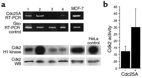
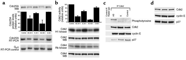
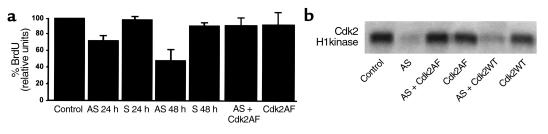
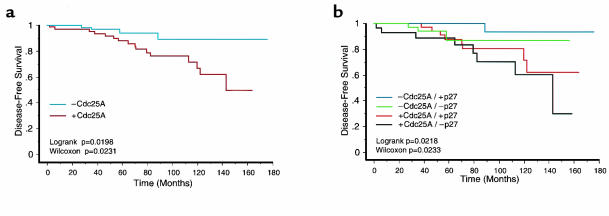
The phosphatase Cdc25A plays an important role in cell cycle regulation by removing inhibitory phosphates from tyrosine and threonine residues of cyclin-dependent kinases, and it has been shown to transform diploid murine fibroblasts in cooperation with activated Ras. Here we show that Cdc25A is overexpressed in primary breast tumors and that such overexpression is correlated with higher levels of cyclin-dependent kinase 2 (Cdk2) enzymatic activity in vivo. Furthermore, in the breast cancer cell line MCF-7, Cdc25A activity is necessary for both the activation of Cdk2 and the subsequent induction of S-phase entry. Finally, in a series of small (< 1 cm) breast carcinomas, overexpression of Cdc25A was found in 47% of patients and was associated with poor survival. These data suggest that overexpression of Cdc25A contributes to the biological behavior of primary breast tumors and that both Cdc25A and Cdk2 are suitable therapeutic targets in early-stage breast cancer.
Figures
Similar articles
-
Reduction of Cdc25A contributes to cyclin E1-Cdk2 inhibition at senescence in human mammary epithelial cells.Oncogene. 2000 Nov 9;19(47):5314-23. doi: 10.1038/sj.onc.1203908. Oncogene. 2000. PMID: 11103932
-
A rate limiting function of cdc25A for S phase entry inversely correlates with tyrosine dephosphorylation of Cdk2.Oncogene. 1999 Jan 21;18(3):573-82. doi: 10.1038/sj.onc.1202362. Oncogene. 1999. PMID: 9989807
-
Inhibition of cyclin-dependent kinase 2 by the Chk1-Cdc25A pathway during the S-phase checkpoint activated by fludarabine: dysregulation by 7-hydroxystaurosporine.Mol Pharmacol. 2002 Sep;62(3):680-8. doi: 10.1124/mol.62.3.680. Mol Pharmacol. 2002. PMID: 12181445
-
The role of Cdc25A in the regulation of cell proliferation and apoptosis.Anticancer Agents Med Chem. 2012 Jul;12(6):631-9. doi: 10.2174/187152012800617678. Anticancer Agents Med Chem. 2012. PMID: 22263797 Free PMC article. Review.
-
CDC25A: a rebel within the CDC25 phosphatases family?Anticancer Agents Med Chem. 2008 Dec;8(8):825-31. doi: 10.2174/187152008786847684. Anticancer Agents Med Chem. 2008. PMID: 19075564 Review.
Cited by
-
The cell cycle-regulatory CDC25A phosphatase inhibits apoptosis signal-regulating kinase 1.Mol Cell Biol. 2001 Jul;21(14):4818-28. doi: 10.1128/MCB.21.14.4818-4828.2001. Mol Cell Biol. 2001. PMID: 11416155 Free PMC article.
-
Cdk2-null mice are resistant to ErbB-2-induced mammary tumorigenesis.Neoplasia. 2011 May;13(5):439-44. doi: 10.1593/neo.101704. Neoplasia. 2011. PMID: 21532884 Free PMC article.
-
Regulation of G(2)/M events by Cdc25A through phosphorylation-dependent modulation of its stability.EMBO J. 2002 Nov 1;21(21):5911-20. doi: 10.1093/emboj/cdf567. EMBO J. 2002. PMID: 12411508 Free PMC article.
-
Overexpression of CDC25B and LAMC2 mRNA and protein in esophageal squamous cell carcinomas and premalignant lesions in subjects from a high-risk population in China.Cancer Epidemiol Biomarkers Prev. 2008 Jun;17(6):1424-35. doi: 10.1158/1055-9965.EPI-06-0666. Cancer Epidemiol Biomarkers Prev. 2008. PMID: 18559558 Free PMC article.
-
Ubiquitin-Specific Protease 29 Regulates Cdc25A-Mediated Tumorigenesis.Int J Mol Sci. 2021 May 28;22(11):5766. doi: 10.3390/ijms22115766. Int J Mol Sci. 2021. PMID: 34071237 Free PMC article.
References
-
- Lees E. Cyclin dependent kinase regulation. Curr Opin Cell Biol. 1995;7:773–780. - PubMed
-
- Sherr CJ, Roberts JM. CDK inhibitors: positive and negative regulators of G1-phase progression. Genes Dev. 1999;13:1501–1512. - PubMed
-
- DelSal G, Loda M, Pagano M. Cell cycle and cancer: critical events at the G1 restriction point. Crit Rev Oncog. 1996;7:127–142. - PubMed
-
- Pardee AB. G1 events and regulation of cell proliferation. Science. 1989;246:603–614. - PubMed
-
- Draetta G, Eckstein J. Cdc25 protein phosphatases in cell proliferation. Biochim Biophys Acta. 1997;1332:M53–M63. - PubMed
