. 2000 Sep;106(6):R59-67.
doi: 10.1172/jci10763.

Functional human T-cell immunity and osteoprotegerin ligand control alveolar bone destruction in periodontal infection

Affiliations

Functional human T-cell immunity and osteoprotegerin ligand control alveolar bone destruction in periodontal infection

Y T Teng et al. J Clin Invest. 2000 Sep.

Abstract

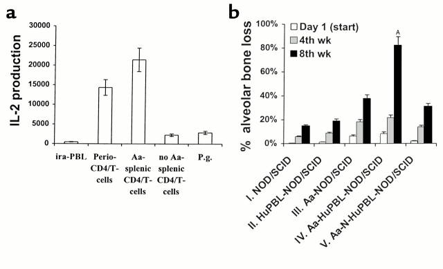
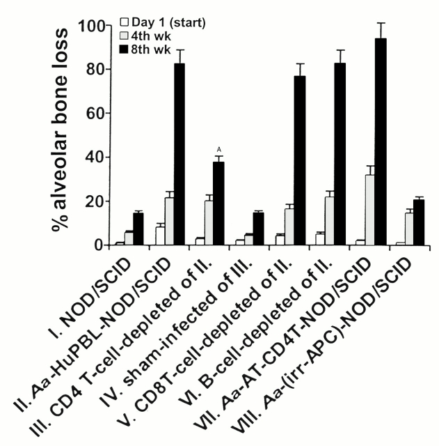
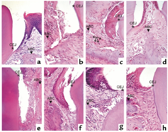
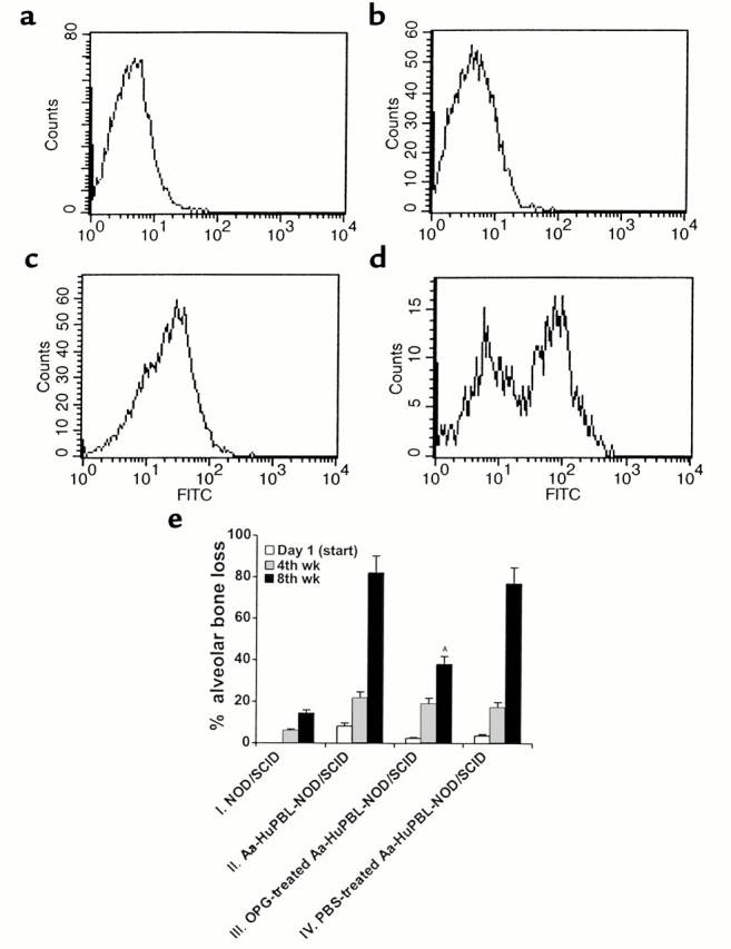
Periodontitis, a prime cause of tooth loss in humans, is implicated in the increased risk of systemic diseases such as heart failure, stroke, and bacterial pneumonia. The mechanisms by which periodontitis and antibacterial immunity lead to alveolar bone and tooth loss are poorly understood. To study the human immune response to specific periodontal infections, we transplanted human peripheral blood lymphocytes (HuPBLs) from periodontitis patients into NOD/SCID mice. Oral challenge of HuPBL-NOD/SCID mice with Actinobacillus actinomycetemcomitans, a well-known Gram-negative anaerobic microorganism that causes human periodontitis, activates human CD4(+) T cells in the periodontium and triggers local alveolar bone destruction. Human CD4(+) T cells, but not CD8(+) T cells or B cells, are identified as essential mediators of alveolar bone destruction. Stimulation of CD4(+) T cells by A. actinomycetemcomitans induces production of osteoprotegerin ligand (OPG-L), a key modulator of osteoclastogenesis and osteoclast activation. In vivo inhibition of OPG-L function with the decoy receptor OPG diminishes alveolar bone destruction and reduces the number of periodontal osteoclasts after microbial challenge. These data imply that the molecular explanation for alveolar bone destruction observed in periodontal infections is mediated by microorganism-triggered induction of OPG-L expression on CD4(+) T cells and the consequent activation of osteoclasts. Inhibition of OPG-L may thus have therapeutic value to prevent alveolar bone and/or tooth loss in human periodontitis.


Figures

Figure 1
Figure 1
(a) Immunocompetence of CD4+ T cells in Aa-HuPBL-NOD/SCID mice. IL-2 production of periodontal CD4+ T cells (Perio-CD4/T-cells) and splenic CD4+ T cells (Aa-splenic CD4/T-cells) isolated from A. actinomycetemcomitans–immunized HuPBL-NOD/SCID mice is shown. Irradiated (25 Gy) HuPBL-derived autologous monocytes/macrophages (irr-PBL) and CD4+ T cells isolated from non–Aa-immunized HuPBL-NOD/SCID mice (no Aa-splenic CD4/T-cells) were included as the negative controls. Restimulation of Aa-immunized splenic CD4+ T cells with a third-party antigen, P. gingivalis sonicate antigens served as the specificity control (P.g.). Serial dilution of Aa sonicate antigens did not result in significantly different patterns of the IL-2 responses measured. Differences in IL-2 production between the Perio-CD4/T-cell and Aa-splenic CD4/T-cell groups as compared with the irr-PBL, no Aa-splenic CD4/T-cell, and P.g. groups were statistically significant (P < 0.008, paired t test). Values shown are mean IL-2 production of triplicate samples ± SD. One result representative of three independent experiments is shown. (b) Increased alveolar bone loss in Aa-HuPBL-NOD/SCID mice. Groups of mice as indicated were either sham-infected or inoculated with A. actinomycetemcomitans (Aa). Data shown are the relative amounts of alveolar bone loss at the day of the first Aa inoculation (Day 1), by the end of 4 weeks (4th wk), and by the end of 8 weeks (8th wk), expressed as a percentage of the amount of bone loss in the positive control, Aa-infected BALB/c mice (100% = 0.6 ± 0.12 mm per tooth in 8 weeks). Group I, sham-infected NOD/SCID mice (n = 10); group II, sham-infected chimeric NOD/SCID mice engrafted with HuPBL from four LJP patients (n = 12); group III, NOD/SCID mice infected with Aa (n = 16); group IV, Aa-infected chimeric NOD/SCID mice engrafted with HuPBL from four LJP patients (n = 32); group V, Aa-infected chimeric NOD/SCID mice engrafted with HuPBL (N-HuPBL) from two healthy donors (n = 12). Data shown are mean values ± SD pooled from four independent experiments involving HuPBL engraftment from four LJP subjects, each of which gave comparable results. Alveolar bone loss was determined as described in Methods. AThe extent of bone loss in group IV at 8 weeks was significantly increased as compared with all other groups (P < 0.01).
Figure 2
Figure 2
Regulation of alveolar bone destruction by A. actinomycetemcomitans–reactive CD4+ T cells. Groups of mice as indicated were either left untreated or inoculated with A. actinomycetemcomitans. CD4+ T, CD8+ T, or B cells were depleted from mice using specific Ab’s and human complement as described in Methods. Data shown are the relative amounts (± SD) of alveolar bone loss accumulated by the end of 8 weeks, expressed as a percentage of the bone loss in the positive control, Aa-infected BALB/c mice (100%). Group I, sham-infected, nondepleted NOD/SCID mice (n = 10); group II, Aa-infected, nondepleted HuPBL-NOD/SCID mice (n = 32); group III, Aa-infected, CD4+ T cell–depleted NOD/SCID mice (n = 20); group IV, sham-infected, CD4+ T cell–depleted NOD/SCID mice (n = 9); group V, Aa-infected, CD8+ T cell–depleted NOD/SCID mice (n = 12); group VI, Aa-infected, B cell–depleted NOD/SCID mice (n = 16); group VII, Aa-infected NOD/SCID mice bearing adoptively transferred Aa-reactive CD4+ T cells (AT-CD4T) plus irradiated autologous monocytes/macrophages as APCs (n = 10); group VIII, Aa-infected NOD/SCID mice bearing adoptively transferred irradiated autologous monocytes/macrophages as APCs (irr-APC; n = 6). AStatistically significant difference in bone loss between group III and groups II, IV, V, and VI (P < 0.005).
Figure 3
Figure 3
Histopathological lesions and bone loss in Aa-HuPBL-NOD/SCID mice. CEJ, cemento-enamel junction, a landmark for tissue loss; ABC, alveolar bone crest. The distance between CEJ and ABC reflects the amount of tissue or alveolar bone loss measured as described in Methods. (a) A representative tissue section from sham-infected HuPBL-NOD/SCID mice demonstrating the normal periodontal and alveolar bone structures (as in Figure 1b, group II). (b, c) Typical histopathological lesions show significant inflammatory infiltration, connective tissue loss below CEJ (in b), and alveolar bone loss with apical growth of sulcular epithelium below CEJ and into alveolar bone area (in c) in Aa-HuPBL-NOD/SCID mice (as in Figure 2, group II) by the end of 8 weeks. Some multinucleated giant cells (arrows in c) can be observed along the surface of alveolar crestal bone. (d) Mild inflammatory infiltrates without obvious alveolar bone loss, as evidenced by sulcular epithelium located at CEJ, in the periodontal tissues of CD4+ T cell–depleted Aa-HuPBL-NOD/SCID mice (as in Figure 2, group III) by the end of 8 weeks. (e, f) In vivo depletion of CD8+ T cells (e) (as in Figure 2, group V) or B cells (f) (as in Figure 2, group VI) has no apparent effects on existing periodontal inflammation accompanied by tissue loss below CEJ (in e) or alveolar bone loss with apical growth of sulcular epithelium below CEJ and ABC (in f). These findings are consistent with those observed in nondepleted Aa-HuPBL-NOD/SCID mice in b and c. (g, h) In vivo inhibition of OPG-L via the decoy receptor OPG abrogates alveolar bone destruction, as evidenced by sulcular epithelium located at CEJ and above ABC (in both panels), in Aa-HuPBL-NOD/SCID mice by the end of 8 weeks (as in Figure 4e, group III). Note that OPG treatment does not affect periodontal inflammation observed in g and h. Aa-infected HuPBL-NOD/SCID mice were treated with an OPG-Fc fusion protein as described in Methods. Parts g and h are representative of eight mice studied in this group. Magnifications: a, b, dh, ×200; c, ×100.
Figure 4
Figure 4
(ad) A. actinomycetemcomitans stimulates OPG-L expression on periodontal CD4+ T cells. (a) Unstimulated HuPBL-derived CD4+ T cells stained with OPG-FITC; negative control. (b) HuPBL-derived CD4+ T cells restimulated with anti-TCR plus CD28 mAb’s, followed by staining with isotypic control Ab; background control. (c) HuPBL-derived CD4+ T cells restimulated with anti-TCR plus CD28 mAb’s, followed by staining with OPG-FITC; positive control. (d) Periodontal CD4+ T cells derived from Aa-HuPBL-NOD/SCID mice stimulated with A. actinomycetemcomitans sonicate antigens, followed by staining with OPG-FITC. Periodontal CD4+ T cells restimulated with P. gingivalis sonicate antigens did not induce significant membrane OPG-L expression over the background level (data not shown). OPG-L membrane expression was determined by FACS analyses 48 hours later. (e) Reduction of alveolar bone destruction in OPG-Fc treated Aa-HuPBL-NOD/SCID mice. Groups of mice as indicated were infected with A. actinomycetemcomitans followed by in vivo treatment with soluble human OPG-Fc fusion protein. Alveolar bone loss (mean values ± SD) was assessed at 8 weeks and normalized to the positive control, Aa-infected BALB/c mice (100%). Group I, sham-infected NOD/SCID mice (n = 10); group II, Aa-infected HuPBL-NOD/SCID mice not injected with OPG-Fc (n = 16); group III, Aa-infected HuPBL-NOD/SCID mice injected with OPG-Fc (n = 10); group IV, Aa-infected HuPBL-NOD/SCID mice injected with PBS (n = 8). AThe differences in alveolar bone loss between group III and groups II and IV are statistically significant (P < 0.002).

Comment in

References

    1. Clark WB, Loe H. Mechanisms of initiation and progression of periodontal disease. Periodontol 2000. 1993;2:72–82. - PubMed
    1. Brown LJ, Oliver RC, Loe H. Evaluating periodontal status of US employed adults. J Am Dent Assoc. 1990;121:226–232. - PubMed
    1. Eklund SA, Burt BA. Risk factors for total tooth loss in the United States; longitudinal analysis of national data. J Public Health Dent. 1994;54:5–14. - PubMed
    1. Genco R. Research news. Healthy gums for a happy heart. Science. 1997;276:203.
    1. Mattila KL, Valle MS, Nieminen MS, Valtonen VV, Hietaniemi KL. Dental infections and coronary atherosclerosis. Atherosclerosis. 1993;103:205–211. - PubMed

Publication types

MeSH terms