. 2000 Sep 15;20(18):6974-82.
doi: 10.1523/JNEUROSCI.20-18-06974.2000.

New features of connectivity in piriform cortex visualized by intracellular injection of pyramidal cells suggest that "primary" olfactory cortex functions like "association" cortex in other sensory systems

Affiliations

New features of connectivity in piriform cortex visualized by intracellular injection of pyramidal cells suggest that "primary" olfactory cortex functions like "association" cortex in other sensory systems

D M Johnson et al. J Neurosci. .

Abstract

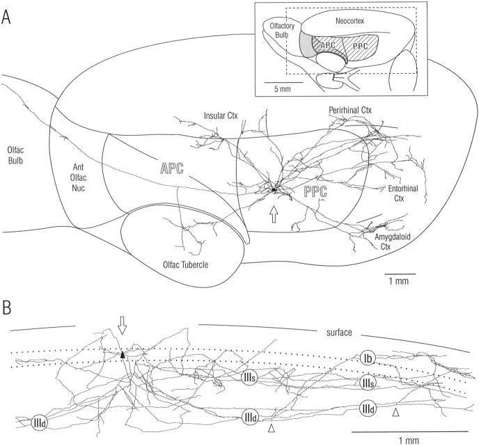
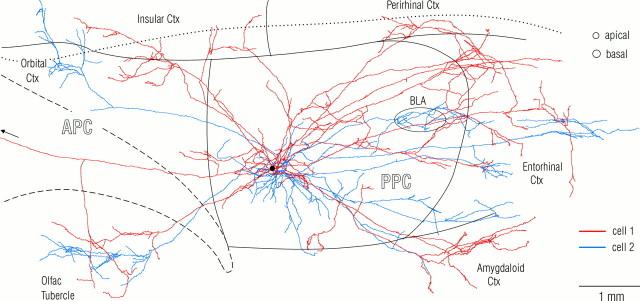
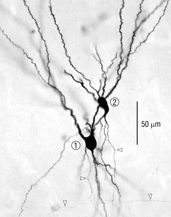
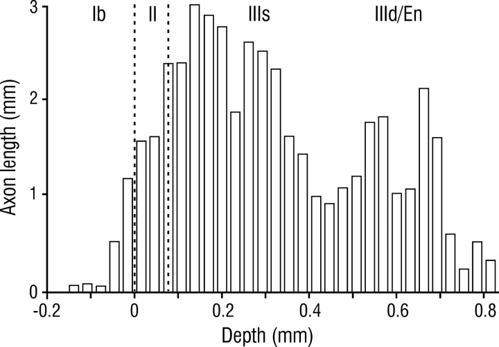
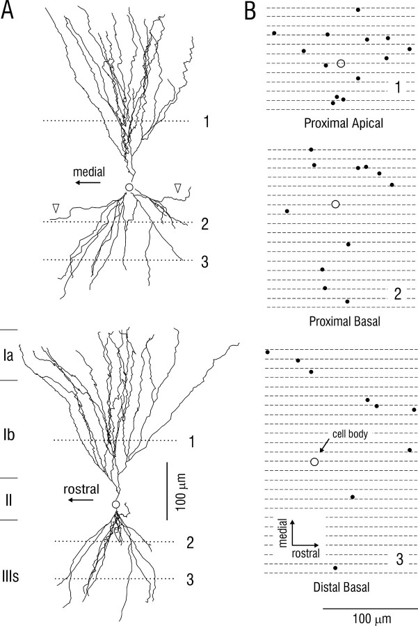
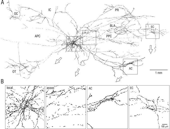
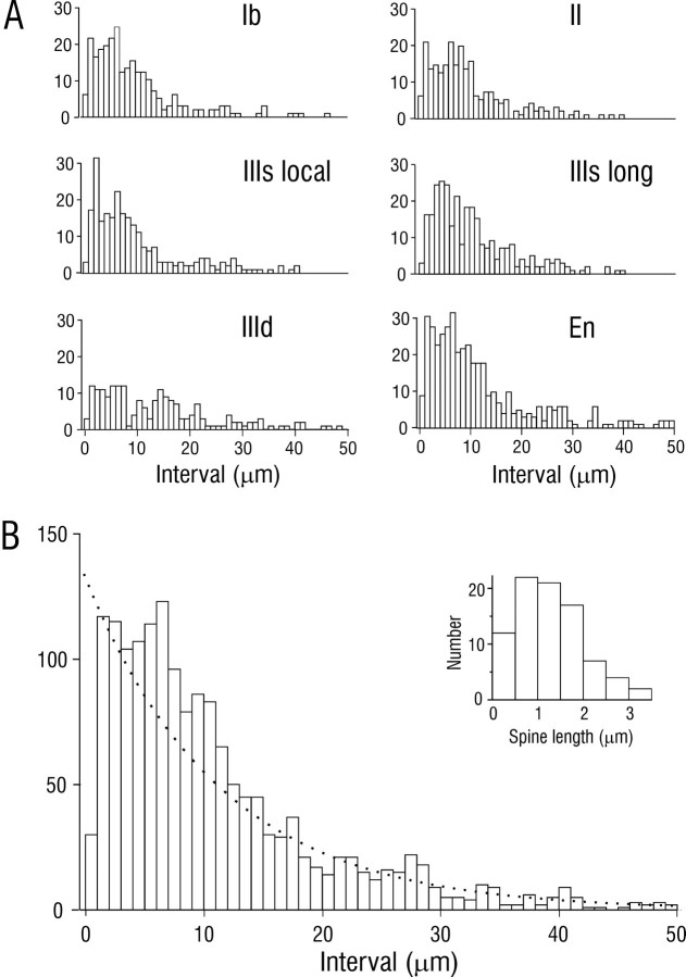
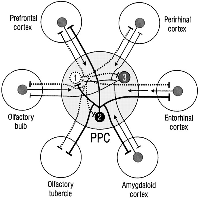
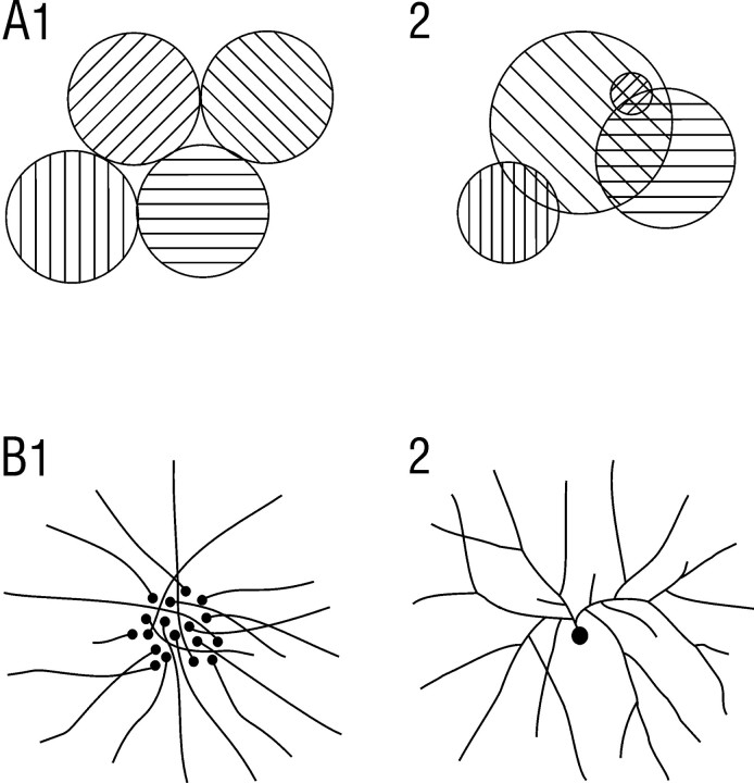
Associational connections of pyramidal cells in rat posterior piriform cortex were studied by direct visualization of axons stained by intracellular injection in vivo. The results revealed that individual cells have widespread axonal arbors that extend over nearly the full length of the cerebral hemisphere. Within piriform cortex these arbors are highly distributed with no regularly arranged patchy concentrations like those associated with the columnar organization in other primary sensory areas (i.e., where periodically arranged sets of cells have common response properties, inputs, and outputs). A lack of columnar organization was also indicated by a marked disparity in the intrinsic projection patterns of neighboring injected cells. Analysis of axonal branching patterns, bouton distributions, and dendritic arbors suggested that each pyramidal cell makes a small number of synaptic contacts on a large number (>1000) of other cells in piriform cortex at disparate locations. Axons from individual pyramidal cells also arborize extensively within many neighboring cortical areas, most of which send strong projections back to piriform cortex. These include areas involved in high-order functions in prefrontal, amygdaloid, entorhinal, and perirhinal cortex, to which there are few projections from other primary sensory areas. Our results suggest that piriform cortex performs correlative functions analogous to those in association areas of neocortex rather than those typical of primary sensory areas with which it has been traditionally classed. Findings from other studies suggest that the olfactory bulb subserves functions performed by primary areas in other sensory systems.


Figures

Fig. 1.
Fig. 1.
Axon from a single pyramidal cell in layer II of rat piriform cortex. Note that axon branches extend over nearly the entire length of the cerebral hemisphere and are widely distributed within piriform cortex and other olfactory and nonolfactory areas. The SP cell in posterior piriform cortex was stained by intracellular injection of biotinylated dextran amine in vivo, and the axon was reconstructed through serial sections with a computer microscope system. A, Spatial distribution of axon branches in surface view. The inset at top right shows the illustrated portion of the rat brain (dashed rectangle) and orientation (45° upward rotation); the hatched area is piriform cortex, and theshaded area is lateral olfactory tract.APC, Anterior piriform cortex; PPC,posterior piriform cortex. B, Depth distribution of same axon within PPC. View is parallel to layers after 90° rotation; rostral is at left as in A; branches outside PPC have been removed; size scale is expanded relative to that in A. Roman numerals indicate layers: Ib, association fiber zone in layer I (molecular layer); IIIs, IIId,superficial and deep portions of layer III; dotted lines, superficial and deep borders of layer II (compact cell body layer). Open arrowheads mark branch points for axon collaterals that ascend to layer I. Open arrows inA and B indicate cell body; dendritic tree is not illustrated (Fig. 3). Ant,Anterior; ctx, cortex; nuc, nucleus;olfac, olfactory.
Fig. 2.
Fig. 2.
Association (cortico-cortical) axons from a pair of neighboring superficial pyramidal cells in posterior piriform cortex. Note the minimal overlap of the two axonal arbors outside the ∼1 mm diameter local collateral region that surrounds SP somata. The axon drawn in red (Fig. 3, cell 1) is the same as in Figure 1. The arborizations from the second cell (blue) in the orbital cortex (top left) and basolateral amygdala (BLA, oval) are deep to piriform cortex. The black spot indicates the position of the cell bodies. The circles at top right denote typical diameters of pyramidal cell dendritic trees at the depths where they are contacted by association fibers (proximal apical dendrites in layer Ib and basal dendrites in layer III). The borders of piriform cortex and the insular-perirhinal border are indicated by solid lines; the dashed line outlines the lateral olfactory tract; the dotted line is the rhinal sulcus.
Fig. 3.
Fig. 3.
Photomicrograph of the neighboring pyramidal cells reconstructed in Figure 2. Arrowheads indicate axons.
Fig. 4.
Fig. 4.
Depth distribution of superficial pyramidal cell axons in piriform cortex. Histogram shows axon length as a function of depth for five intracellularly injected cells. En, Endopiriform nucleus.
Fig. 5.
Fig. 5.
Dendritic morphology of superficial pyramidal cell in posterior piriform cortex. A, Top, Reconstruction in frontal plane (perpendicular to predominant course of associational axons); bottom, same cell after 90° rotation. B, Positions of dendrites from same cell at depths indicated by the dotted lines in A. Black spots indicate points of intersection of dendrites with horizontal planes (parallel to surface) at depths of proximal apical, proximal basal, and distal basal segments where SP cells receive association fiber synapses. Circles indicate cell body position, orthogonally projected onto the three horizontal planes. Diameters of black spots in B correspond to area that contains ∼90% of the dendritic spines (sites of association fiber synapses). Comparison of dendritic and axonal morphologies suggests that each SP cell makes a small number of contacts on a large number of other SP cells; see Results.
Fig. 6.
Fig. 6.
A, Surface plot of the locations of putative synaptic boutons for the axons illustrated in Figure 2. Eachdot represents a bouton visualized by light microscopy.B, Enlargements of regions indicated byboxes in A. First panel (left) shows local axon collaterals; second panel shows longer association (assoc) fibers; upward arrowheads indicate an axon in layer IIIs with a relatively high bouton density; downward arrowheads indicate an axon in layer IIId with a lower density. Third andfourth panels show boutons in amygdaloid cortex (AC) and entorhinal cortex (EC).Oval in A indicates a cluster of boutons in the basolateral amygdala (BLA), deep to piriform cortex. IC, Insular cortex; OC, orbital cortex; OT, olfactory tubercle; PR,perirhinal cortex.
Fig. 7.
Fig. 7.
Distribution of interbouton intervals for association axons in PPC. Data were derived by random sampling of sections from five injected cells. A, Interval histograms from individual layers in PPC and the subjacent endopiriform nucleus (En). IIIs local, Local axon collaterals in the superficial part of layer III; IIIs long, long association axons in superficial III.B, Composite histogram from individual plots.Dotted line is an exponential distribution with identical mean. Inset is a histogram of dendritic spine length for SP cells; mean = 1.21 ± 0.08 μm (SEM);n = 85.
Fig. 8.
Fig. 8.
Summary of intrinsic and extrinsic cortical connections of superficial pyramidal cells in PPC. Each SP cell (numbered circles) has an extensively branching axon that contacts a large number of other SP cells in piriform cortex at disparate locations (flat end bars are synapses). Axons from each cell also arborize in adjoining cortical areas, including those involved in the highest order brain functions. Axonal branches from each cell extend to most, but not all of the target areas (e.g., cell 1 projects to all areas with the exception of amygdaloid cortex; cell 2 does not project to the olfactory bulb or perirhinal cortex). Reciprocal projections from target areas are indicated by arrows. Connections with cortical areas in the olfactory peduncle are not illustrated.
Fig. 9.
Fig. 9.
Alternative schemes for modular organization (A), and spatially distributed associational connections (B) in PPC. A1,Modular organization as observed in primary neocortical areas where discrete columns of cells defined by response properties and connections are regular in size and arrangement, and do not overlap.A2, Organization in which cells with similar response properties are spatially grouped, but in an irregular overlapping manner. B1, Distributed connectivity in which each cell projects to a specific spot, but neighboring cells project to spatially disparate spots. B2, Distributed connectivity in which individual cells have highly branched, spatially divergent axons.

References

    1. Buck LB. Information coding in the vertebrate olfactory system. Annu Rev Neurosci. 1999;19:517–544. - PubMed
    1. Burkhalter A, Charles V. Organization of local axon collaterals of efferent projection neurons in rat visual cortex. J Comp Neurol. 1990;302:920–934. - PubMed
    1. Burwell RD, Amaral DG. Cortical afferents of the perirhinal, postrhinal, and entorhinal cortices of the rat. J Comp Neurol. 1998;398:179–205. - PubMed
    1. Chapin JK, Sadeq M, Guise JL. Corticocortical connections within the primary somatosensory cortex of the rat. J Comp Neurol. 1987;263:326–346. - PubMed
    1. Cheng K, Saleem KS, Tanaka K. Organization of corticostriatal and corticoamygdalar projections arising from the anterior inferotemporal area TE of the macaque monkey: a Phaseolus vulgaris leucoagglutinin study. J Neurosci. 1997;17:7902–7925. - PMC - PubMed

Publication types

LinkOut - more resources