. 2000 Sep 15;20(18):7087-95.
doi: 10.1523/JNEUROSCI.20-18-07087.2000.

Origin of transient and sustained responses in ganglion cells of the retina

Affiliations

Origin of transient and sustained responses in ganglion cells of the retina

G B Awatramani et al. J Neurosci. .

Abstract

Phasic and tonic light responses provide a fundamental division of visual information that is thought to originate in the inner retina. However, evidence presented here indicates that this duality originates in the outer retina. In response to a steady light stimulus, the temporal responses of On-bipolar cells fell into two groups. In one group, the light response peaked and then rapidly declined (tau approximately 400 msec) close to the resting membrane potential. At light offset, these cells exhibited a transient afterhyperpolarization. In the second group of On-bipolar cells, the light response declined 10-fold more slowly and reached a steady depolarization that was approximately 40% of the peak response. These neurons had a slowly decaying afterhyperpolarization at light offset. A metabotropic glutamate antagonist, (RS)-alpha-cyclopropyl-4-phosphonophenylyglycine (CPPG), blocked light responses in both types of On-bipolar cell. CPPG only slightly depolarized transient On-bipolar cells, whereas sustained On-bipolar cells were significantly depolarized. Inorganic calcium channel blockers disclosed that these distinct On-bipolar responses were inherent to the bipolar cell and not attributable to synaptic feedback. CPPG had distinct effects on sustained and transient ganglion cells, similar to its action on bipolar cells. The antagonist depolarized and blocked the light responses of sustained ganglion cells. In transient ganglion cells, CPPG suppressed the On light response but did not depolarize the cell or block the Off light response. These results suggest that transient and sustained light responses in ganglion cells result from selective bipolar cell input and that these two fundamental visual channels originate at the dendritic terminals of bipolar cells.


Figures
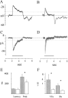
Fig. 1.
Fig. 1.
On-bipolar cells fall into two groups based on the temporal characteristics of their response to step illumination.A, B, Current-clamp records of the light-evoked EPSPS in sustained (A) and transient (B) On-bipolar cells. C, D, Light-evoked excitatory currents in the same two bipolar cells, voltage-clamped at −70 mV (Cl reversal). The solid barrepresents the light stimulus. E, The latencies for the two types of On-bipolar cells were similar, but the sustained On-bipolar cells had longer times to peak (337.5 ± 56.8 vs 233.4 ± 45 msec). The mean + SD of the latency and time to peak of the light-evoked EPSPs are plotted. (*p < 0.005). Using PClamp software, the voltage and current responses were fit to single exponential functions (as shown by the dark curve overlaying the data traces inA–D). F, The mean τ values for the decay of sustained versus transient voltage responses (VRs) were 4019 and 409 msec, respectively. The mean τ values for the decay of sustained versus transient current responses (IRs) were 802 and 320 msec, respectively. Sustained bipolar cells data are represented by gray bars; transient bipolar cells are represented by white bars.
Fig. 2.
Fig. 2.
Normalizing On-bipolar light responses revealed two nonoverlapping sets of responses. A, The responses at light onset of 12 On-bipolar cells were normalized to their peak voltages, and the responses were superimposed. B, Similarly, the responses at light offset were normalized and superimposed. C, The ratio of the voltage amplitudes at the points marked by the dotted lines inA and B relative to the peak amplitudes was plotted for each On-bipolar cell.D, The mean tonic-to-phasic ratios at light onset and offset are graphed for the sustained (dark bars) and transient (light bars) cells for both the On and Off responses.
Fig. 3.
Fig. 3.
Effects of CPPG on the light-evoked EPSPs in the outer retina. A, CPPG suppressed the light response of an On-bipolar cell. CPPG has no significant effect on the light-evoked potentials of an Off-bipolar cell (B) or a rod photoreceptor (C). Whole-cell recordings of neurons in control Ringer's solution (left), then in the presence of CPPG (middle), and after the mGluR antagonist has been washed out (right) are shown. Thesolid bar represents a 5 sec light stimulus.
Fig. 4.
Fig. 4.
CPPG antagonized and reversed the effects of AP4 on On-bipolar cells. A, Light-evoked EPSPs in an On-bipolar cell in control, in the presence of 2 μm AP4, in the presence of 2 μm AP4 + 200 μm CPPG, and after the drugs had been washed out (left toright). The solid bar represents a 4 sec light stimulus. B, The same cell was voltage-clamped from −120 to −20 mV in 25 mV steps for 50 msec. Each trace was separated by 1 sec during which the cell was held at −70 mV. Membrane currents in this cell are shown in control Ringer's solution (left), in response to AP4 (middle), and in the presence of AP4 + CPPG (right). Current–voltage relationships shown in C were calculated by subtracting the control current–voltage relationship from those obtained in the presence of the drugs (squares, AP4 data; circles, CPPG + AP4 data). Linesare a linear fit to the data points using PClamp software.
Fig. 5.
Fig. 5.
AP4 and CPPG suppress the b wave of the ERG.A, The ERG recorded in response to full-field light stimulation in control Ringer's solution (left) and in the presence of CPPG (right) is shown in the top panel. An ERG recorded in another eyecup in control (left) and in the presence of AP4 (right) is shown in the bottom panel. The solid bar represents a 2 sec light stimulus. B, Dose–response curve for CPPG. Each point represents the mean ± SEM percentage of the maximal suppression of the b wave by CPPG (from 4 preparations). The data were fit to the logistic equation using Origin software.
Fig. 6.
Fig. 6.
Differential effect of CPPG on sustained and transient On-bipolar cells. Responses of a sustained On-bipolar cell (A) and transient On-bipolar cell (B) in control, CPPG, and after the drug had been washed out (left to right) are shown.
Fig. 7.
Fig. 7.
Similar effects produced by blocking glutamate transmission presynaptically and postsynaptically. A, Plot of light response of a transient On bipolar cell under control conditions, during application of 50 μm cadmium, after recovery from cadmium, during application of 200 μm CPPG, and after recovery from CPPG. The dark bars under the voltage traces indicate the timing of a 5 sec light stimulus.B, Same protocol as in A but while recording from a sustained On-bipolar cell. C, Plot of the depolarization produced by CPPG and cadmium versus the sustained depolarization during the light response of On-bipolar cells.
Fig. 8.
Fig. 8.
Light responses of transient and sustained ganglion cells. A, C, Voltage and current recordings from a transiently responding ganglion cell. Note the responses return to resting levels while the light is still on.B, D, Voltage and current responses in a ganglion cell that responds in a more sustained manner (Vhold, −70 mV).
Fig. 9.
Fig. 9.
Effect of CPPG on a transient ganglion cell.A, B, The light-evoked EPSP and EPSCs (Vhold, −70 mV) of a transient ganglion cell in control solution, in the presence of 200 μm CPPG, and after the drug had been washed out. The three barsbetween A and B indicate the timing of the light stimuli. C, Effect of CPPG (bar) on the membrane potential of a transiently responding ganglion cell. D, Effect of CPPG on the membrane current of the same cell (Vhold, −70 mV).
Fig. 10.
Fig. 10.
Effect of CPPG effect on a sustained ganglion cell. A, B, The light-evoked EPSPs and EPSCs of a sustained ganglion in control solution, in the presence of 200 μm CPPG, and after the drug has been washed out. The three bars between A and Bindicate the timing of the light stimuli. C, Effect of CPPG (bar) on the membrane potential of a sustained responding ganglion cell. D, Effect of CPPG on the membrane current of the same cell (Vhold, −70 mV).
Fig. 11.
Fig. 11.
Model. Two separate channels, originating in bipolar cells, generate transient and sustained responses. The light responses of bipolar cells and ganglion cells are shown; thedark traces show voltage responses under control conditions, and the light traces show the responses in the presence of CPPG.

References

    1. Barnes S, Werblin F. Direct excitatory and lateral inhibitory synaptic inputs to amacrine cells in the tiger salamander retina. Brain Res. 1987;406:233–237. - PubMed
    1. Bieda M, Copenhagen DR. Inhibition is not required for the production of transient spiking responses form retinal ganglion cells. Vis Neurosci. 2000;17:243–254. - PubMed
    1. Boycott B, Wassle H. Parallel processing in the mammalian retina: the Proctor Lecture. Invest Ophthalmol Vis Sci. 1999;40:1313–1327. - PubMed
    1. Cook PB, McReynolds JS. Lateral inhibition in the inner retina is important for spatial tuning of ganglion cells. Nat Neurosci. 1998;1:714–719. - PubMed
    1. Dacheux RF, Frumkes TE, Miller RF. Pathways and polarities of synaptic interactions in the inner retina of the mudpuppy: I. Synaptic blocking studies. Brain Res. 1979;161:1–12. - PubMed

Publication types

MeSH terms

Substances

LinkOut - more resources