. 2000 Oct;232(4):466-79.
doi: 10.1097/00000658-200010000-00002.

Endovascular grafts and other image-guided catheter-based adjuncts to improve the treatment of ruptured aortoiliac aneurysms

Affiliations

Endovascular grafts and other image-guided catheter-based adjuncts to improve the treatment of ruptured aortoiliac aneurysms

T Ohki et al. Ann Surg. 2000 Oct.

Abstract

Objective: To report a new management approach for the treatment of ruptured aortoiliac aneurysms.

Methods: This approach includes hypotensive hemostasis, minimizing fluid resuscitation, and allowing the systolic blood pressure to fall to 50 mmHg. Under local anesthesia, a transbrachial guidewire was placed under fluoroscopic control in the supraceliac aorta. A 40-mm balloon catheter was inserted over this guidewire and inflated only if the blood pressure was less than 50 mmHg, before or after the induction of anesthesia. Fluoroscopic angiography was used to determine the suitability for endovascular graft repair. When possible, a prepared, "one-size-fits-most" endovascular aortounifemoral stented PTFE graft was used, combined with occlusion of the contralateral common iliac artery and femorofemoral bypass. If the patient's anatomy was unsuitable for endovascular graft repair, standard open repair was performed using proximal balloon control as needed.

Results: Twenty-five patients with ruptured aortoiliac aneurysms (18 aortic, 7 iliac) were managed using this approach. Balloon inflation for proximal control was required in nine of the 25 patients. Twenty patients were treated with endovascular grafts. Five patients required open repair. The ruptured aneurysm was excluded in all 25 patients; 23 survived. Two deaths occurred in patients who received endovascular grafts with serious comorbidities. The surviving patients who received endovascular grafts had a median hospital stay of 6 days, and the preoperative symptoms resolved in all patients.

Conclusions: Hypotensive hemostasis is usually an effective means to provide time for balloon placement and often for endovascular graft insertion. With appropriate preparation and planning, many if not most patients with ruptured aneurysms can be treated by endovascular grafts. Proximal balloon control is not required often but may, when needed, be an invaluable adjunct to both endovascular graft and open repairs. The use of endovascular grafts and this approach using other image-guided catheter-based adjuncts appear to improve treatment outcomes for patients with ruptured aortoiliac aneurysms.


Figures
None
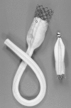
Figure 1. The main graft and the occluder device of the Montefiore Endovascular Grafting System. G, gold bead denoting cranial end of expanded polytetrafluoroethylene graft; P, Palmaz stent; L, ligature.
None
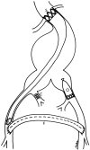
Figure 2. An endovascular graft (EVG) repair of a complex aneurysm repair using the Montefiore Endovascular Grafting System. The cranial end of the graft is fixed within the proximal neck with a large Palmaz stent (S). In this example, the bare portion of the stent is deployed across the orifice of the renal arteries. The distal end of the EVG is secured to the femoral artery by a hand-sewn endoluminal anastomosis (E). The occluder device (O) is deployed in the contralateral common iliac artery to preserve at least one internal iliac artery. C, embolization coils; F, femorofemoral bypass.
None
Figure 3. Tailoring the proximal stent to varying neck diameters by changing the inflation pressure of the balloon. (A) The proximal stent is expanded to 20 mm when the deployment balloon is inflated to 2 atm. (B) At 6 atm of inflation pressure, the stent is expanded to 28 mm.
None
Figure 4. Tailoring the length of the endovascular graft (EVG). (A) The EVG is always made long enough so that its distal end (D) emerges from the femoral arteriotomy site. (B) The distal end of the EVG is cut to appropriate length and a hand-sewn endoluminal anastomosis (E) is performed within the femoral artery. This arteriotomy site is used as the anastomosis site for the following femorofemoral bypass.
None
Figure 5. The endovascular operating room equipped with a portable digital fluoroscope (F). Note the insertion of the brachial wire (B) before induction.
None
Figure 6. A 64-year-old man was found unconscious at home. On arrival in the emergency room, his systolic blood pressure was 40 mmHg, the abdomen was distended, and there was a pulsatile mass. A diagnosis of presumed ruptured aortoiliac aneurysm (AIA) was made, and he was taken to the operating room without obtaining a preoperative computed tomography scan. (A) A brachial wire (B) and subsequently an occlusion balloon (O) were placed in the abdominal aorta under local anesthesia. On inflating the occlusion balloon in the supraceliac aorta, the systolic blood pressure immediately rose to 110 mmHg. Note the curled brachial wire, suggestive of a large AIA. (B) A diagnostic aortogram was obtained with the occlusion balloon inflated. The aortogram confirmed the presence (diagnosis) of an abdominal aortic aneurysm with a well-defined infrarenal neck, fulfilling the inclusion criteria for endovascular graft repair. Note the underfilled visceral and renal arteries resulting from proximal occlusion and lack of prograde flow. (C) After the femoral arteries were exposed and the sheath was inserted into the abdominal aorta, the supraceliac occlusion balloon (O) was deflated and exchanged for an infrarenal occlusion balloon (I) inserted from the femoral artery. (D) An aortogram was repeated by means of the brachial catheter to confirm perfusion of the visceral and renal arteries. (E) Completion angiogram revealed complete exclusion of the large aneurysm with no evidence of an endoleak. P, proximal Palmaz stent. (F) Aneurysm-sac-gram was performed by means of a catheter inserted from the left femoral artery after the deployment of the main graft system. The intrasac pressure measured through the catheter was 30 mmHg; the radial artery pressure was 120 mmHg. The contrast remained in the sac after injection, suggesting lack of an endoleak. (G, H, I) Postoperative contrast-enhanced computed tomography scan. The aneurysm is completely excluded from the circulation and the contrast is confined within the endovascular graft (E). Although this patient underwent evacuation of the hematoma for abdominal compartment syndrome, a considerable amount of hematoma (H) remained in the retroperitoneal space. (J) A limited transabdominal incision was made to evacuate the large hematoma. Five thousand milliliters of blood and clot were evacuated from both the intraperitoneal and retroperitoneal spaces, confirming overt rupture into the peritoneal cavity. The retroperitoneum was opened, and the anterior wall of the aneurysm (A) was exposed and the lack of hemorrhage was confirmed.
None
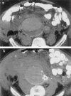
Figure 7. (A) Preoperative computed tomography (CT) scan of a 85-year-old man who came to the emergency room with severe hypotension, syncope, and excruciating abdominal pain. A CT scan revealed a large right common iliac aneurysm and a large retroperitoneal hematoma (H). (B) Postoperative CT scan shows a proximal stent (P) deployed within the proximal neck. The retroperitoneal hematoma (H) was subsequently evacuated through a limited retroperitoneal incision.
None
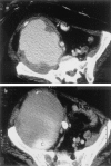
Figure 8. This patient came to the emergency room after developing abdominal pain and experiencing syncope. (A) Preoperative computed tomography (CT) scan showed a large ruptured iliac pseudoaneurysm of unknown cause. (B) Postoperative contrast-enhanced CT scan revealed exclusion of the large aneurysm. The intravenously injected contrast was confined within the endovascular graft (E). C, contrast remaining from intraoperative angiogram. This large hematoma was subsequently evacuated through a limited retroperitoneal incision.

References

    1. Gerbode F. Ruptured abdominal aortic aneurysm: a surgical emergency. Surg Gynecol Obstet 1954; 98: 759–754.
    1. Ernst CB. Abdominal aortic aneurysms. N Engl J Med 1993; 328: 1167–1172. - PubMed
    1. Wakefield TW, Whitehouse WM, Wu SC. Abdominal aortic aneurysm rupture: statistical analysis of factors affecting outcome of surgical treatment. Surgery 1982; 91: 586–595. - PubMed
    1. Donaldson MC, Rosenberg JM, Bucknam CA. Factors affecting survival after ruptured abdominal aortic aneurysm. J Vasc Surg 1985; 2: 564–570. - PubMed
    1. Shackleton CR, Schechter MT, Bianco R, Hildebrand KD. Preoperative predictors of mortality risk in ruptured abdominal aortic aneurysm. J Vasc Surg 1987; 6: 583–589. - PubMed

Publication types

Substances

LinkOut - more resources