

Intrinsic susceptibility of rhesus macaque peripheral CD4(+) T cells to simian immunodeficiency virus in vitro is predictive of in vivo viral replication
- PMID: 11000207
- PMCID: PMC112367
- DOI: 10.1128/jvi.74.20.9388-9395.2000
Intrinsic susceptibility of rhesus macaque peripheral CD4(+) T cells to simian immunodeficiency virus in vitro is predictive of in vivo viral replication
Abstract
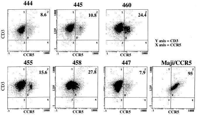
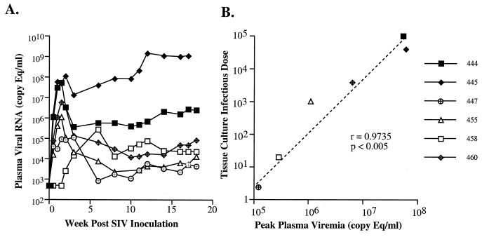
Previous studies with simian immunodeficiency virus (SIV) infection of rhesus macaques suggested that the intrinsic susceptibility of peripheral blood mononuclear cells (PBMC) to infection with SIV in vitro was predictive of relative viremia after SIV challenge. The present study was conducted to evaluate this parameter in a well-characterized cohort of six rhesus macaques selected for marked differences in susceptibility to SIV infection in vitro. Rank order relative susceptibility of PBMC to SIVsmE543-3-infection in vitro was maintained over a 1-year period of evaluation. Differential susceptibility of different donors was maintained in CD8(+) T-cell-depleted PBMC, macrophages, and CD4(+) T-cell lines derived by transformation of PBMC with herpesvirus saimiri, suggesting that this phenomenon is an intrinsic property of CD4(+) target cells. Following intravenous infection of these macaques with SIVsmE543-3, we observed a wide range in plasma viremia which followed the same rank order as the relative susceptibility established by in vitro studies. A significant correlation was observed between plasma viremia at 2 and 8 weeks postinoculation and in vitro susceptibility (P < 0.05). The observation that the two most susceptible macaques were seropositive for simian T-lymphotropic virus type 1 may suggests a role for this viral infection in enhancing susceptibility to SIV infection in vitro and in vivo. In summary, intrinsic susceptibility of CD4(+) target cells appears to be an important factor influencing early virus replication patterns in vivo that should be considered in the design and interpretation of vaccine studies using the SIV/macaque model.
Figures
Similar articles
-
Rhesus macaque resistance to mucosal simian immunodeficiency virus infection is associated with a postentry block in viral replication.J Virol. 2002 Jun;76(12):6016-26. doi: 10.1128/jvi.76.12.6016-6026.2002. J Virol. 2002. PMID: 12021334 Free PMC article.
-
Dynamic Modulation of Expression of Lentiviral Restriction Factors in Primary CD4+ T Cells following Simian Immunodeficiency Virus Infection.J Virol. 2017 Mar 13;91(7):e02189-16. doi: 10.1128/JVI.02189-16. Print 2017 Apr 1. J Virol. 2017. PMID: 28100613 Free PMC article.
-
nef gene is required for robust productive infection by simian immunodeficiency virus of T-cell-rich paracortex in lymph nodes.J Virol. 2003 Apr;77(7):4169-80. doi: 10.1128/jvi.77.7.4169-4180.2003. J Virol. 2003. PMID: 12634375 Free PMC article.
-
Comparison of susceptibility to SIVmac239 infection between CD4(+) and CD4(+)8(+) T cells.Arch Virol. 2005 Aug;150(8):1517-28. doi: 10.1007/s00705-005-0536-7. Epub 2005 Apr 21. Arch Virol. 2005. PMID: 15841338
-
CD4+ CCR5+ T-cell dynamics during simian immunodeficiency virus infection of Chinese rhesus macaques.J Virol. 2007 Dec;81(24):13865-75. doi: 10.1128/JVI.00452-07. Epub 2007 Sep 26. J Virol. 2007. PMID: 17898067 Free PMC article.
Cited by
-
Infectious molecular clones from a simian immunodeficiency virus-infected rapid-progressor (RP) macaque: evidence of differential selection of RP-specific envelope mutations in vitro and in vivo.J Virol. 2006 Feb;80(3):1463-75. doi: 10.1128/JVI.80.3.1463-1475.2006. J Virol. 2006. PMID: 16415023 Free PMC article.
-
The TRIM5 gene modulates penile mucosal acquisition of simian immunodeficiency virus in rhesus monkeys.J Virol. 2011 Oct;85(19):10389-98. doi: 10.1128/JVI.00854-11. Epub 2011 Jul 20. J Virol. 2011. PMID: 21775457 Free PMC article.
-
In vitro characterization of primary SIVsmm isolates belonging to different lineages. In vitro growth on rhesus macaque cells is not predictive for in vivo replication in rhesus macaques.Virology. 2007 Jun 5;362(2):257-70. doi: 10.1016/j.virol.2006.12.037. Epub 2007 Feb 15. Virology. 2007. PMID: 17303205 Free PMC article.
-
Cell surface expression of CCR5 and other host factors influence the inhibition of HIV-1 infection of human lymphocytes by CCR5 ligands.Virology. 2007 Aug 1;364(2):281-90. doi: 10.1016/j.virol.2007.02.022. Epub 2007 Apr 10. Virology. 2007. PMID: 17428518 Free PMC article.
-
Enhanced SIV replication and accelerated progression to AIDS in macaques primed to mount a CD4 T cell response to the SIV envelope protein.Proc Natl Acad Sci U S A. 2004 Aug 31;101(35):13026-31. doi: 10.1073/pnas.0404739101. Epub 2004 Aug 23. Proc Natl Acad Sci U S A. 2004. PMID: 15326293 Free PMC article.
References
-
- Allan J S. Pathogenic properties of simian immunodeficiency viruses in nonhuman primates. Annu Rev AIDS Res. 1991;1:191–206.
-
- Baskin G B, Murphey-Corb M, Watson E A, Martin L N. Necropsy findings in rhesus monkeys experimentally infected with cultured simian immunodeficiency virus (SIV/Delta) Vet Pathol. 1988;25:456–467. - PubMed
-
- Cao Y, Qin L, Zhang L, Safrit J, Ho D. Virologic and immunologic characterization of long-term survivors of human immunodeficiency virus type 1 infection. N Engl J Med. 1995;332:201–208. - PubMed
-
- Carrington M, Nelson G W, Martin M P, Kissner T, Vlahov D, Goedert J J, Kaslow R, Buchbinder S, Hoots K, O'Brien S J. HLA and HIV-1: heterozygote advantage and B*35-Cw*04 disadvantage. Science. 1999;283:1748–1752. - PubMed
Publication types
MeSH terms
Substances
Grants and funding
LinkOut - more resources
Full Text Sources
Research Materials
