

Adeno-associated virus type 2 Rep78 induces apoptosis through caspase activation independently of p53
- PMID: 11000213
- PMCID: PMC112373
- DOI: 10.1128/jvi.74.20.9441-9450.2000
Adeno-associated virus type 2 Rep78 induces apoptosis through caspase activation independently of p53
Abstract
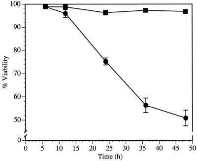
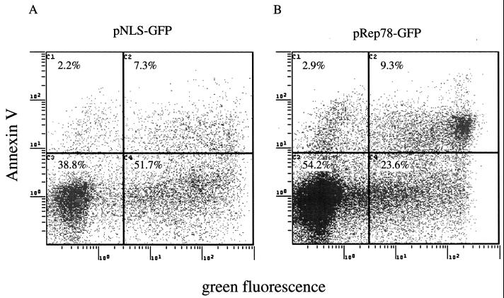
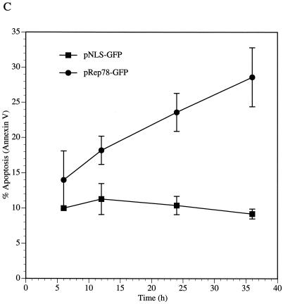
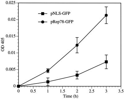
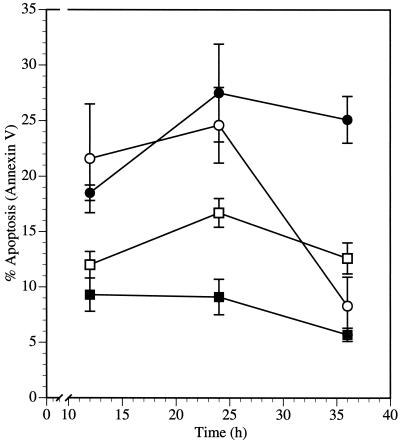
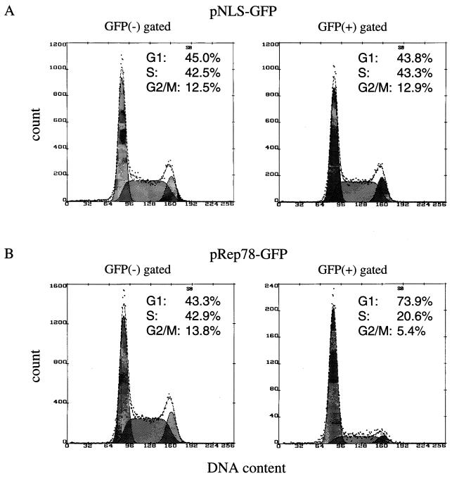
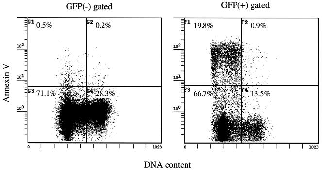
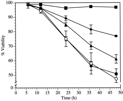
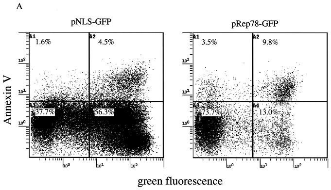
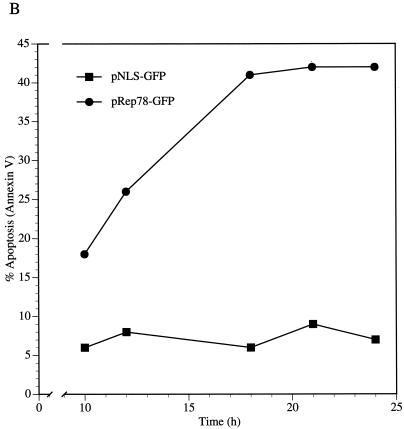
Adeno-associated virus (AAV) type 2 Rep78 is a multifunctional protein required for AAV DNA replication, integration, and gene regulation. The biochemical activities of Rep78 have been described, but the effects of Rep proteins on the cell have not been characterized. We have analyzed Rep-mediated cytotoxicity. We demonstrated that Rep78 expression is sufficient to induce cell death and disruption of the cell cycle. Cell death was found to be mediated by apoptosis. Rep78 expression resulted in the activation of caspase-3, a terminal caspase directly involved in the execution of cell death. A peptidic inhibitor of caspase-3, Z-Asp-Glu-Val-Asp-fluoromethylketone (Z-DEVD-FMK), abrogated Rep78-induced apoptosis, indicating that Rep78-mediated apoptosis is caspase-3 dependent. Rep78 induced apoptosis in wild-type p53-containing human embryonal carcinoma NT-2 cells and in p53-null promyelocytic human HL-60 cells, indicating that at least one pathway of Rep78-induced apoptosis is p53 independent. Apoptosis was shown to occur during the G(1) and early S phases of the cell cycle. By analyzing the effects of Rep78 mutations on cell viability, the cause of cell death was attributed in part to two biochemical activities of Rep78, DNA binding and ATPase/helicase activity. The endonuclease activity of Rep78 did not contribute to apoptosis induction.
Figures
Similar articles
-
Adeno-associated virus type 2 infection activates caspase dependent and independent apoptosis in multiple breast cancer lines but not in normal mammary epithelial cells.Mol Cancer. 2011 Aug 9;10:97. doi: 10.1186/1476-4598-10-97. Mol Cancer. 2011. PMID: 21827643 Free PMC article.
-
Roles of adeno-associated virus Rep protein and human chromosome 19 in site-specific recombination.J Virol. 2000 May;74(9):3953-66. doi: 10.1128/jvi.74.9.3953-3966.2000. J Virol. 2000. PMID: 10756007 Free PMC article.
-
Charge-to-alanine mutagenesis of the adeno-associated virus type 2 Rep78/68 proteins yields temperature-sensitive and magnesium-dependent variants.J Virol. 1999 Nov;73(11):9433-45. doi: 10.1128/JVI.73.11.9433-9445.1999. J Virol. 1999. PMID: 10516052 Free PMC article.
-
p53, caspase 8, and regulation of apoptosis after ionizing radiation.J Pediatr Hematol Oncol. 2001 Mar-Apr;23(3):185-8. doi: 10.1097/00043426-200103000-00014. J Pediatr Hematol Oncol. 2001. PMID: 11305724 Review. No abstract available.
-
Regulation of apoptosis by viral gene products.J Virol. 1997 Mar;71(3):1739-46. doi: 10.1128/JVI.71.3.1739-1746.1997. J Virol. 1997. PMID: 9032302 Free PMC article. Review. No abstract available.
Cited by
-
Identification of rep-associated factors in herpes simplex virus type 1-induced adeno-associated virus type 2 replication compartments.J Virol. 2010 Sep;84(17):8871-87. doi: 10.1128/JVI.00725-10. Epub 2010 Jun 23. J Virol. 2010. PMID: 20573815 Free PMC article.
-
Targeting site-specific chromosome integration.Acta Biochim Pol. 2005;52(2):285-91. Epub 2005 Jun 3. Acta Biochim Pol. 2005. PMID: 15940345 Free PMC article. Review.
-
Adeno-associated virus type 2 infection activates caspase dependent and independent apoptosis in multiple breast cancer lines but not in normal mammary epithelial cells.Mol Cancer. 2011 Aug 9;10:97. doi: 10.1186/1476-4598-10-97. Mol Cancer. 2011. PMID: 21827643 Free PMC article.
-
Intensification of rAAV Production Based on HEK293 Cell Transient Transfection.Biotechnol J. 2025 Jun;20(6):e70020. doi: 10.1002/biot.70020. Biotechnol J. 2025. PMID: 40491022 Free PMC article.
-
Through its nonstructural protein NS1, parvovirus H-1 induces apoptosis via accumulation of reactive oxygen species.J Virol. 2010 Jun;84(12):5909-22. doi: 10.1128/JVI.01797-09. Epub 2010 Apr 7. J Virol. 2010. PMID: 20375165 Free PMC article.
References
-
- Bagchi D, Joshi S S, Bagchi M, Balmoori J, Benner E J, Kuszynski C A, Stohs S J. Cadmium- and chromium-induced oxidative stress, DNA damage, and apoptotic cell death in cultured human chronic myelogenous leukemic K562 cells, promyelocytic leukemic HL-60 cells, and normal human peripheral blood mononuclear cells. J Biochem Mol Toxicol. 2000;14:33–41. - PubMed
-
- Banki K, Hutter E, Gonchoroff N J, Perl A. Molecular ordering in HIV-induced apoptosis. Oxidative stress, activation of caspases, and cell survival are regulated by transaldolase. J Biol Chem. 1998;273:11944–11953. - PubMed
-
- Barry M, McFadden G. Apoptosis regulators from DNA viruses. Curr Opin Immunol. 1998;10:422–430. - PubMed
-
- Batchu R B, Shammas M A, Wang J Y, Munshi N C. Interaction of adeno-associated virus Rep78 with p53: implications in growth inhibition. Cancer Res. 1999;59:3592–3595. - PubMed
MeSH terms
Substances
LinkOut - more resources
Full Text Sources
Other Literature Sources
Research Materials
Miscellaneous
