. 2000 Oct;74(20):9471-8.
doi: 10.1128/jvi.74.20.9471-9478.2000.

The binding site of transcription factor YY1 is required for intramolecular recombination between terminally repeated sequences of linear replicative hepatitis B virus DNA

Affiliations

The binding site of transcription factor YY1 is required for intramolecular recombination between terminally repeated sequences of linear replicative hepatitis B virus DNA

Y Hayashi et al. J Virol. 2000 Oct.

Abstract

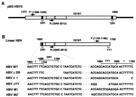
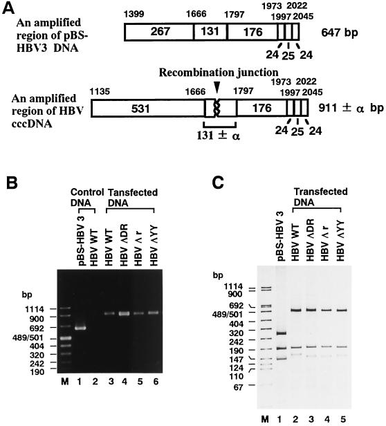
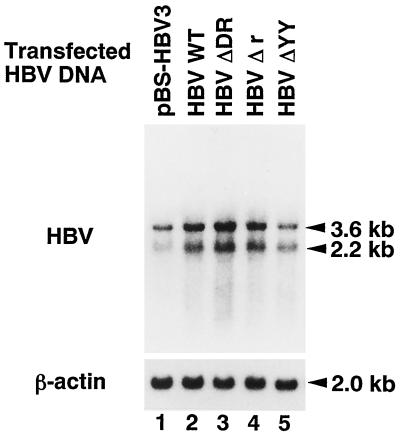
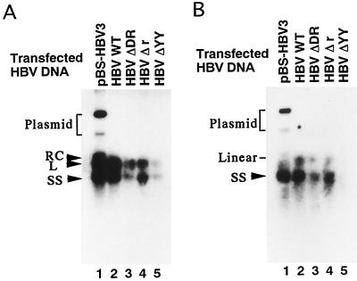
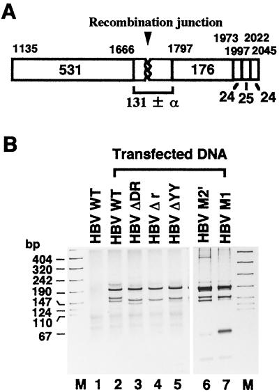
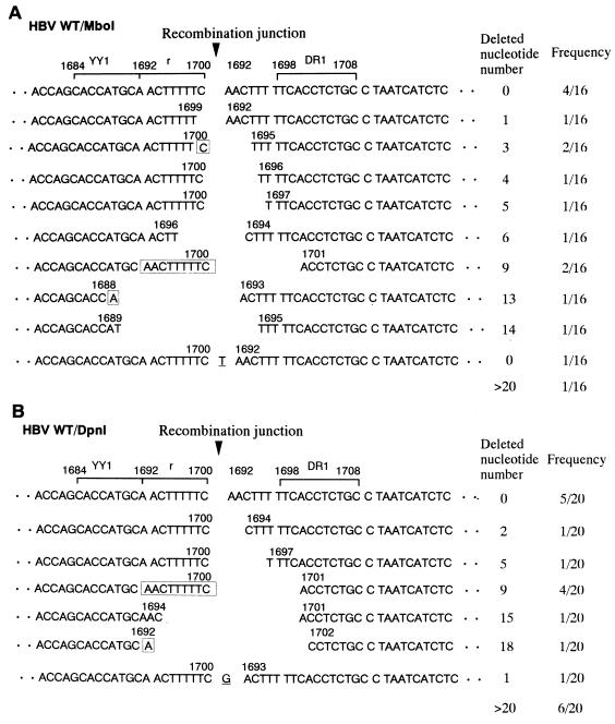
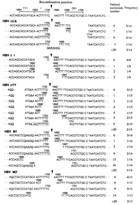
In the replication cycle of hepadnavirus DNA, the double-stranded linear form of viral DNA is generated as a minor replicative intermediate, which is efficiently converted to covalently closed circular DNA (cccDNA) by intramolecular recombination (W. Yang and J. Summers, J. Virol. 69:4029-4036, 1995). We previously found a binding site of transcription factor Yin and Yang 1 (YY1) in one terminal region of the double-stranded linear replicative hepatitis B virus (HBV) DNA (M. Nakanishi-Matsui, Y. Hayashi, Y. Kitamura, and K. Koike, J. Virol. 74:5562-5568, 2000). However, it is not known whether the YY1-binding site is required for the intramolecular recombination of HBV DNA. In this study, we established an HBV-producing system in which the cccDNA appeared to be generated from the transfected linear DNA or the linear replicative DNA by nonhomologous end joining (NHEJ) or by both NHEJ and homologous recombination between terminally repeated sequences, respectively. When the YY1-binding site in the terminal region of transfected linear viral DNA was mutated, the cccDNA was generated merely by NHEJ. Results suggest that the YY1-binding site in the terminal region of linear replicative HBV DNA is required for intramolecular recombination between terminally repeated sequences.


Figures

FIG. 1
FIG. 1
Schematic representation of pBS-HBV3 and linear HBV DNA. (A) Schematic representation of pBS-HBV3 DNA. The open rectangles show the inserted whole HBV genome (3,215 bp) with the 584-bp overlapping DNA region (nt 1275 to 1858). Numbers above the diagram show the nucleotide numbers of the HBV subtype adr sequence (6). The shaded regions indicate DR1 (nt 1698 to 1708). C indicates the open reading frame of HBcAg. The arrows indicate the locations of the PCR primers F* (nt 1399 to 1429) and R (nt 2045 to 2013). The solid bars indicate the vector sequence. (B) Schematic representation of linear HBV DNA. Numbers and a region referred as C are shown as in panel A. Arrows indicate the locations of the PCR primers F (nt 1135 to 1166) and R. The DNA sequences of both terminal regions of WT HBV, HBV ΔDR, HBV Δr, HBV ΔYY, HBV M1, and HBV M2′ are enlarged; the deleted sequences are missing, the mutated sequences are underlined, and the dots indicate the common internal HBV DNA sequence. The terminally repeated r sequence (nt 1692 to 1700), the YY1-binding site (nt 1684 to 1692), and DR1 are also mapped. Note that the promoter region for the 3.6-kb mRNA is located at the terminal region distant from the open reading frame of HBcAg (2).
FIG. 2
FIG. 2
Detection of the recombination junction in MboI-resistant cccDNA. (A) Schematic representation of PCR products (911 ± α bp: nt 1135 to 2045), containing the recombination junction (arrowhead), amplified from MboI-resistant cccDNA with the F and R primers. The DNA region (647 bp; nt 1399 to 2045) amplified from pBS-HBV3 DNA, using the F* and R primers, is also shown on the top. The locations of the three primers are shown in Fig. 1. Numbers above the open rectangles show the nucleotide numbers of Tsp509I recognition sites in the HBV DNA sequence (6), and the numbers in or under the open rectangles indicate the sizes (in base pairs) of Tsp509I-digested DNA fragments. (B) Detection of PCR products. Shown are PCR products generated by 2% agarose gel electrophoresis (lanes 3 to 6) from MboI-resistant cccDNA in cells transfected with WT HBV, HBV ΔDR, HBV Δr, or HBV ΔYY (Fig. 1B). Products generated with pBS-HBV3 (lane 1) and WT HBV DNA (lane 2) by the same treatment are also shown as controls. Lane M and numbers at the left indicate the size markers in base pairs. (C) Detection of the DNA fragment containing the recombination junction. The PCR products obtained in panel B were digested with Tsp509I and subjected to 15 to 25% gradient polyacrylamide gel electrophoresis. Lanes 2 to 5 contain the samples from MboI-resistant cccDNA in WT-HBV-, HBV ΔDR-, HBV Δr-, and HBV ΔYY-transfected cells, respectively. The PCR product generated from pBS-HBV3 DNA by the same treatment was also loaded as the size marker control (lane 1).
FIG. 3
FIG. 3
Northern blot analysis of HBV mRNA. Total RNA obtained from the cells transfected with pBS-HBV3, WT HBV, HBV ΔDR, HBV Δr, or HBV ΔYY (lanes 1 to 5, respectively) was subjected to 1% agarose gel electrophoresis, transferred to nitrocellulose filter paper, and then hybridized with an HBV DNA probe. Two major transcripts (3.6 and 2.2 kb) were detected. The filter paper was rehybridized with a β-actin DNA probe to show the 2.0-kb transcript.
FIG. 4
FIG. 4
Southern blot analysis of HBV DNA in viral or core particles. (A) HBV DNA in viral particles. HBV particles secreted into the culture medium of the cells transfected with pBS-HBV3, WT HBV, HBV ΔDR, HBV Δr, or HBV ΔYY (lanes 1 to 5) were treated with 1 mg of proteinase K per ml and 1% SDS and then directly subjected to 1% agarose gel electrophoresis. The resultant DNA was blotted to the filter paper and hybridized with an HBV DNA probe. Arrowheads indicate the positions corresponding to three different forms of HBV DNA (RC, L, and SS) and the bracket shows the position of transfected plasmid DNA. (B) HBV DNA in core particles was treated as described for panel A. Lanes 1 to 5 contain the samples from pBS-HBV3-, WT-HBV-, HBV ΔDR-, HBV Δr-, and HBV ΔYY-transfected cells, respectively. The arrowhead indicates the position of the SS form of HBV DNA. The positions of the transfected plasmid and linear DNA are also indicated.
FIG. 5
FIG. 5
Detection of the recombination junction in DpnI-resistant cccDNA. (A) Schematic representation of the PCR product (nt 1135 to 2045) amplified from DpnI-resistant cccDNA with the F and R primers. Numbers above the open rectangles show the Tsp509I recognition sites in the HBV DNA sequence (6), and the numbers in or under the open rectangles indicate the sizes (in base pairs) of Tsp509I-digested fragments. (B) Detection of the DNA fragment containing the recombination junction. PCR products amplified from DpnI-resistant cccDNA in the cells transfected with WT HBV, HBV ΔDR, HBV Δr, HBV ΔYY, HBV M2′, or HBV M1 (lanes 2 to 7, respectively) were digested with Tsp509I and subjected to 15 to 25% gradient polyacrylamide gel electrophoresis. The bands that migrated around 176 and 131 bp are shown, but the 531-bp band is omitted. The PCR product generated from WT HBV DNA (lane 1) by the same treatment is shown as a control. Lane M and the numbers at the left indicate the size markers in base pairs.
FIG. 6
FIG. 6
Sequence alignment of the recombination junction of cccDNA formed in WT-HBV-transfected cells. (A) Sequence alignment of the recombination junction of MboI-resistant cccDNA. The 16 junction clones obtained were sequenced and aligned in order of the number of nucleotides deleted. When exactly the same sequence was obtained, the numbers of clones are written in the frequency column. The sequence with more than 20 nt deleted is omitted. The numbers above the DNA sequences correspond to the nucleotide numbers of the HBV DNA sequence (6). The possible nucleotide sequence involved in the joining of two terminal regions of viral DNA is boxed. Underlined nucleotides are not derived from the HBV DNA sequence. The YY1-binding site (nt 1684 to 1692), the r sequence (nt 1692 to 1700), and DR1 (nt 1698 to 1708) are indicated with brackets. The arrowhead indicates the recombination junction. (B) Sequence alignment of the recombination junction of DpnI-resistant cccDNA. The 20 junction clones obtained were sequenced and aligned as described for panel A.
FIG. 7
FIG. 7
Sequence alignment of the recombination junctions of DpnI-resistant cccDNAs formed in cells transfected with HBV ΔDR, HBV Δr, HBV ΔYY, HBV M1, and HBV M2′. The 14, 9, 15, 13, or 14 junction clones of DpnI-resistant cccDNA obtained in HBV ΔDR-, HBV Δr-, HBV ΔYY, HBV M1-, or HBV M2′-transfected cells, respectively, were sequenced and aligned as described for Fig. 6.

Similar articles

Cited by

References

    1. Chang C, Jeng K-S, Hu C-P, Lo S J, Su T-S, Ting L-P, Chou C-K, Han S-H, Pfaff E, Salfeld J, Schaller H. Production of hepatitis B virus in vitro by transient expression of cloned HBV DNA in a hepatoma cell line. EMBO J. 1987;6:675–680. - PMC - PubMed
    1. Ganem D. Hepadnaviridae and their replication. In: Fields B N, Knipe P M, Howley P M, editors. Fields virology. Philadelphia, Pa: Lippincott-Raven Publishers; 1996. pp. 2703–2737.
    1. Havert M B, Loeb D D. cis-acting sequences in addition to donor and acceptor sites are required for template switching during synthesis of plus-strand DNA for duck hepatitis B virus. J Virol. 1997;71:5336–5344. - PMC - PubMed
    1. Hayashi Y, Koike K. Interferon inhibits hepatitis B virus replication in a stable expression system of transfected viral DNA. J Virol. 1989;63:2936–2940. - PMC - PubMed
    1. Knowles B B, Howe C C, Aden D P. Human hepatocellular carcinoma cell lines secrete the major protein and hepatitis B surface antigen. Science. 1980;209:497–499. - PubMed

LinkOut - more resources