. 2000 Oct;74(20):9553-61.
doi: 10.1128/jvi.74.20.9553-9561.2000.

Passage of classical swine fever virus in cultured swine kidney cells selects virus variants that bind to heparan sulfate due to a single amino acid change in envelope protein E(rns)

Affiliations

Passage of classical swine fever virus in cultured swine kidney cells selects virus variants that bind to heparan sulfate due to a single amino acid change in envelope protein E(rns)

M M Hulst et al. J Virol. 2000 Oct.

Abstract

Infection of cells with Classical swine fever virus (CSFV) is mediated by the interaction of envelope glycoprotein E(rns) and E2 with the cell surface. In this report we studied the role of the cell surface glycoaminoglycans (GAGs), chondroitin sulfates A, B, and C (CS-A, -B, and -C), and heparan sulfate (HS) in the initial binding of CSFV strain Brescia to cells. Removal of HS from the surface of swine kidney cells (SK6) by heparinase I treatment almost completely abolished infection of these cells with virus that was extensively passaged in swine kidney cells before it was cloned (clone C1.1.1). Infection with C1.1.1 was inhibited completely by heparin (a GAG chemically related to HS but sulfated to a higher extent) and by dextran sulfate (an artificial highly sulfated polysaccharide), whereas HS and CS-A, -B, and -C were unable to inhibit infection. Bound C1.1.1 virus particles were released from the cell surface by treatment with heparin. Furthermore, C1.1.1 virus particles and CSFV E(rns) purified from insect cells bound to immobilized heparin, whereas purified CSFV E2 did not. These results indicate that initial binding of this virus clone is accomplished by the interaction of E(rns) with cell surface HS. In contrast, infection of SK6 cells with virus clones isolated from the blood of an infected pig and minimally passaged in SK6 cells was not affected by heparinase I treatment of cells and the addition of heparin to the medium. However, after one additional round of amplification in SK6 cells, infection with these virus clones was affected by heparinase I treatment and heparin. Sequence analysis of the E(rns) genes of these virus clones before and after amplification in SK6 cells showed that passage in SK6 cells resulted in a change of an Ser residue to an Arg residue in the C terminus of E(rns) (amino acid 476 in the polyprotein of CSFV). Replacement of the E(rns) gene of an infectious DNA copy of C1.1.1 with the E(rns) genes of these virus variants proved that acquisition of this Arg was sufficient to alter an HS-independent virus to a virus that uses HS as an E(rns) receptor.


Figures

FIG. 1
FIG. 1
Inhibition of infection of SK6 cells with CSFV by GAGs. The numbers of plaques in 2-cm2 tissue culture wells were measured in a plaque assay; SK6 cells were preincubated for 30 min with 100 μl of medium with different concentrations of GAGs. Subsequently, 100 μl of a virus dilution containing about 1,000 PFU of CSFV strain Brescia clone C1.1.1 was added to the wells. The cells were incubated for 30 min, washed, and supplied with overlay medium. After incubation for 24 h the cells were immunostained and the plaques were counted. The x axis represents the concentration of GAGs present during the 30 min of virus adsorption (a twofold-lower concentration of GAGs than in the preincubation solution). Plot symbols represent the mean of two independent observations, and the error bars represent the variation between these two observations.
FIG. 2
FIG. 2
Removal of GAGs from the cell surface. SK6 cells grown in 2-cm2 tissue culture wells were digested for 2 h at 20°C with different concentrations of heparinase I or chondroitinase ABC. After the enzymes were removed from the cells, wells were infected for 30 min at 37°C with 500 PFU of CSFV strain Brescia clone C1.1.1. After infection the virus was removed, and the cells were washed, supplied with overlay medium, and further treated as described in the legends to Fig. 1. Plot symbols represent the mean of two independent observations, and the error bars represent the variation between these two observations.
FIG. 3
FIG. 3
Heparin inhibits infection by cell-bound virus particles. Six 2-cm2 tissue culture wells with SK6 cells were infected with about 1,200 PFU of CSFV strain Brescia clone C1.1.1 per well at 4°C. After 30 min of infection, the virus was removed and the cells were washed twice with medium. Subsequently, at 4°C, three wells were chased with 100 μl of EMEM (V) and three wells were chased with 100 μl of EMEM containing 100 μg of heparin per ml (H). After incubation for 30 min, chase media were collected and cells were washed twice with EMEM. Then, 200 μl of fresh EMEM was added, and the cells were incubated for 1 h at 37°C. The medium was removed, and the wells were supplied with overlay medium. After incubation for 24 h at 37°C, the wells were immunostained and plaques were counted. Open bars represent the average number of plaques of three wells. After 10-fold dilution in EMEM supplemented with 10% FBS, the collected chase media were used to infect 2-cm2 tissue culture wells. After 90 min of infection, the virus was removed and overlay medium was added. After 24 h of growth, wells were stained and plaques were counted (shaded bars, average of three observations). The error bars represent the standard deviation (n − 1).
FIG. 4
FIG. 4
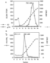
(A) Heparin-Sepharose chromatography of Erns and E2. In a separate experiment, purified Erns or E2 was loaded on the column at a concentration of 0 mM NaCl. Proteins were eluted with a stepwise NaCl gradient (0 to 1,000 mM). The fractions were assayed for Erns or E2 in an ELISA as described in Materials and Methods. The results of Erns and E2 are presented in a single graph. The NaCl concentration is shown for the Erns fractions. The concentration NaCl of the E2 fractions did not differ significantly from the concentration of the corresponding Erns fractions. (B) Heparin-Sepharose chromatography of CSFV strain Brescia C1.1.1. Cell-free, partially purified virus was loaded on the column at a concentration of 100 mM NaCl and eluted with a stepwise NaCl gradient of 100 to 1,000 mM. Fractions were assayed for virus in a plaque assay as described in Materials and Methods. The NaCl concentration of fractions was determined by measuring the osmolarity.
FIG. 5
FIG. 5
Characterization of virus clones. The percent inhibition of infection of SK6 cells by 200 μg of heparin per ml and the percentage reduction of infection after treatment of SK6 cells with 12.5 mIU of heparinase I per ml, as measured in a plaque assay as described in the legends of Fig. 1 and Fig. 2 respectively, is shown. In these experiments, wells were infected with about 200 PFU of virus. Each bar is the mean of two independent observations. < and >, relative plaque size of virus clones observed in wells to which no heparin was added (control wells) after 2 days of growth under methyl cellulose.
FIG. 6
FIG. 6
Erns amino acid sequence of Brescia virus clones with different passage numbers. Differences compared to the published sequence of Erns of Brescia clone C1.1.1 (29) are listed. Identical amino acid sequences were obtained for clones Ap3, Bp2, and Ep2 and for clones Ap4, Bp3, and Ep3. RNase domains are underlined (16, 36).

References

    1. Anonymous. Current protocols in molecular biology: a laboratory manual. 3, suppl. 27. New York, N.Y: John Wiley & Sons, Inc.; 1994.
    1. Becher P, Shannon A D, Tautz N, Thiel H-J. Molecular characterization of border disease virus, a pestivirus from sheep. Virology. 1994;198:542–551. - PubMed
    1. Bruschke C J, Hulst M M, Moormann R J M, van Rijn P A, van Oirschot J T. Glycoprotein Erns of pestiviruses induces apoptosis in lymphocytes of several species. J Virol. 1997;71:6692–6696. - PMC - PubMed
    1. Byrnes A P, Griffin D E. Binding of Sindbis virus to cell surface heparan sulfate. J Virol. 1998;72:7349–7356. - PMC - PubMed
    1. Carbrey E A, Stewart W C, Kresse J L, Snijder M L. Natural infection of pigs with bovine diarrhoea virus and its differential diagnosis from hog cholera. J Am Vet Med Assoc. 1976;169:1217–1219. - PubMed

LinkOut - more resources