

Three-dimensional structure of the human herpesvirus 8 capsid
- PMID: 11000237
- PMCID: PMC112397
- DOI: 10.1128/jvi.74.20.9646-9654.2000
Three-dimensional structure of the human herpesvirus 8 capsid
Abstract
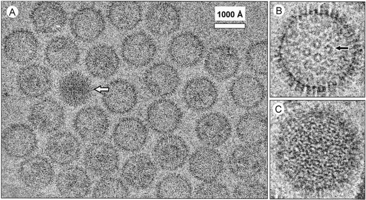
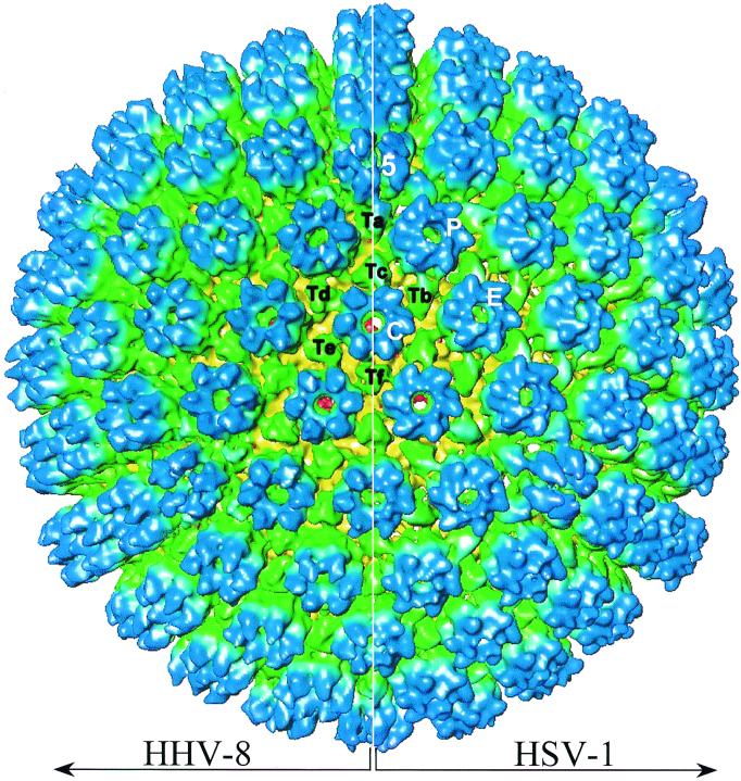
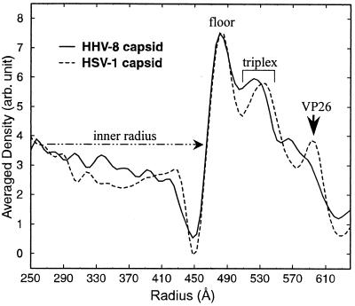
Human herpesvirus 8 (HHV-8), or Kaposi's sarcoma-associated herpesvirus, is a gammaherpesvirus implicated in all forms of Kaposi's sarcoma and certain lymphomas. HHV-8 has been extensively characterized, both biochemically and immunologically, since its first description in 1994. However, its three-dimensional (3D) structure remained heretofore undetermined largely due to difficulties in viral purification. We have used log-phase cultures of body cavity-based lymphoma 1 cells induced with 12-O-tetradecanoylphorbol-13-acetate to obtain HHV-8 capsids for electron cryomicroscopy and computer reconstruction. The 3D structure of the HHV-8 capsids revealed a capsid shell composed of 12 pentons, 150 hexons, and 320 triplexes arranged on a T=16 icosahedral lattice. This structure is similar to those of herpes simplex virus type 1 (HSV-1) and human cytomegalovirus (HCMV), which are prototypical members of alpha- and betaherpesviruses, respectively. The inner radius of the HHV-8 capsid is identical to that of the HSV-1 capsid but is smaller than that of the HCMV capsid, which is consistent with the relative sizes of the genomes they enclose. While the HHV-8 capsid exhibits many structural similarities to the HSV-1 capsid, our reconstruction shows two major differences: its hexons lack the "horn-shaped" VP26 densities bound to the HSV-1 hexon subunits, and the HHV-8 triplexes appear smaller and less elongated than those of HSV-1. These differences are in excellent agreement with our sequence comparisons of HHV-8 and HSV-1 capsid proteins. This gammaherpesvirus capsid structure complements previous structural studies on alpha- and betaherpesviruses in providing an account of structural similarities and differences among capsids representing all human herpesvirus subfamilies.
Figures


Similar articles
-
Lytic replication of Kaposi's sarcoma-associated herpesvirus results in the formation of multiple capsid species: isolation and molecular characterization of A, B, and C capsids from a gammaherpesvirus.J Virol. 2001 Mar;75(6):2866-78. doi: 10.1128/JVI.75.6.2866-2878.2001. J Virol. 2001. PMID: 11222712 Free PMC article.
-
Three-dimensional localization of pORF65 in Kaposi's sarcoma-associated herpesvirus capsid.J Virol. 2003 Apr;77(7):4291-7. doi: 10.1128/jvi.77.7.4291-4297.2003. J Virol. 2003. PMID: 12634386 Free PMC article.
-
Structure of the human cytomegalovirus B capsid by electron cryomicroscopy and image reconstruction.J Struct Biol. 1998 Dec 1;124(1):70-6. doi: 10.1006/jsbi.1998.4055. J Struct Biol. 1998. PMID: 9931275
-
Herpesvirus Capsid Assembly and DNA Packaging.Adv Anat Embryol Cell Biol. 2017;223:119-142. doi: 10.1007/978-3-319-53168-7_6. Adv Anat Embryol Cell Biol. 2017. PMID: 28528442 Free PMC article. Review.
-
Human herpes virus 8 (Kaposi's sarcoma herpesvirus).Oral Oncol. 1998 Jan;34(1):5-14. doi: 10.1016/s1368-8375(97)00038-9. Oral Oncol. 1998. PMID: 9659514 Review.
Cited by
-
Handedness of the herpes simplex virus capsid and procapsid.J Virol. 2002 Aug;76(15):7855-9. doi: 10.1128/jvi.76.15.7855-7859.2002. J Virol. 2002. PMID: 12097597 Free PMC article.
-
Virion-wide protein interactions of Kaposi's sarcoma-associated herpesvirus.J Virol. 2008 May;82(10):4742-50. doi: 10.1128/JVI.02745-07. Epub 2008 Mar 5. J Virol. 2008. PMID: 18321973 Free PMC article.
-
Unique structures in a tumor herpesvirus revealed by cryo-electron tomography and microscopy.J Struct Biol. 2008 Mar;161(3):428-38. doi: 10.1016/j.jsb.2007.10.010. Epub 2007 Nov 20. J Struct Biol. 2008. PMID: 18096403 Free PMC article.
-
Molecular biology of KSHV in relation to AIDS-associated oncogenesis.Cancer Treat Res. 2007;133:69-127. doi: 10.1007/978-0-387-46816-7_3. Cancer Treat Res. 2007. PMID: 17672038 Free PMC article. Review.
-
Mass spectrometric analyses of purified rhesus monkey rhadinovirus reveal 33 virion-associated proteins.J Virol. 2006 Feb;80(3):1574-83. doi: 10.1128/JVI.80.3.1574-1583.2006. J Virol. 2006. PMID: 16415032 Free PMC article.
References
-
- Arvanitakis L, Geras-Raaka E, Varma A, Gershengorn M C, Cesarman E. Human herpesvirus KSHV encodes a constitutively active G-protein-coupled receptor linked to cell proliferation. Nature. 1997;385:347–350. - PubMed
-
- Bhella D, Rixon F J, Dargan D J. Cryomicroscopy of human cytomegalovirus virions reveals more densely packed genomic DNA than in herpes simplex virus type 1. J Mol Biol. 2000;295:155–161. - PubMed
Publication types
MeSH terms
Grants and funding
LinkOut - more resources
Full Text Sources
