. 2000 Oct;74(20):9655-67.
doi: 10.1128/jvi.74.20.9655-9667.2000.

Internalization of adenovirus by alveolar macrophages initiates early proinflammatory signaling during acute respiratory tract infection

Affiliations

Internalization of adenovirus by alveolar macrophages initiates early proinflammatory signaling during acute respiratory tract infection

Z Zsengellér et al. J Virol. 2000 Oct.

Abstract

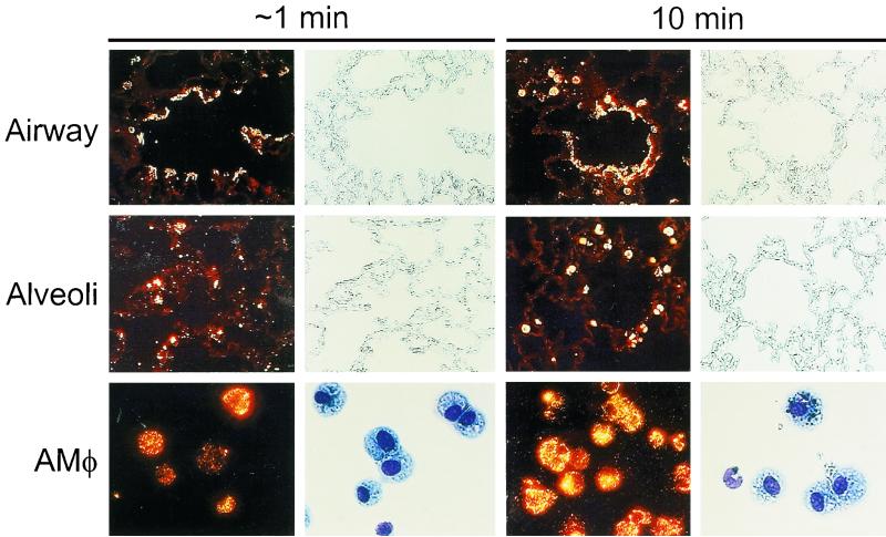
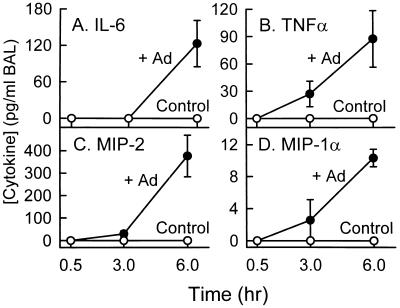
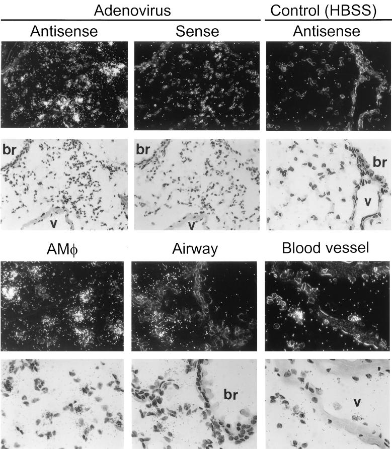
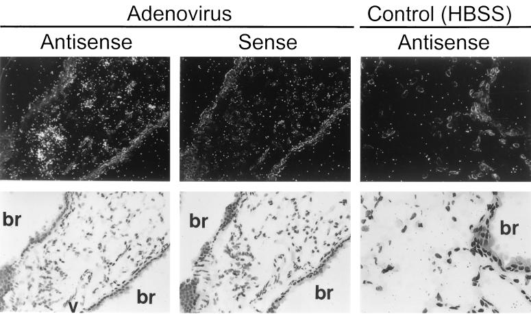
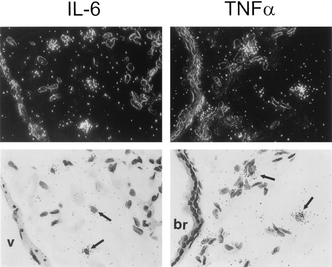
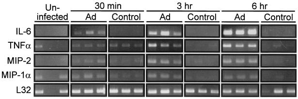
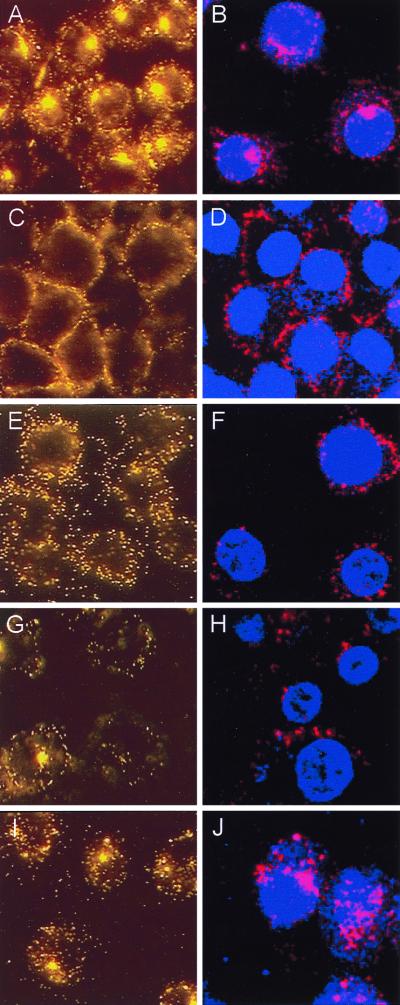
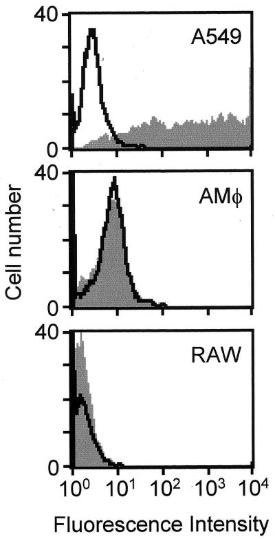
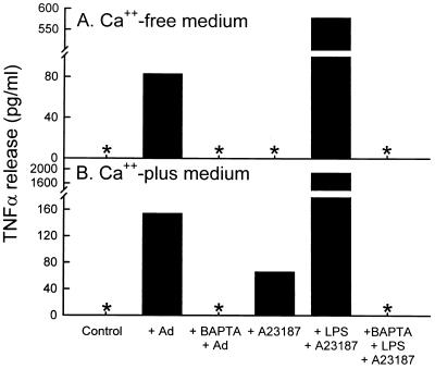
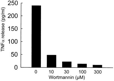
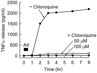
Adenovirus is a common respiratory pathogen which causes a broad range of distinct clinical syndromes and has recently received attention for its potential for in vivo gene delivery. Although adenovirus respiratory tract infection (ARTI) results in dose-dependent, local inflammation, the pathogenesis of this remains unclear. We hypothesized that alveolar macrophages (AMphi) rapidly internalize adenovirus following in vivo pulmonary administration and then initiate inflammatory signaling within the lung. To evaluate the role of AMphi in the induction of lung inflammation during ARTI in vivo, we directly assessed adenovirus uptake by murine AMphi and correlated uptake with the initiation of proinflammatory gene expression. Stimulation of cytokine (tumor necrosis factor alpha [TNF-alpha], interleukin-6 [IL-6], macrophage inflammatory protein-2 [MIP-2], and MIP-1alpha) expression in the lung was evaluated at the level of mRNA (by reverse transcription-PCR [RT-PCR]) and protein (by enzyme-linked immunosorbent assay) and by identification of cells expressing TNF-alpha and IL-6 mRNA in lung tissues (by in situ hybridization) and isolated lung lavage cells (by RT-PCR). Adenovirus, labeled with the fluorescent dye (Cy3), was rapidly and widely distributed on epithelial surfaces of airways and alveoli and was very rapidly ( approximately 1 min) localized within AMphi. At 30 min after infection AMphi but not airway epithelial or vascular endothelial cells expressed mRNA for TNF-alpha and IL-6, thus identifying AMphi as the cell source of initial cytokine signaling. IL-6, TNF-alpha, MIP-2, and MIP-1alpha levels progressively increased in bronchoalveolar lavage fluid after pulmonary adenovirus infection, and all were significantly elevated at 6 h (P < 0.05). To begin to define the molecular mechanism(s) by which adenovirus initiates the inflammatory signaling in macrophages, TNF-alpha expression from adenovirus-infected RAW264.7 macrophages was evaluated in vitro. TNF-alpha expression was readily detected in adenovirus-infected RAW cell supernatant with kinetics similar to AMphi during in vivo infection. Blockage of virus uptake at specific cellular sites, including internalization (by wortmannin), endosome acidification and/or lysis (by chloroquine) or by Ca(2+) chelation (by BAPTA) completely blocked TNF-alpha expression. In conclusion, results showed that during ARTI, (i) AMphi rapidly internalized adenovirus, (ii) expression of inflammatory mediators was initiated within AMphi and not airway epithelial or other cells, and (iii) the initiation of inflammatory signaling was linked to virion uptake by macrophages occurring at a point after vesicle acidification. These results have implications for our understanding of the role of the AMphi in the initiation of inflammation following adenovirus infection and adenovirus-mediated gene transfer to the lung.


Figures

FIG. 1
FIG. 1
Distribution of adenovirus during acute respiratory tract infection. Infectious, fluorescently labeled adenovirus (Ad-Cy3) or HBSS, as a sham control, was administered by intratracheal instillation into the lungs of mice. Mice were then sacrificed after either 1 min (left panels) or 10 min (right panels), and the lungs were removed and processed for tissue sections or the mice were subjected to BAL, followed by recovery and cytospin preparation of cells as described in Materials and Methods. Shown are fluorescence and corresponding phase photomicrographs for tissue sections (top and middle panels, ×114) and fluorescence and bright-field photomicrographs for BAL cells (bottom panels, ×232). For BAL cells, separate slides were prepared for fluorescence and bright-field photomicroscopy because Diff-Quick staining partially quenched Cy3 fluorescence.
FIG. 2
FIG. 2
Stimulation of proinflammatory cytokine and chemotactic chemokine levels in lung during acute adenovirus respiratory tract infection. Infectious adenovirus (+Ad) or HBSS (Control) was administered by intratracheal instillation into the lungs of mice (n = 3/time point). At the subsequent times indicated, mice were sacrificed and the lung epithelial lining fluid was recovered by BAL. Proinflammatory cytokine (IL-6 and TNFα) and chemoattractive chemokine (MIP-2 and MIP-1α) levels were measured in unconcentrated BAL fluid by ELISA. The entire experiment was performed twice, and representative data from one experiment are shown.
FIG. 3
FIG. 3
Localization of IL-6 mRNA expression by in situ hybridization in lung tissues 6 h after adenovirus respiratory tract infection. Infectious adenovirus or HBSS was administered by intratracheal instillation into the lungs of mice. After 6 h, mice were sacrificed, and the lungs were removed, inflation fixed, and subjected to in situ hybridization analysis using IL-6-specific 35S-labeled antisense and sense cRNA probes as described in Materials and Methods. (Upper panels) Dark-field (above) and bright-field (below) views of the hybridizations of lung tissues from adenovirus-infected or sham control (HBSS)-exposed mice with antisense or sense probes (indicated). The bronchial epithelium (br) and vascular endothelium (v) are indicated. The sense probe did not show specific hybridization in HBSS-exposed mouse lung (not shown but similar to results for sense probe [middle panels] in adenovirus-infected mice). (Left and middle panels, ×143; right panels, ×286). (Lower panels) High-power dark-field (above) and bright-field (below) views demonstrating hybridization of the antisense probe to lung tissues from adenovirus-infected mice. Bronchial and vascular tissues are indicated for upper panels (×343).
FIG. 4
FIG. 4
Localization of TNF-α mRNA expression by in situ hybridization in lung tissues 6 h after adenovirus respiratory tract infection. Mice were exposed to adenovirus or HBSS and then sacrificed and prepared for in situ hybridization as described in the legend to Fig. 3. Tissues were hybridized with TNF-α-specific 35S-labeled antisense and sense cRNA probes. Dark-field (above) and bright-field (below) views of the hybridizations of lung tissues from adenovirus-infected or sham control (HBSS)-exposed mice with antisense or sense probes (indicated) are shown. The bronchial epithelium (br) and vascular endothelium (v) are indicated. The sense probe did not show specific hybridization in HBSS-exposed mouse lung (not shown but similar to sense probe [middle panel] in adenovirus-infected mice). (Left and middle panels, ×143; right panels, ×286).
FIG. 5
FIG. 5
Localization of IL-6 and TNF-α mRNA expression by in situ hybridization in lung tissues 30 min after adenovirus respiratory tract infection. Mice were exposed to adenovirus or HBSS and then sacrificed and prepared as described in the legend to Fig. 3 except that lung tissues were recovered 30 min after initiation of infection. In situ hybridization was done using TNF-α or IL-6-specific 35S-labeled antisense or sense cRNA probes as described above. Results for mice infected with adenovirus for 30 min after hybridization to antisense probes for IL-6 (left panels) or TNF-α (right panels) are shown. For each field, both dark-field (above) and bright-field (below) views are shown. Note the hybridization in AMφ (arrows) but not in the bronchial (br) epithelium or the vascular (v) endothelium. The results for the sense probe and HBSS sham infection controls are shown in Fig. 3 and 4 (experiments conducted simultaneously). Magnification, ×430.
FIG. 6
FIG. 6
Stimulation of proinflammatory cytokine mRNA levels in AMφ recovered after adenovirus respiratory tract infection. Mice were exposed to adenovirus (Ad) or HBSS (Control) and sacrificed at subsequent times (indicated), and RT-PCR analysis was done as described in Materials and Methods. All experiments were conducted twice and results from experiment 1 (IL-6) or experiment 2 (TNF-α, MIP-2, MIP-1α, and L32) were chosen so as to permit adequate photographic reproduction of the data at 30 min. Each lane represents data from a separate mouse.
FIG. 7
FIG. 7
Direct visualization of uptake of adenovirus virions by cultured macrophages in vitro and blockage of uptake at distinct stages. Ad-Cy3 was incubated with RAW264.7 cells under various conditions, and the results were evaluated by fluorescence (A, C, E, G, and I) and confocal (B, D, F, H, and J) microscopy as described in Materials and Methods. (A and B) Infection at 37°C. (C and D) Infection at 4°C. (E and F) Effect of the Ca2+ chelator, BAPTA, on the cellular distribution of virions during infection at 37°C. (G and H) Effect of wortmannin, a PI3K inhibitor known to block adenovirus endocytosis. (I and J) Effect of chloroquine, a lysosomotropic agent know to block endosome acidification. All panels, ×706.
FIG. 8
FIG. 8
Analysis of adenovirus-mediated transgene delivery and expression. AMφ, RAW264.7 macrophages, or A549 cells, as an epithelial-like cell positive control, were infected with Av1GFP, an adenovirus vector similar in structure to Av1nBg except expressing a mammalianized GFP encoding transgene. After 48 h, cells were evaluated for GFP expression by quantifying fluorescence using flow cytometry (gray profiles). Uninfected control cell samples were also evaluated (black profiles).
FIG. 9
FIG. 9
Adenovirus-stimulated macrophage TNF-α expression in vitro does not occur in the presence of the Ca2+ chelator, BAPTA. RAW264.7 cells were evaluated for TNF-α expression and release 2 h after a 30-min in vitro exposure to adenovirus. All experiments were carried out in the absence (A) or presence (B) of Ca2+ in the culture medium. Cells were infected with adenovirus alone (+Ad) or in the presence of the intracellular Ca2+ chelator, BAPTA (+BAPTA +Ad) to demonstrate the effect of blocking intracellular calcium fluxes on adenovirus-stimulated TNF-α expression. Cells were exposed to HBSS (Control) alone as a negative control for spontaneous TNF-α release. Cells were treated with the calcium ionophore, A23187 in Ca2+-free or Ca2+-containing medium as controls to demonstrate the role of the extracellular Ca2+ in stimulation of TNF-α expression in RAW264.7 cells. As an additional positive control, cells were exposed to lipopolysaccharide and A23187 (+ LPS + A23187), a potent stimulus for TNF-α expression in RAW264.7 cells. As an additional control to demonstrate the blocking effect of BAPTA, LPS-A23187 stimulation was done in the presence of BAPTA (+BAPTA +LPS +A23187).
FIG. 10
FIG. 10
Adenovirus-stimulated macrophage TNF-α expression does not occur when virion internalization is blocked by wortmannin. RAW264.7 cells were infected by adenovirus in vitro in the absence or presence of various concentrations of the PI3K inhibitor, wortmannin, and TNF-α expression was measured at 2 h as described in Materials and Methods.
FIG. 11
FIG. 11
Adenovirus-stimulated macrophage TNF-α expression does not occur when endosome acidification is blocked by chloroquine. RAW264.7 cells were infected by adenovirus in vitro in the absence or presence of two concentrations of chloroquine to block endosome-lysosome acidification, and TNF-α release into the culture medium was measured at various subsequent times as described in Materials and Methods.

Similar articles

Cited by

References

    1. Allen R D, Staley T A, Sidman C L. Differential cytokine expression in acute and chronic murine graft- versus-host-disease. Eur J Immunol. 1993;23:333–337. - PubMed
    1. Andiman W A, Miller G. Persistent infection with adenovirus types 5 and 6 in lymphoid cells from humans and woolly monkeys. J Infect Dis. 1982;145:83–88. - PubMed
    1. Araki N, Johnson M T, Swanson J A. A role for phosphoinositide 3-OH kinase in the completion of macropinocytosis and phagocytosis by macrophages. J Cell Biol. 1996;135:1249–1260. - PMC - PubMed
    1. Becroft D M. Histopathology of fatal adenovirus infection of the respiratory tract in young children. J Clin Pathol. 1967;20:561–569. - PMC - PubMed
    1. Bergelson J M, Krithivas A, Celi L, Droguett G, Horwitz M S, Wickham T, Crowell R L, Finberg R W. The murine CAR homolog is a receptor for coxsackie B viruses and adenoviruses. J Virol. 1998;72:415–419. - PMC - PubMed

Publication types

LinkOut - more resources