. 2000 Oct;74(20):9701-11.
doi: 10.1128/jvi.74.20.9701-9711.2000.

The vaccinia virus A9L gene encodes a membrane protein required for an early step in virion morphogenesis

Affiliations

The vaccinia virus A9L gene encodes a membrane protein required for an early step in virion morphogenesis

W W Yeh et al. J Virol. 2000 Oct.

Abstract

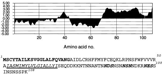
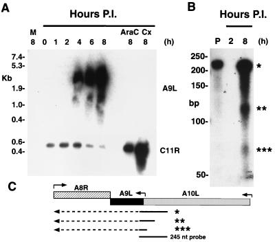
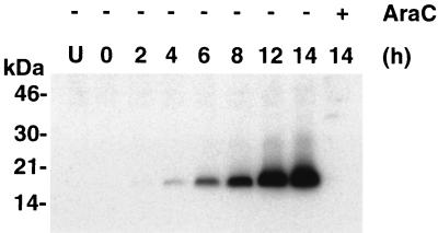
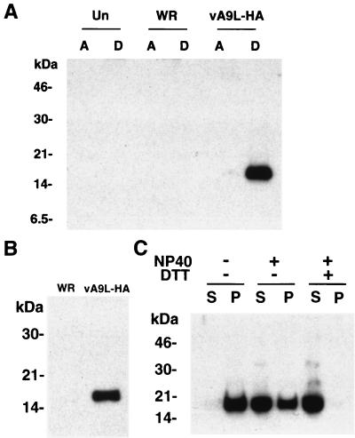
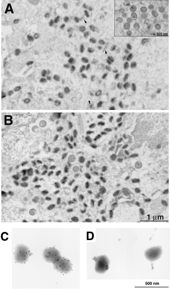
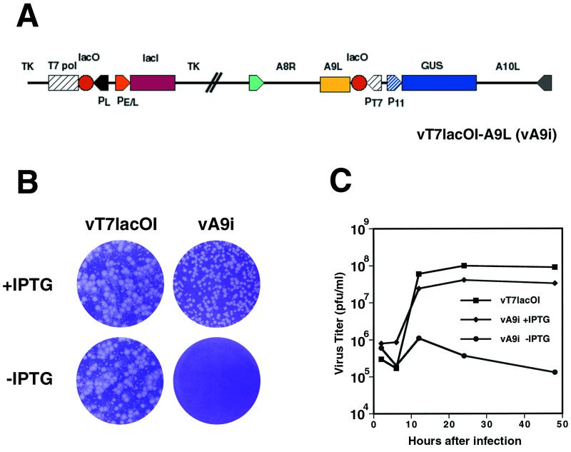
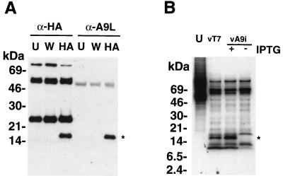
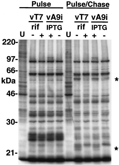
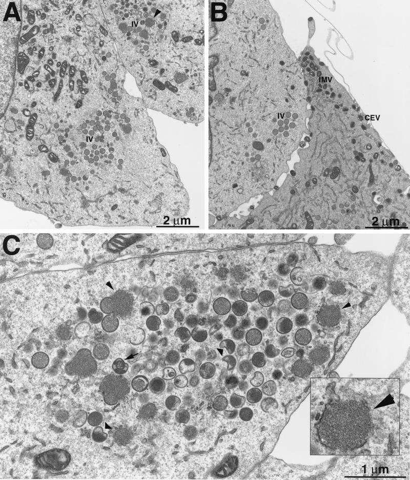
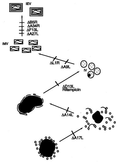
The A9L open reading frame of vaccinia virus was predicted to encode a membrane-associated protein. A transcriptional analysis of the A9L gene indicated that it was expressed at late times in vaccinia virus-infected cells. Late expression, as well as virion membrane association, was demonstrated by the construction and use of a recombinant vaccinia virus encoding an A9L protein with a C-terminal epitope tag. Immunoelectron microscopy revealed that the A9L protein was associated with both immature and mature virus particles and was oriented in the membrane with its C terminus exposed on the virion surface. To determine whether the A9L protein functions in viral assembly or infectivity, we made a conditional-lethal inducible recombinant vaccinia virus. In the absence of inducer, A9L expression and virus replication were undetectable. Under nonpermissive conditions, viral late protein synthesis occurred, but maturational proteolytic processing was inhibited, and there was an accumulation of membrane-coated electron-dense bodies, crescents, and immature virus particles, many of which appeared abnormal. We concluded that the product of the A9L gene is a viral membrane-associated protein and functions at an early stage in virion morphogenesis.


Figures

FIG. 1
FIG. 1
Sequence analysis of the A9L protein. A hydrophilicity plot and the corresponding sequence of the A9L ORF are shown. The predicted signal peptide is boldfaced, the transmembrane domain is italicized and underlined, and the potential sites of N-linked glycosylation are indicated by bold italics.
FIG. 2
FIG. 2
Transcriptional analysis of the A9L gene. (A) Northern blot. BS-C-1 cells were infected with vaccinia virus and harvested after 1 to 8 h. Additional cells were harvested after 8 h of infection in the presence of araC or cycloheximide (Cx). RNAs were resolved by electrophoresis on a denaturing agarose gel, transferred to a nylon membrane, and hybridized with RNA (upper blot) or DNA (lower blot) probes specific for the A9L and C11R ORFs, respectively. The sizes of RNA markers are shown on the left. Abbreviations: M, mock-infected cells; P.I., postinfection. (B) RNase protection assay of RNAs from infected cells harvested at 2 and 8 h after infection. RNAs were incubated with a uniformly 32P-labeled RNA probe complementary to a sequence overlapping the 5′ end of the A9L ORF and the 3′ end of the A10L ORF. After digestion with a mixture of RNases, the material was resolved on a polyacrylamide gel and autoradiographed. Sample P is the full-length undigested probe. Asterisks indicate protected products. (C) Schematic showing the probe and RNA species that could generate the protected products. Arrows above the ORFs indicate the approximate locations of promoters and the direction of transcription. Arrows below the ORFs represent RNAs, with the solid parts protecting the probe and the dashed parts extending beyond. Asterisks correspond to the bands in panel B.
FIG. 3
FIG. 3
Late expression of the HA-tagged A9L protein. Uninfected cells or cells infected with vA9L-HA in the presence (+) or absence (−) of araC were harvested between 0 and 14 h. Proteins in total-cell extracts were resolved by SDS-PAGE and detected by Western blotting with a MAb to the HA tag (MAb HA.11). The positions of migration and molecular masses (in kilodaltons) of marker proteins are indicated on the left.
FIG. 4
FIG. 4
Hydrophobicity and virion association of the A9L protein. (A) Uninfected BS-C-1 cells or cells infected with either vaccinia virus WR or vA9L-HA were harvested 24 h after infection. Triton X-114 extracts were prepared and subjected to phase separation. The A9L protein in the aqueous (A) and detergent (D) phases was analyzed by SDS-PAGE and Western blotting using MAb HA.11. (B) Sucrose gradient-purified vaccinia virus WR and vA9L-HA virions were solubilized directly in Laemmli gel-loading buffer and separated by SDS-PAGE on a 10% polyacrylamide gel. Proteins were transferred to nitrocellulose and subjected to Western blot analysis using MAb HA.11. (C) Purified vA9L-HA virions were incubated at 4°C for 1 h in 50 mM Tris-HCl buffer (pH 7.4) or in the same buffer containing either 1% NP-40 or 1% NP-40 with 50 mM DTT. Soluble material (S) and insoluble material (P) were collected by centrifugation and mixed with Laemmli sample buffer containing DTT, and the proteins were separated by electrophoresis on an SDS–16% polyacrylamide gel. Separated proteins were transferred to nitrocellulose and detected using MAb HA.11. The positions of migration and molecular masses (in kilodaltons) of marker proteins are indicated on the left.
FIG. 5
FIG. 5
Localization of the A9L-HA protein by immunoelectron microscopy. BS-C-1 cells were infected with either vA9L-HA (A) or vT7lacOI (B) for 22 h, fixed in paraformaldehyde, cryosectioned, and incubated with MAb HA.11 followed by rabbit anti-mouse IgG and then protein A conjugated to 10-nm-diameter colloidal gold. Electron micrographs of these samples are shown with a 1-μm and a 500-nm marker (inset). Arrowheads point to unidentified structures with associated gold grains. Grids containing purified intact vA9L-HA (C) or WR (D) virions were stained as in panels B and C.
FIG. 6
FIG. 6
(A) Diagram of the vA9i genome. The expression of the A9L ORF is under the control of the T7 promoter and is IPTG inducible. Abbreviations: PT7, T7 promoter; P11, vaccinia virus late promoter; PE/L, vaccinia early/late promoter; TK, thymidine kinase locus; T7 pol, T7 polymerase ORF; GUS, β-glucuronidase ORF; lacI, E. coli lac repressor ORF; lacO, E. coli lac operator element. (B) Effect of IPTG on formation of vA9i plaques. Monolayers of BS-C-1 cells were infected with either vT7lacOI or vA9i in the presence or absence of 100 μM IPTG as indicated. At 48 h after infection, plaques were visualized by staining with crystal violet. (C) Effect of IPTG on virus yields over time. BS-C-1 cells were infected with vaccinia virus T7LacOI or vA9i at a multiplicity of 5 in the presence or absence of 100 μM IPTG and were harvested at 6, 12, 24, and 48 h after infection. Virus titers for each sample were determined by plaque assay on BS-C-1 cells in the presence of IPTG.
FIG. 7
FIG. 7
Production of the A9L protein in vA9i-infected cells is dependent on inducer. (A) To test the specificity of the anti-A9L antiserum, uninfected BS-C-1 cells (U) or cells infected for 24 h with vaccinia virus WR (W) or vA9L-HA (HA) were lysed in radioimmunoprecipitation assay buffer. Lysates were incubated with either MAb HA.11 (α-HA) or anti-MBP-A9L (α-A9L) followed by protein A-Sepharose. The immune complexes were collected by centrifugation and washed, and the proteins were resolved by SDS-PAGE. The proteins were transferred to nitrocellulose membranes and probed with MAb HA.11. (B) BS-C-1 cells were infected with vaccinia virus vT7LacOI (vT7) or vA9i in the presence or absence of 100 μM IPTG at a multiplicity of 10. After 6 h, infected and uninfected (U) cells were labeled for 18 h with [35S]methionine. Lysates of labeled cells were incubated with anti-MBP-A9L followed by protein A-Sepharose, and immunoprecipitated proteins were separated by SDS-PAGE and visualized by autoradiography. The positions of migration and molecular masses (in kilodaltons) of marker proteins are indicated on the left. The position of migration of the A9L protein is indicated by an asterisk.
FIG. 8
FIG. 8
Late protein synthesis was normal in cells infected with vA9i, but maturational processing was inhibited. Uninfected BS-C-1 cells (U) or cells infected either with vT7lacOI (vT7) in the presence (+) or absence (−) of rifampin (rif; 100 μg/ml) or with vA9Li in the presence (+) or absence (−) of 100 μM IPTG were labeled for 1 h with [35S]methionine at 12 h after infection. Cells were either harvested immediately into sample buffer and analyzed by SDS-PAGE (Pulse) or incubated in medium with excess cold methionine for 12 h, harvested, and then analyzed (Pulse/Chase). Asterisks mark positions of processed forms of proteins. The masses of marker proteins (in kilodaltons) are shown on the left.
FIG. 9
FIG. 9
Only immature and abnormal viral particles were made when A9L was repressed. BS-C-1 cells were infected with vA9i in the presence (B) or absence (A and C) of IPTG and were prepared for electron microscopy. All viral structures were seen in the presence of IPTG (B), whereas in the absence of inducer only electron-dense viroplasm with associated membranes and immature forms developed (A). With some enlargement (C), it can be seen that many of the IV are abnormal. (Inset) Higher-magnification view of electron-dense viroplasm with associated membranes.
FIG. 10
FIG. 10
Diagram indicating the effects of deleting or repressing the expression of specific membrane proteins on virion morphogenesis. In each case, the mutant with a deleted or repressed gene is indicated by a Δ next to the ORF; morphogenesis is blocked at the stage before the slash. The stage at which morphogenesis is blocked by rifampin is also indicated.

Similar articles

Cited by

References

    1. Appleyard G, Hapel A J, Boulter E A. An antigenic difference between intracellular and extracellular rabbitpox virus. J Gen Virol. 1971;13:9–17. - PubMed
    1. Baldick C J, Moss B. Resistance of vaccinia virus to rifampicin conferred by a single nucleotide substitution near the predicted NH2 terminus of a gene encoding an Mr 62,000 polypeptide. Virology. 1987;156:138–145. - PubMed
    1. Betakova T, Wolffe E J, Moss B. Regulation of vaccinia virus morphogenesis: phosphorylation of the A14L and A17L membrane proteins and C-terminal truncation of the A17L protein are dependent on the F10L kinase. J Virol. 1999;73:3534–3543. - PMC - PubMed
    1. Betakova T, Wolffe E J, Moss B. The vaccinia virus A14.5L gene encodes a hydrophobic 53-amino-acid virion membrane protein that enhances virulence in mice and is conserved among vertebrate poxviruses. J Virol. 2000;74:4085–4092. - PMC - PubMed
    1. Blasco R, Moss B. Extracellular vaccinia virus formation and cell-to-cell virus transmission are prevented by deletion of the gene encoding the 37,000-Dalton outer envelope protein. J Virol. 1991;65:5910–5920. - PMC - PubMed

Publication types

Substances

LinkOut - more resources