. 2000 Sep;21(8):1423-33.

Plasticity of the human motor cortex in patients with arteriovenous malformations: a functional MR imaging study

Affiliations

Plasticity of the human motor cortex in patients with arteriovenous malformations: a functional MR imaging study

H Alkadhi et al. AJNR Am J Neuroradiol. 2000 Sep.

Abstract

Background and purpose: The capacity of the human brain to recover from damage has been explained on the basis of plasticity, according to which remaining areas assume functions that would normally have been performed by the damaged brain. Patients with cerebral arteriovenous malformations (AVMs) involving primary motor areas may present without significant neurologic deficits. We used functional MR imaging to investigate the organization of cortical motor areas in patients with AVMs.

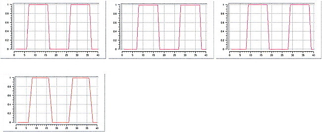
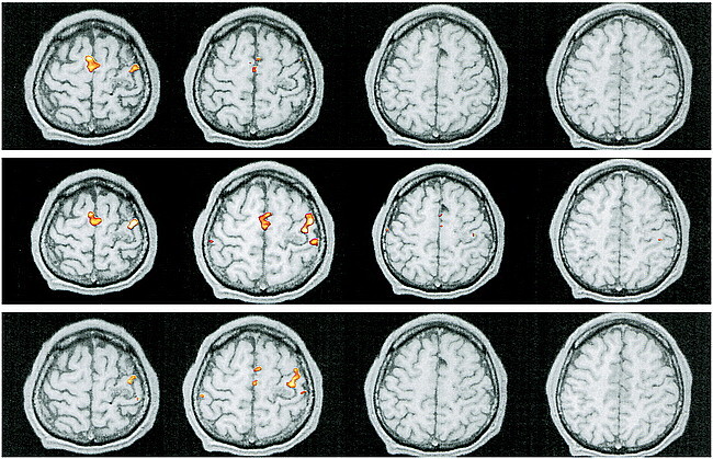
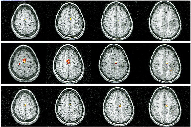
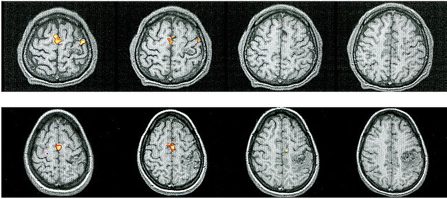
Methods: Cortical motor hand and foot representations were mapped in nine right-handed patients harboring AVMs occupying the hand (n = 6) or foot (n = 3) region of the primary motor cortex (M1). None of the patients exhibited motor deficits. Simple movements of the hand and foot were performed. In eight patients, both right and left extremities were tested; in one patient, only the hand contralateral to the AVM was examined. Localization of activation in the affected hemisphere was compared with that in the unaffected hemisphere and evaluated with respect to the normal M1 somatotopic organization shown in earlier functional MR imaging investigations.

Results: Cortical activation showed three patterns: 1) functional displacement within the affected M1 independent of the structural distortion induced by the AVM (n = 4), 2) presence of activation within the unaffected M1 ipsilateral to the moving extremity without activation in the affected M1 (n = 3), and 3) prominent activation in nonprimary motor areas without activation in either the affected or unaffected M1 (n = 2).

Conclusion: Preliminary evidence suggests that brain AVMs lead to reorganization within the somatotopic representation in M1 and to occasional abnormal expansion into nonprimary motor areas.


Figures

<sc>fig</sc> 1.
fig 1.
Patient 1. A, Sagittal T1-weighted image (500/14/4) shows the AVM in the left precentral gyrus and central sulcus, thereby involving the expected primary representation area of the left hand. B and C, Four adjacent axial sections show cortical maps related to finger movements of the left (B) and right (C) hand. Compared with the right hemisphere M1 representation, which is in the expected anatomic location (B), activation in the left hemisphere is displaced laterally. This displacement does not follow the structural distortion of the rolandic region induced by the AVM. Note further activation in bilateral parietal and SMAs. fig A1. Plotted signal models of the boxcar functions with different time delays (one, two, and three TRs; upper row), and the trapezoidal function with a one-TR delay and a rise time of two TRs (lower image)
<sc>fig</sc> 2.
fig 2.
Patient 3. A, Sagittal T1-weighted image (500/14/4) shows an AVM in the right precentral gyrus, involving the anatomically expected area of hand representation. B and C, As compared with the unaffected left hemisphere (B), the hand representation in the affected right hemisphere (C) is displaced medially within the precentral gyrus. Additional activation within the ipsilateral M1 is detected. The functional displacement does not follow the structural distortion induced by the underlying disorder. Note additional activation in bilateral supplementary motor (arrows, upper right image in C) and parietal areas. fig A2. Patient 1. Reprocessed functional MR imaging experiment using a boxcar function with delays of one TR (upper row), two TRs (middle row), and three TRs (lower row)
<sc>fig</sc> 3.
fig 3.
Patient 6. A, Sagittal T1-weighted image (500/14/4) shows the relationship between the left-sided, central AVM and the precentral knob. B, Movements of the right foot show the expected response in the left paracentral lobule (contralateral M1) with additional activation in bilateral SMAs. C, Right-hand movements do not elicit activation in M1 but there is prominent signal in bilateral SMAs. Note additional activation in ipsilateral dorsal premotor areas and in the contralateral parietal areas and CMA. fig A3. Patient 6. Reprocessed functional MR imaging experiment using a boxcar function with delays of one TR (upper row), two TRs (middle row), and three TRs (lower row)
<sc>fig</sc> 4.
fig 4.
Patient 7. A, T1-weighted sagittal image (500/14/4) shows an extensive AVM in the right paracentral lobule extending to the precuneus, thereby involving the expected primary representation of the left foot. B, Left-hand movements show the expected activation in the contralateral M1. C, Flexion and extension of the left foot reveal absent contralateral activation but prominent activation in the ipsilateral M1, SMA, dorsal premotor area, and CMA, and in the parietal areas bilaterally. fig A4. Reprocessed functional MR imaging experiment using a trapezoidal function with a delay of one TR and a rise time of two TRs in patients 1 (upper row) and 6 (lower row)

Similar articles

Cited by

References

    1. Kaplan HA, Aronson SM, Browder EJ. Vascular malformations of the brain: an anatomical study. J Neurosurg 1961;18:630-635 - PubMed
    1. Berenstein A, Lasjaunias P. Classification of brain arteriovenous malformations. In: Surgical Neuroangiography. Berlin: Springer 1991;4:1-88
    1. Valavanis A. The role of angiography in the evaluation of cerebral vascular malformations. Neuroimaging Clin N Am 1996;6:679-704 - PubMed
    1. Valavanis A, Yasargil MG. The endovascular treatment of brain arteriovenous malformations. In: Advances and Technical Standards in Neurosurgery. Berlin: Springer 1998;24:131-214 - PubMed
    1. Lasjaunias P. Vascular remodelling and the congenital nature of arteriovenous shunts. In: Vascular Diseases in Neonates, Infants and Children. Berlin: Springer 1997;53-65

Publication types