

Case Reports
Granulomatous hypophysitis due to Wegener's granulomatosis
Affiliations
- PMID: 11003280
- PMCID: PMC7974053
Item in Clipboard
Case Reports
Granulomatous hypophysitis due to Wegener's granulomatosis
AJNR Am J Neuroradiol.
2000 Sep.
Abstract
We describe the MR image findings in a case of granulomatous hypophysitis due to Wegner's granulomatosis. A high index suspicion of hypophysitis based on imaging findings allowed successful medical management and helped avoid surgery. The MR imaging features included a thickened stalk, a diffusely and uniformly enlarged gland, a normal size or minimally enlarged sella, and enhancement of the optic chiasm.
Figures
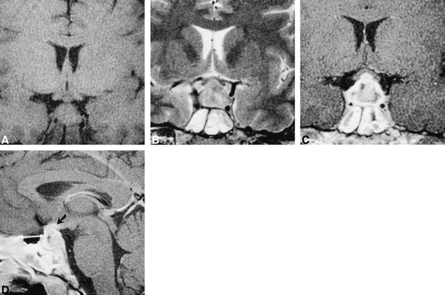
MR images obtained through the pituitary gland show diffuse enlargement of the pituitary gland. The gland shows diffuse heterogeneous enhancement that extends to the optic chiasm (arrow). Also note the presence of mucosal thickening in the bilateral sphenoid sinus. A, T1-weighted coronal (450/15/4 [TR/TE/excitations]) MR image. B, T2-weighted coronal (4000/90/3; echo train length, 8) MR image. C, Contrast-enhanced T1-weighted coronal (450/15/4) MR image. D, Contrast-enhanced T1-weight sagittal (450/15/4) MR image.
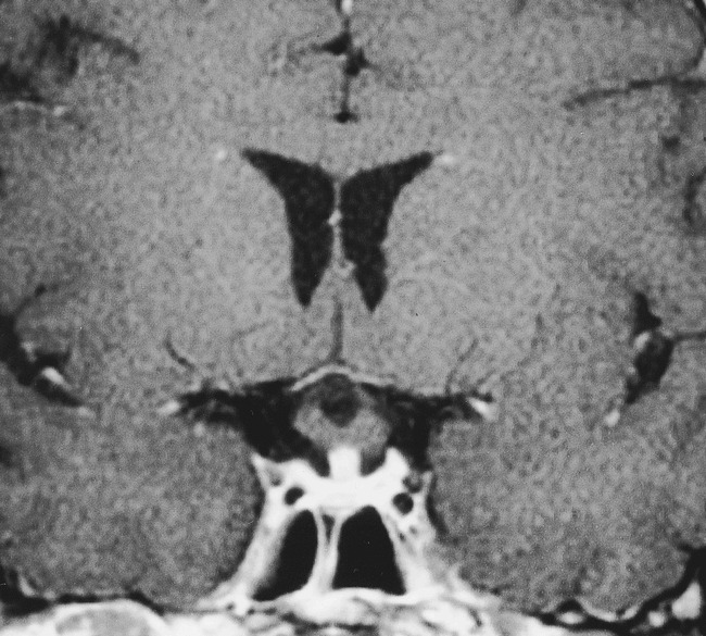
Follow-up MR examination after 6-week administration of steroids. Contrast-enhanced T1-weighted coronal (450/15/4) MR image shows dramatic resolution in the size of the pituitary gland. No enhancement is seen in the optic chiasm
References
-
- Tóth M, Szabó P, Rácz K, et al: Granulomatous hypophysitis associated with Takayasu's disease. Clin Endocrinol (Oxf) 1996;45:499-503 - PubMed
-
- Albini CH, MacGillivray MH, Fisher JE, Voorhess ML, Klein DM. Triad of hypopituitarism, granulomatous hypophysitis, and ruptured Rathke's cleft cyst. Neurosurgery 1988;22:133-136 - PubMed
-
- Roncaroli F, Bacci A, Frank G, Calbucci F:, Granulomatous hypophysitis caused by a ruptured intrasellar Rathke's cleft cyst: report of a case and review of the literature. Neurosurgery 1998;43:146-149 - PubMed
-
- De Bruin WI, van ‘t Verlaat JW, Graamans K, de Bruin TWA:, Sellar granulomatous mass in a pregnant woman with active Crohn's disease. Neth J Med 1991;39:136-141 - PubMed
-
- Scanarini M, D'Avella D, Rotilio A, Kitromilis N, Mingrino S:, Giant-cell granulomatous hypophysitis: a distinct clinicopathological entity. J Neurosurg 1989;71:681-686 - PubMed
Publication types
MeSH terms
LinkOut - more resources
Full Text Sources
Medical