

MR imaging and proton MR spectroscopy in adult Krabbe disease
- PMID: 11003282
- PMCID: PMC7974033
MR imaging and proton MR spectroscopy in adult Krabbe disease
Abstract
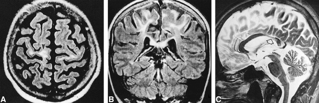
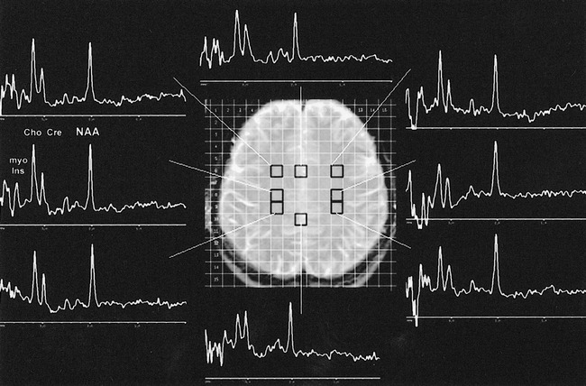
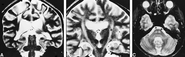
We present the MR imaging findings in four patients (two pairs of siblings from two unrelated families) with adult Krabbe disease. In the first family, clinical presentation mimicked familial spastic paraplegia. Their MR images showed selective, increased signal intensity on T2-weighted sequences along the corticospinal tracts, most prominently in the proband and barely detectable in her brother. Proton MR spectroscopy showed increased choline and myo-inositol in the affected white matter. In the second family, the clinical presentation differed in that the signs of pyramidal tract involvement were asymmetrical, with concomitant asymmetry on MR images in one. In adults, Krabbe disease may present on MR imaging with selective pyramidal fiber involvement.
Figures
References
-
- Wenger DA, Rafi MA, Luzi P. Molecular genetics of Krabbe disease (globoid cell leukodystrophy): diagnostic and clinical implications. Hum Mutat 1997;10:268-279 - PubMed
-
- Luzi P, Rafi MA, Wenger DA. Characterization of the large deletion in the GALC gene found in patients with Krabbe disease. Hum Mol Genet 1995;4:2335-2338 - PubMed
-
- Van der Knaap MS, Valk J. Magnetic Resonance of Melin, Myelination, and Myelin Disorders.. 2nd ed. Berlin: Springer 1995:22-30 68–75-129–139
-
- Percy AK, Odrezin GT, Knowles PD, Rouah E, Armstrong DD. Globoid cell leukodystrophy: comparison of neuropathology with magnetic resonance imaging. Acta Neuropathol 1994;88:26-32 - PubMed
Publication types
MeSH terms
Grants and funding
LinkOut - more resources
Full Text Sources
Medical