. 2000 Sep;21(8):1511-9.

Cerebellar cortical dysplasia: MR findings in a complex entity

Affiliations

Cerebellar cortical dysplasia: MR findings in a complex entity

G Soto-Ares et al. AJNR Am J Neuroradiol. 2000 Sep.

Abstract

Background and purpose: MR imaging findings of cerebellar cortical dysplasia have been described as a new cerebellar malformation. The purpose of this study was to assess the association of cerebellar cortical dysplasia with other cerebral malformations.

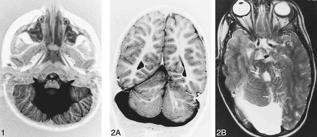
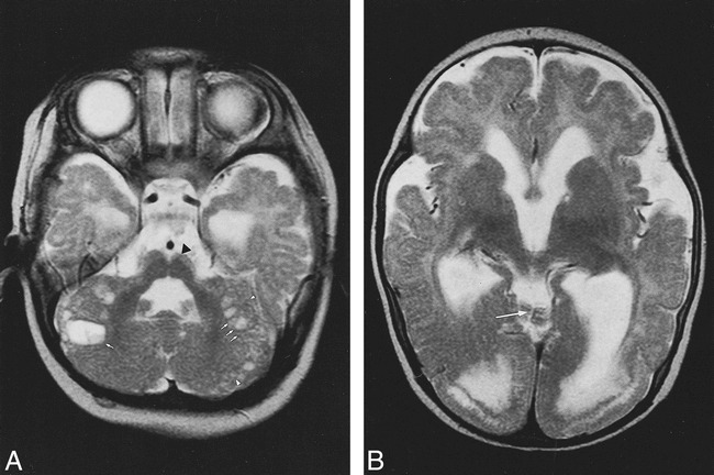
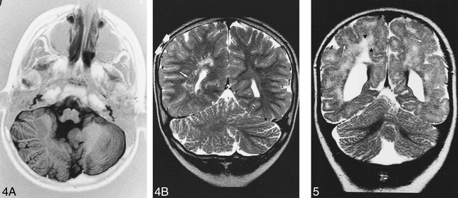
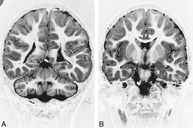
Methods: We retrospectively reviewed 46 MR examinations of patients presenting with developmental delay, hypotonia, and facial deformities to identify abnormal folia or fissures or both within cerebellar hemispheres or vermis suggesting cortical dysplasia.

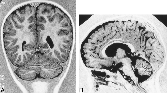
Results: Cerebellar cortical dysplasia was diagnosed in 17 patients. In two patients, it was isolated. In the remaining 15 patients, the malformation was associated with vermian malformation (n=11), cerebral cortical dysplasias (n=8), dysplasia of corpus callosum (n=6), and heterotopia (n=5). A widespread malformation of the posterior fossa was observed in eight patients (Dandy-Walker, Chiari II and III, and hypoplasia of brain stem). One patient with hypertrophied cerebellar hemisphere had minor enlargement of the right cerebral hemisphere and lateral ventricle. He also had nodular heterotopia, suggesting unilateral megalencephaly.

Conclusion: Our study suggests that cerebellar cortical dysplasias are common in cases with more widespread cerebral malformations. Technical progress providing high-quality tridimensional MR imaging of the cerebellum may explain its recent descriptions.


Figures

<sc>fig</sc> 1.
fig 1.
Patient 15, Dandy-Walker complex. Axial inversion-recovery (IR) T1-weighted image (11520/60/400/2 [TR/TE/TI/excitations]) shows bilateral, vertical, orientated folia of the cerebellar cortex associated with an enlarged fourth ventricle.
<sc>fig</sc> 2.
fig 2.
Patient 8, possible Joubert malformation. A, Coronal IR T1-weighted image (11520/60/400/2) shows rostral vermian dysgenesis (black arrowhead), bilateral abnormal arborization of the white matter (double black arrowhead), and linear heterotopia (white arrow). B, Axial T2-weighted image (5000/120/2 [TR/TE/excitations]) shows vermian dysgenesis (short arrow) associated with mega cisterna magna and prominent appearance of the superior cerebellar peduncles (long arrow).
<sc>fig</sc> 3.
fig 3.
Patient 1. Axial T2-weighted images (5000/120/2) show cystlike inclusions (arrows), cortical dysplasia (arrowhead), and pons hypoplasia (black arrowhead) (A) associated with medial vermian fissure (long arrow), occipital polymicrogyria, white matter hyperintensity, and ventricular enlargement (B)
<sc>fig</sc> 4.
fig 4.
Patient 4. A, Axial IR T1-weighted image (11520/60/400/2) shows right cerebellar hypertrophy with vertical folia in an 8-year-old boy with ataxia and psychomotor retardation. B, Coronal T2-weighted image (5000/120/2) shows associated malformations: minor parietal lobe (double arrow) and ventricle enlargement, nodular heterotopia (long arrow) and white matter hyperintensity (short arrows).
<sc>fig</sc> 5.
fig 5.
Patient 5. Ten-month-old boy with spastic tetraparesia, developmental delay, and difficulty in swallowing. Coronal T2-weighted image (5000/120/2) shows right cerebellar cortical disorganization without normal fissures associated with hyperplasia of the right cerebellar lobe and cortical thickening. Note white matter hyperintensities (black arrowheads) and cortical dysplasia (white arrows) in supratentorial images
<sc>fig</sc> 6.
fig 6.
Patient 16. MR imaging findings in a 4-year-old boy with microcephalia, motor delay, and facial deformities. A, Coronal IR T1-weighted image (11520/60/400/2) shows bilateral cerebellar defective foliation and irregularity of the gray/white matter junction (black arrowheads). Note subependymal heterotopia along the floor of the left temporal horn (white arrow). B, Coronal IR T1-weighted images (11520/60/400/2) shows heterotopia (white arrow) and associated cortical dysplasia of parahippocampal gyrus (asterisk).
<sc>fig</sc> 7.
fig 7.
Patient 17. MR imaging findings in a 1 year-old boy with facial deformities, hypotonia, and developmental delay. A, Coronal IR T1-weighted image (11520/60/400/2) shows medial vermian fissure (arrow). B, Sagittal IR T1-weighted image (same parameters) shows lack of normal fissures of the vermis associated with inferior vermis hypoplasia.

References

    1. Rorke LB, Fogelson MH, Riggs HE. Cerebellar heterotopia in infancy. Dev Med Child Neurol 1968;10:644-650 - PubMed
    1. Yachnis AT, Rorke LB, Trojanowki JQ. Cerebellar dysplasias in humans: Development and possible relationship to glial and primitive neuroectodermal tumors of the cerebellar vermis. J Neuropathol Exp Neurol 1994;53:61-71 - PubMed
    1. Jay V. Coexistence of cerebellar primitive neuroectodermal tumor and cerebellar dysplasia: case report. Pediatr Pathol Lab Med 1996;16:837-843 - PubMed
    1. Friede RL. Developmental Neuropathology. 2nd ed. Berlin Heidelberg New York: Springer 1989;361-371
    1. Barkovich AJ. Neuroimaging manifestations and classification of congenital muscular dystrophies. AJNR Am J Neuroradiol 1998;19:1389-1396 - PMC - PubMed

MeSH terms