. 2000 Oct;20(20):7419-26.
doi: 10.1128/MCB.20.20.7419-7426.2000.

Retroviral expression in embryonic stem cells and hematopoietic stem cells

Affiliations

Retroviral expression in embryonic stem cells and hematopoietic stem cells

S R Cherry et al. Mol Cell Biol. 2000 Oct.

Abstract

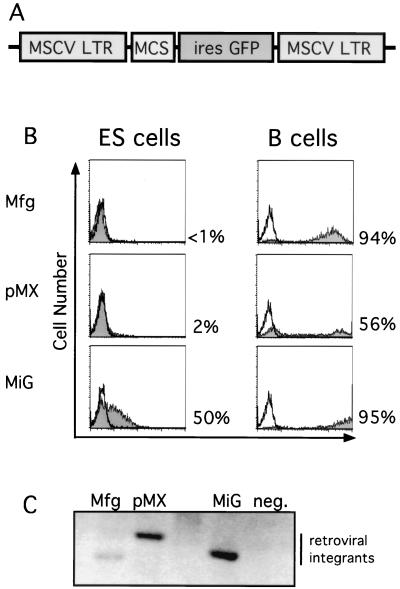
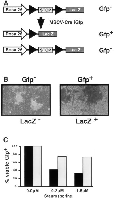
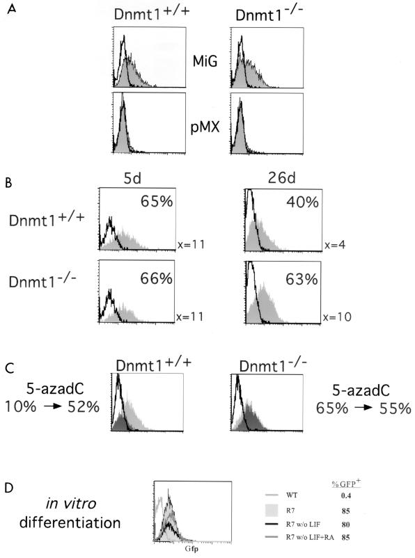
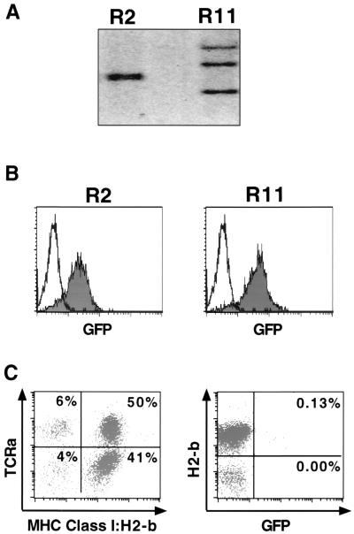
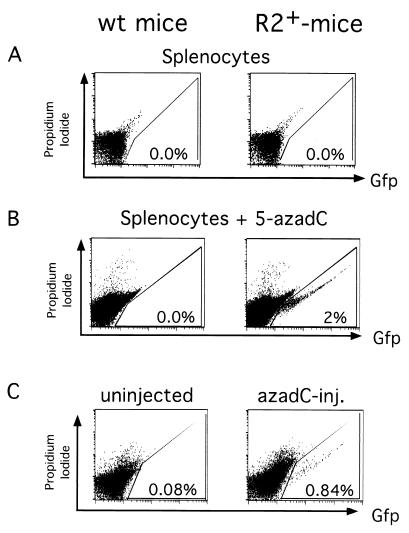
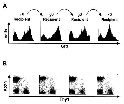
Achieving long-term retroviral expression in primary cells has been problematic. De novo DNA methylation of infecting proviruses has been proposed as a major cause of this transcriptional repression. Here we report the development of a mouse stem cell virus (MSCV) long terminal repeat-based retroviral vector that is expressed in both embryonic stem (ES) cells and hematopoietic stem (HS) cells. Infected HS cells and their differentiated descendants maintained long-term and stable retroviral expression after serial adoptive transfers. In addition, retrovirally infected ES cells showed detectable expression level of the green fluorescent protein (GFP). Moreover, GFP expression of integrated proviruses was maintained after in vitro differentiation of infected ES cells. Long-term passage of infected ES cells resulted in methylation-mediated silencing, while short-term expression was methylation independent. Tissues of transgenic animals, which we derived from ES cells carrying the MSCV-based provirus, did not express GFP. However, treatment with the demethylating agent 5-azadeoxycytidine reactivated the silent provirus, demonstrating that DNA methylation is involved in the maintenance of retroviral repression. Our results indicate that retroviral expression in ES cells is repressed by methylation-dependent as well as methylation-independent mechanisms.


Figures

FIG. 1
FIG. 1
Efficient retroviral infection of ES cells. (A) Schematic diagram of MiG vector containing the MSCV LTR followed by a multiple cloning site (MCS) and an IRES-GFP cassette. (B) MSCV-based (MiG) but not Moloney virus-based (Mfg and pMX) retroviruses express in ES cells. B cells or ES cells were infected by the indicated retroviruses and assayed by flow cytometry 2 days postinfection. Uninfected cells (unshaded) and infected cells (shaded area) were electronically gated for live cells and subsequently analyzed for GFP fluorescence and for cell number. Percentages of GFP-positive cells are indicated. (C) Comparable levels of integration of different retroviruses into ES cells, determined by Southern blot analysis of genomic DNA purified from infected and uninfected ES cells 2 days postinfection, digested with KpnI, a restriction site present within the LTRs, and probed with the GFP coding sequence.
FIG. 2
FIG. 2
Expression from the MSCV LTR is sufficient to drive functional gene expression. (A) Schematic diagram of the Rosa 26 locus in Cre reporter ES cells. Before Cre-mediated recombination, LacZ expression is prevented by the presence of a stop fragment. Retroviral infection with a Cre-expressing retrovirus with a GFP reporter results in two populations of cells. Cells that are GFP+ become LacZ+ due to efficient Cre-mediated recombination of the stop fragment. In contrast, cells that are GFP were not infected and thus remained LacZ. (B) ES cells were infected with the MSCVCreiresGFP retrovirus and sorted for either Gfp or Gfp+ as indicated. The cells were subsequently cultured and stained for LacZ expression. Gfp cells are white (and therefore LacZ) while Gfp+ cells are blue (and therefore LacZ+). More than 99% of the Gfp+ cells were LacZ+ in multiple experiments. (C) ES cells were infected with either MSCViresGFP (■) or MSCVBcl-2iresGFP (□) and treated with the indicated amounts of staurosporine. The percentage of viable, Gfp+ (infected) cells was determined by flow cytometry. The results are shown as a percentage of Gfp+ cells before treatment. The results are from one representative experiment of three performed.
FIG. 3
FIG. 3
Long-term expression of retroviruses is repressed by methylation. (A) MSCV-based (MiG) but not Moloney virus-based (pMX) retroviruses express in ES cells independent of the methylation status of the cells. Dnmt1+/+ or Dnmt1−/− ES cells were not infected (unshaded) or infected by the indicated retroviruses (shaded) and assayed by flow cytometry 2 days postinfection as for Fig. 1B. (B) Long-term expression of GFP in ES cells is decreased in Dnmt1+/+ cells but not Dnmt1−/− cells. The ES cells were infected with MiG, passaged for 5 or 26 days postinfection, and assayed by flow cytometry as above. The mean fluorescent intensity for the population and the percentage of GFP-positive cells are indicated. (C) Treatment with 5-azadC rescues the expression of retroviruses silenced by long-term passage. Dnmt1+/+ or Dnmt1−/− ES cells were infected by MiG and passaged for >40 days. The cells were divided, and half were treated with 5-azadC. Then uninfected ES cells (unshaded), MiG-infected untreated ES cells (dark shading), and MiG-infected 5-azadC-treated ES cells (light shading) were assayed by FACS analysis. Numbers below the FACS plots are percentages of GFP-positive cells before and after 5-azadC treatment. (D) In vitro differentiation of ES cells does not affect retroviral expression. A clonal ES cell line (R7) infected with MiG or an uninfected wild-type (WT) ES cell control was in vitro differentiated by passage without feeders and LIF, with or without retinoic acid (RA) as indicated. The cells were assayed by flow cytometry, and the percentage of GFP-positive cells is indicated.
FIG. 4
FIG. 4
Retrovirally infected, GFP-expressing ES cells generated nonexpressing mice. (A) Two retrovirally infected clones sorted for GFP expression were analyzed for proviral integrants by Southern blot analysis. R2 contained two integrants, while R11 contained three. Uninfected cells are negative. (B) The clones were passaged after sorting for GFP-expressing cells by flow cytometry and reanalyzed for GFP expression. Both R2 and R11 express GFP (shaded) compared to uninfected controls (unshaded). (C) PBMCs from the R2 chimera (more than 50% contribution by coat color) were analyzed by flow cytometry. ES cell contribution to the chimera was determined by phycoerythrin-H2-b staining and cyc-TCRa staining and demonstrated contribution to the T-cell compartment. The percentage of cells in each quadrant is listed. The cells were also monitored for GFP expression. The percentage of GFP+ cells that are either major histocompatibility complex (MHC) class I H2-b+ (ES cell derived) or H2-b (blastocyst derived) is listed in the quadrant.
FIG. 5
FIG. 5
Silenced retroviruses can be reactivated with 5-azadC. (A) Splenocytes from an R2+ or R2 littermate do not express GFP by flow cytometry. Cells were stained with propidium iodide to exclude dead cells, and the percentage of GFP+ cells is indicated. (B) The splenocytes from panel B were induced to proliferate with anti-CD3 and treated with 5-azadC. Cells were stained with propidium iodide to exclude dead cells and analyzed by flow cytometry. The percentage of GFP+ cells is indicated. (C) Flow cytometric analysis of the splenocytes of littermates that were either uninjected or injected with 5-azadC at passage 5 and analyzed at passage 14 for GFP expression. The percentage of GFP+ splenocytes is indicated.
FIG. 6
FIG. 6
Serial adoptive transfers maintain expression of the MSCV-based retrovirus. (A) Bone marrow was infected with MiG and used to reconstitute multiple lethally irradiated mice to generate the 10 recipient. The spleen of the 10 recipient was analyzed for GFP expression by flow cytometry. The bone marrow of the 10 recipient was used to reconstitute lethally irradiated 20 recipients. The spleen of a 20 recipient was analyzed for GFP expression, and the bone marrow was used to reconstitute lethally irradiated 30 recipients. The spleen of a 30 recipient was analyzed for GFP expression, and the bone marrow was used to reconstitute lethally irradiated 40 recipients. A representative analysis is shown. (B) splenocytes from panel A, stained with pan-B-cell (B220) and pan-T-cell (Thy-1) antibodies and electronically gated for GFP+ cells, are shown below the GFP histogram they are derived from. The FACS diagrams are shown for these serially reconstituted spleens, demonstrating that the transferred cells contribute to both B- and T-cell lineages in the appropriate ratios.

References

    1. Asche W, Colletta G, Warnecke G, Nobis P, Pennie S, King R M, Ostertag W. Lack of retrovirus gene expression in somatic cell hybrids of Friend cells and teratocarcinoma cells with a teratocarcinoma phenotype. Mol Cell Biol. 1984;4:923–930. - PMC - PubMed
    1. Cedar H. DNA methylation and gene activity. Cell. 1988;53:3–4. - PubMed
    1. Challita P M, Kohn D B. Lack of expression from a retroviral vector after transduction of murine hematopoietic stem cells is associated with methylation in vivo. Proc Natl Acad Sci USA. 1994;91:2567–2571. - PMC - PubMed
    1. Coligan, J. E., et al. (ed.). Current protocols in immunology. J. Wiley & Sons, New York, N.Y.
    1. Gautsch J W. Embryonal carcinoma stem cells lack a function required for virus replication. Nature. 1980;285:110–112. - PubMed

Publication types

MeSH terms