. 2000 Oct;20(20):7505-15.
doi: 10.1128/MCB.20.20.7505-7515.2000.

Essential roles for ankyrin repeat and transactivation domains in induction of T-cell leukemia by notch1

Affiliations

Essential roles for ankyrin repeat and transactivation domains in induction of T-cell leukemia by notch1

J C Aster et al. Mol Cell Biol. 2000 Oct.

Abstract

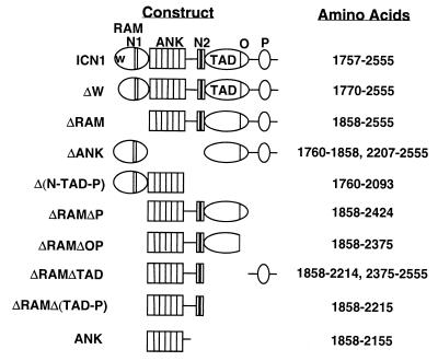
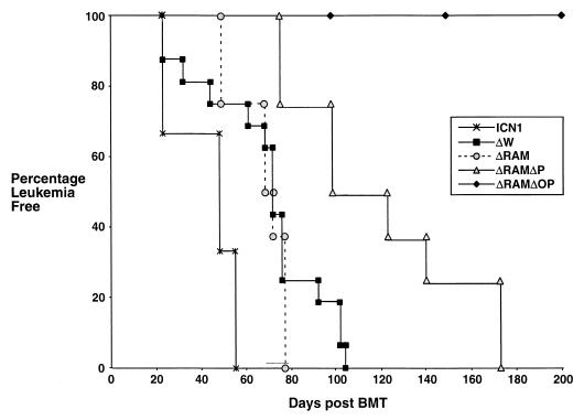
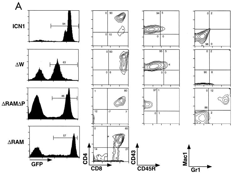
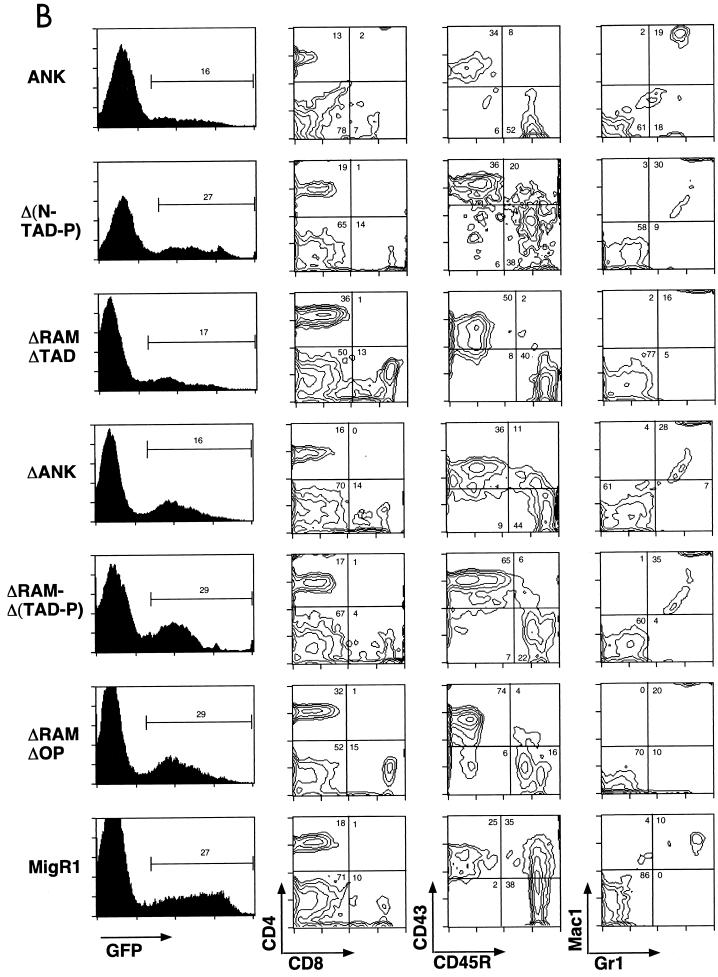
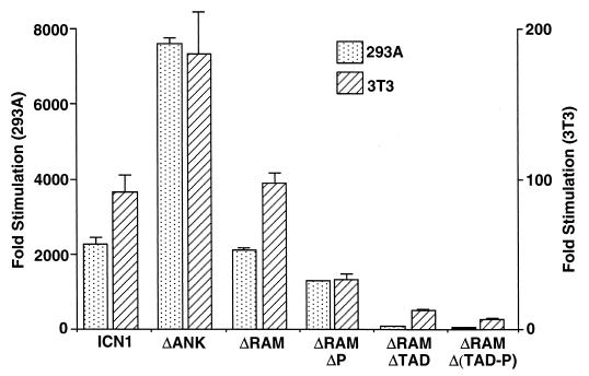
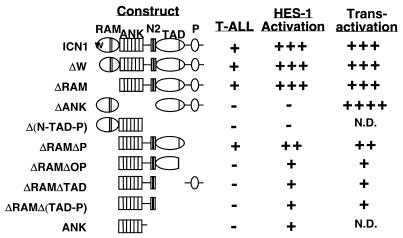
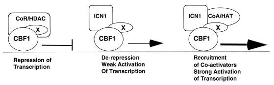
Notch receptors participate in a conserved signaling pathway that controls the development of diverse tissues and cell types, including lymphoid cells. Signaling is normally initiated through one or more ligand-mediated proteolytic cleavages that permit nuclear translocation of the intracellular portion of the Notch receptor (ICN), which then binds and activates transcription factors of the Su(H)/CBF1 family. Several mammalian Notch receptors are oncogenic when constitutively active, including Notch1, a gene initially identified based on its involvement in a (7;9) chromosomal translocation found in sporadic T-cell lymphoblastic leukemias and lymphomas (T-ALL). To investigate which portions of ICN1 contribute to transformation, we performed a structure-transformation analysis using a robust murine bone marrow reconstitution assay. Both the ankyrin repeat and C-terminal transactivation domains were required for T-cell leukemogenesis, whereas the N-terminal RAM domain and a C-terminal domain that includes a PEST sequence were nonessential. Induction of T-ALL correlated with the transactivation activity of each Notch1 polypeptide when fused to the DNA-binding domain of GAL4, with the exception of polypeptides deleted of the ankyrin repeats, which lacked transforming activity while retaining strong transactivation activity. Transforming polypeptides also demonstrated moderate to strong activation of the Su(H)/CBF1-sensitive HES-1 promoter, while polypeptides with weak or absent activity on this promoter failed to cause leukemia. These experiments define a minimal transforming region for Notch1 in T-cell progenitors and suggest that leukemogenic signaling involves recruitment of transcriptional coactivators to ICN1 nuclear complexes.


Figures

FIG. 1
FIG. 1
Schematic representation of Notch1 polypeptides encoded by expression constructs. N1, nuclear localization signal sequence 1; N2, nuclear localization signal sequence 2; O, OPA sequence; P, PEST sequence.
FIG. 2
FIG. 2
Onset of leukemia in mice reconstituted with retrovirally transduced BM cells. Time of leukemia onset is defined by the time post-BMT that CD4+-CD8+ DP GFP+ T cells were identified in the peripheral circulation. Mice typically survived for approximately 1 to 2 months after the onset of leukemia, during which time the fraction and number of DP GFP+ cells continued to increase (data not shown). None of the mice reconstituted with the nontransforming viral constructs or the empty MigRI virus developed circulating DP GFP+ cells or any evidence of neoplasia. A representative survival curve for a cohort of mice reconstituted with a nontransforming virus (ΔRAMΔOP) is shown. Although this figure shows leukemia-free survival to 200 days, these mice remained leukemia free for the duration of this study (see Table 1).
FIG. 3
FIG. 3
Immunophenotypes of splenic WBC from mice receiving transduced BM cells. (A) Presence of GFP+ DP T cells in mice reconstituted with BM transduced by leukemogenic Notch1 retroviruses. CD43, CD45R, Mac1, and Gr1 staining were not performed on cells obtained from ICNΔRAM animals. (B) Lack of GFP+ DP T cells in mice reconstituted with BM transduced by nonleukemogenic Notch1 retroviruses. GFP expression is shown on the left in both panels; gates used to identify the GFP+ population (given as percentages) are indicated. The antibodies used for staining are shown at the bottom adjacent to the relevant axis; numbers correspond to relative percentages within the GFP+ gate. The results shown are representative of individual analyses of each mouse included in the Table 1 summary. Similar results were also found in analyses of cells isolated from the PB, BM, and lymph nodes from each mouse.
FIG. 3
FIG. 3
Immunophenotypes of splenic WBC from mice receiving transduced BM cells. (A) Presence of GFP+ DP T cells in mice reconstituted with BM transduced by leukemogenic Notch1 retroviruses. CD43, CD45R, Mac1, and Gr1 staining were not performed on cells obtained from ICNΔRAM animals. (B) Lack of GFP+ DP T cells in mice reconstituted with BM transduced by nonleukemogenic Notch1 retroviruses. GFP expression is shown on the left in both panels; gates used to identify the GFP+ population (given as percentages) are indicated. The antibodies used for staining are shown at the bottom adjacent to the relevant axis; numbers correspond to relative percentages within the GFP+ gate. The results shown are representative of individual analyses of each mouse included in the Table 1 summary. Similar results were also found in analyses of cells isolated from the PB, BM, and lymph nodes from each mouse.
FIG. 4
FIG. 4
Western blot analyses of tissue extracts. (A) Extracts of nodal masses in ICN1 and ΔRAM animals. (B and C) Extracts of BM (B), splenic cells (S), or (D) thymic cells from animals expressing transforming ΔRAMΔP or nontransforming ΔRAMΔTAD, ΔRAMΔOP, and ΔRAMΔ(TAD-P) polypeptides. Portions of lymph node tumors, spleens, and thymuses were snap frozen in liquid nitrogen, pulverized with a pestle, and then lysed in sodium dodecyl sulfate-polyacrylamide gel electrophoresis loading buffer (∼100 μl/mg of tissue) at 95°C for 10 min. BM cells were flushed from femurs with normal saline, pelleted, and immediately lysed in loading buffer. Blots shown in panels A and B were stained with αTC rabbit serum (12) raised against the TAD of human Notch1, while blots shown in panels C and D were stained with αT6 rabbit serum (2) raised against the human Notch1 ANK and revealed by a chemiluminescent method.
FIG. 5
FIG. 5
Analysis of proviral integration and TCRβ chain rearrangement in leukemias from mice receiving transduced BM cells. (A) Southern blots of EcoRI-digested genomic DNA isolated from ICN1 spleens (mouse 4 and 5), ΔRAM spleens (mouse 1 and 5), and a kidney (KID) obtained from a control BALB/c animal. (B) Southern blots of EcoRI-digested genomic DNA isolated from ΔW spleens (spl) (mouse 67 and 71) and a liver (liv) from a MigRI control animal. (C) Southern blots of either EcoRI-digested (IRES) or HindIII-digested TCRβ (TCR) genomic DNA obtained from ΔRAMΔP BM (mouse 5) or spleen (mouse 3). To determine the number of proviral integration sites, DNAs were digested with EcoRI, which cleaves once within the provirus, and analyzed on Southern blots hybridized with a 592-bp encephalomyelitis virus IRES probe. A second set of digestions with XbaI, which cleaves once in each retroviral long terminal repeat, confirmed the presence of intact proviral DNA in all samples (data not shown). To determine the configuration of TCRβ genes, DNAs digested with either EcoRI or HindIII were analyzed on blots incubated with a TCR-Jβ2-specific DNA probe (10) that hybridizes to a 2.2-kb EcoRI fragment or a 5-kb HindIII fragment in germ line DNA. Two to five micrograms of DNA was loaded in each lane (A to C) except the BALB/c kidney lanes (A), which contained 15 μg. The hybridization probes are listed below the blots. The HindIII-digested lambda size markers are indicated adjacent to each blot. Sizes (in kilobases) are 23.1, 9.4, 6.6, 4.4, 2.3, and 2.0, reading from top to bottom.
FIG. 6
FIG. 6
Transcriptional activation of the HES-1 promoter by Notch1 polypeptides. (A and C) Activation in acutely expressing 293A cells and NIH 3T3 cells, respectively. (B) Dose response in 293A cells. In the experiments shown in panels A and C, six-well dishes were transfected with 250 ng of pcDNA3 plasmids containing the indicated Notch1 cDNAs, 500 ng of HES-AB firefly luciferase plasmid, and 10 ng of pRL-TK Renilla luciferase plasmid. In the experiments shown in panel B, the amounts of pcDNA3 plasmids were varied. Cell lysates were prepared ∼48 h posttransfection. Firefly luciferase activities were normalized to the corresponding Renilla luciferase activities. Transcriptional activities were expressed relative to the normalized activities observed in extracts prepared from cells transfected with empty pcDNA3 plasmid. The results shown represent the means of three experiments.
FIG. 6
FIG. 6
Transcriptional activation of the HES-1 promoter by Notch1 polypeptides. (A and C) Activation in acutely expressing 293A cells and NIH 3T3 cells, respectively. (B) Dose response in 293A cells. In the experiments shown in panels A and C, six-well dishes were transfected with 250 ng of pcDNA3 plasmids containing the indicated Notch1 cDNAs, 500 ng of HES-AB firefly luciferase plasmid, and 10 ng of pRL-TK Renilla luciferase plasmid. In the experiments shown in panel B, the amounts of pcDNA3 plasmids were varied. Cell lysates were prepared ∼48 h posttransfection. Firefly luciferase activities were normalized to the corresponding Renilla luciferase activities. Transcriptional activities were expressed relative to the normalized activities observed in extracts prepared from cells transfected with empty pcDNA3 plasmid. The results shown represent the means of three experiments.
FIG. 7
FIG. 7
Transcriptional activation by GAL4-Notch1 fusion polypeptides. 293A or NIH 3T3 cells grown in six-well plates were transfected with 250 ng of pM plasmids encoding various GAL4-Notch1 fusion polypeptides, 500 ng of GAL4X5 firefly luciferase plasmid, and 10 ng of pRL-TK Renilla luciferase plasmid. Firefly luciferase activities were normalized to the corresponding Renilla luciferase activities. Transcriptional activities were expressed relative to the normalized activities observed in extracts prepared from cells transfected with empty pcDNA3 plasmid. The results shown represent the means of three experiments.
FIG. 8
FIG. 8
Correlation of leukemogenesis with the transcriptional activities of various forms of Notch1. HES-1 activation refers to the capacity to activate transcription from a promoter element derived from the murine HES-1 gene; transactivation refers to the activity of Notch1-GAL4 DNA-binding domain fusion polypeptides on an artificial promoter containing five iterated GAL4 binding sites.
FIG. 9
FIG. 9
Model for leukemogenic Notch1 signaling complex. CBF1, transcription factor Su(H)/CBF1; CoR/HDAC, corepressor-histone deacetylase complex; X, molecule capable of binding to Su(H)/CBF1 and ICN1, of which SKIP and LAG-3 are prototypes; CoA/HAT, coactivator-histone acetylase complex.

References

    1. Artavanis-Tsakonas S, Rand M D, Lake R J. Notch signaling: cell fate control and signal integration in development. Science. 1999;284:770–776. - PubMed
    1. Aster J, Pear W, Hasserjian R, Erba H, Davi F, Luo B, Scott M, Baltimore D, Sklar J. Functional analysis of the TAN-1 gene, a human homolog of Drosophila notch. Cold Spring Harbor Symp Quant Biol. 1994;59:125–136. - PubMed
    1. Aster J C, Robertson E S, Hasserjian R P, Turner J R, Kieff E, Sklar J. Oncogenic forms of NOTCH1 lacking either the primary binding site for RBP-Jkappa or nuclear localization sequences retain the ability to associate with RBP-Jkappa and activate transcription. J Biol Chem. 1997;272:11336–11343. - PubMed
    1. Bailey A M, Posakony J W. Suppressor of hairless directly activates transcription of enhancer of split complex genes in response to Notch receptor activity. Genes Dev. 1995;9:2609–2622. - PubMed
    1. Bigas A, Martin D I, Milner L A. Notch1 and Notch2 inhibit myeloid differentiation in response to different cytokines. Mol Cell Biol. 1998;18:2324–2333. - PMC - PubMed

Publication types

MeSH terms

LinkOut - more resources