. 2000 Oct;20(20):7516-26.
doi: 10.1128/MCB.20.20.7516-7526.2000.

The peroxisome biogenesis factors pex4p, pex22p, pex1p, and pex6p act in the terminal steps of peroxisomal matrix protein import

Affiliations

The peroxisome biogenesis factors pex4p, pex22p, pex1p, and pex6p act in the terminal steps of peroxisomal matrix protein import

C S Collins et al. Mol Cell Biol. 2000 Oct.

Abstract

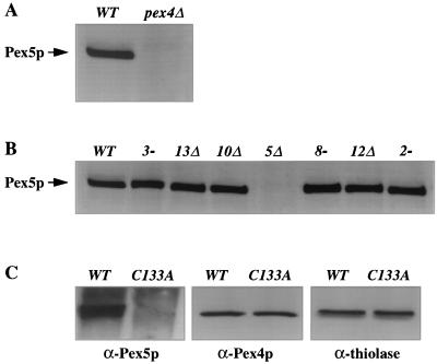
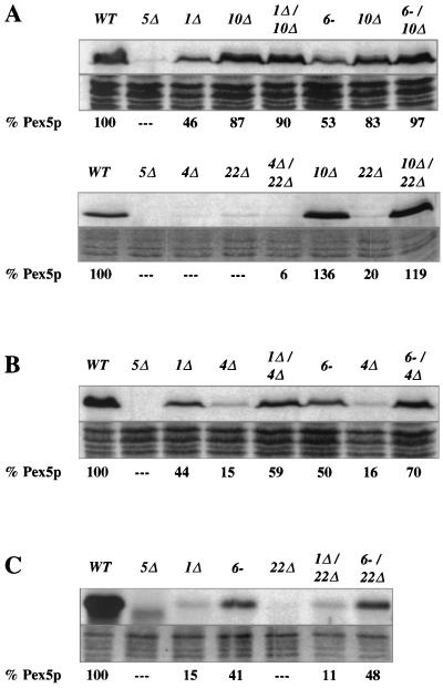
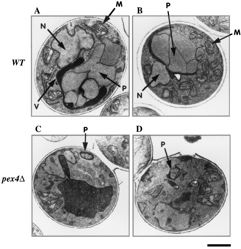
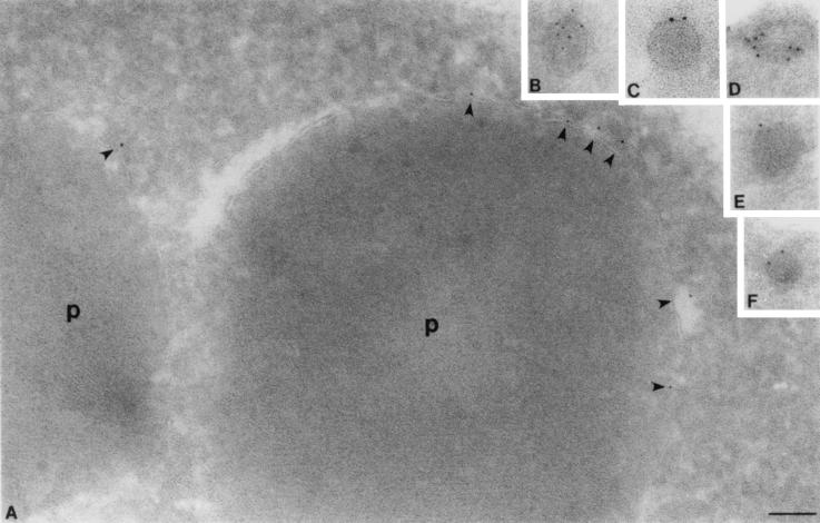
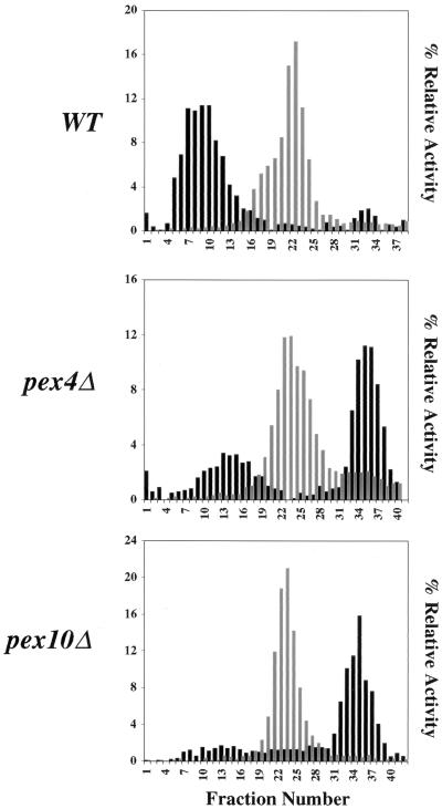
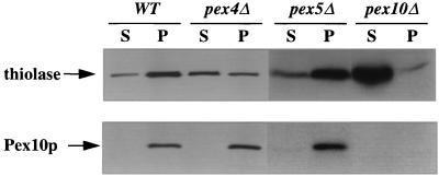
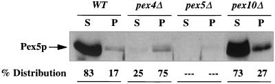
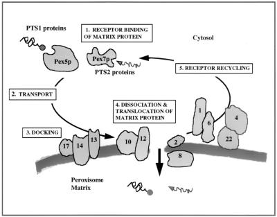
Peroxisomes are independent organelles found in virtually all eukaryotic cells. Genetic studies have identified more than 20 PEX genes that are required for peroxisome biogenesis. The role of most PEX gene products, peroxins, remains to be determined, but a variety of studies have established that Pex5p binds the type 1 peroxisomal targeting signal and is the import receptor for most newly synthesized peroxisomal matrix proteins. The steady-state abundance of Pex5p is unaffected in most pex mutants of the yeast Pichia pastoris but is severely reduced in pex4 and pex22 mutants and moderately reduced in pex1 and pex6 mutants. We used these subphenotypes to determine the epistatic relationships among several groups of pex mutants. Our results demonstrate that Pex4p acts after the peroxisome membrane synthesis factor Pex3p, the Pex5p docking factors Pex13p and Pex14p, the matrix protein import factors Pex8p, Pex10p, and Pex12p, and two other peroxins, Pex2p and Pex17p. Pex22p and the interacting AAA ATPases Pex1p and Pex6p were also found to act after Pex10p. Furthermore, Pex1p and Pex6p were found to act upstream of Pex4p and Pex22p. These results suggest that Pex1p, Pex4p, Pex6p, and Pex22p act late in peroxisomal matrix protein import, after matrix protein translocation. This hypothesis is supported by the phenotypes of the corresponding mutant strains. As has been shown previously for P. pastoris pex1, pex6, and pex22 mutant cells, we show here that pex4Delta mutant cells contain peroxisomal membrane protein-containing peroxisomes that import residual amounts of peroxisomal matrix proteins.


Figures

FIG. 1
FIG. 1
Loss of Pex4p results in reduced steady-state levels of Pex5p. (A and B) Equal amounts of protein were extracted from methanol-induced cells, separated by SDS-PAGE, and blotted with anti-Pex5p antibodies. (A) Levels of Pex5p in wild-type (WT) and pex4Δ cells. (B) Normal Pex5p levels in pex3, pex13, pex10, pex8, pex12, and pex2 strains. (C) Total cellular proteins extracted from pex4Δ strains carrying a wild-type PEX4 expression plasmid and a plasmid that expresses an active-site mutant of PEX4, C133A. Levels of Pex5p (left), Pex4p (middle), and the peroxisomal matrix enzyme thiolase (right) present in these samples were determined by immunoblotting with specific antibodies.
FIG. 2
FIG. 2
PEX5 mRNA and Pex5p are synthesized normally in pex4Δ cells. (A) Northern blot analysis of poly(A)+ RNAs from pex4Δ and pex10Δ cells. The two RNA samples were separated by denaturing gel electrophoresis, transferred to membranes, and sequentially hybridized to radiolabeled probes specific for the PEX5 (top) and ACT1 (bottom) genes. Quantification revealed that the PEX5:ACT1 hybridization signal was 1.3-fold higher in the pex4Δ strain than in the pex10Δ strain. Similar results were observed in two additional independent trials. (B) Synthesis of equivalent levels of Pex5p during pulse-labeling of pex4Δ and pex10Δ cells. Each strain was incubated in the presence of [35S]methionine for 2 min and lysed in alkali, and the Pex5p present in each sample was immunoprecipitated using excess anti-Pex5p antibodies. The immunoprecipitates were separated by SDS-PAGE, and the amount of Pex5p was determined by fluorographic exposure to X-ray film. Similar results were observed in nine additional trials of this experiment.
FIG. 3
FIG. 3
The destabilization of Pex5p in the pex4Δ strain requires Pex3p, Pex13p, Pex14p, and Pex17p. For each panel, the strains were grown side by side and total cellular protein was extracted with alkali, separated by SDS-PAGE, and blotted with antibodies specific for Pex5p (top of each panel). Coomassie blue staining of each protein sample is also shown (bottom of each panel), and the quantity of Pex5p in each sample (as assessed by scanning densitometry of the resulting films) is shown below each lane. (A) Levels of Pex5p in wild-type (WT) cells; the pex5Δ, pex3-1, and pex4Δ mutants; and the pex3-1 pex4Δ double mutant. (B) Levels of Pex5p in wild-type cells; the pex5Δ, pex13Δ, and pex4Δ mutants; and the pex4Δ pex13Δ double mutant. The lower blot in panel B shows levels of Pex5p in wild-type cells; the pex5Δ, pex4Δ, and pex14Δ mutants; the pex4Δ pex14Δ double mutant; the pex4Δ and pex17Δ mutants; and the pex4Δ pex17Δ double mutant.
FIG. 4
FIG. 4
Destabilization of Pex5p in the pex4Δ strain requires Pex10p, Pex12p, Pex8p, and Pex2p. For each panel, the strains were grown side by side and total cellular protein was extracted with alkali, separated by SDS-PAGE, and blotted with antibodies specific for Pex5p (top of each panel). Coomassie blue staining of each protein sample is also shown (bottom of each panel), and the quantity of Pex5p in each sample (as assessed by scanning densitometry of the resulting films) is shown below each lane. (A) Levels of Pex5p in wild-type (WT) cells; the pex5Δ, pex4Δ, and pex10Δ mutants; and the pex4Δ pex10Δ double mutant. The lower blot in panel A shows levels of Pex5p in wild-type cells; the pex5Δ, pex12Δ, and pex4Δ mutants, and the pex4Δ pex12Δ double mutant. (B) Levels of Pex5p in wild-type cells; the pex5Δ, pex8-3, and pex4Δ mutants; the pex4Δ pex8-3 double mutant; the pex2-2 and pex4Δ mutants; and the pex2-2 pex4Δ double mutant.
FIG. 5
FIG. 5
Pex1p and Pex6p act downstream of Pex10p and upstream of Pex4p and Pex22p. For each panel, the strains were grown side by side and total cellular protein was extracted with alkali, separated by SDS-PAGE, and blotted with antibodies specific for Pex5p (top of each panel). Coomassie blue staining of each protein sample is also shown (bottom of each panel), and the quantity of Pex5p in each sample (as assessed by scanning densitometry of the resulting films) is shown below each lane. (A) Levels of Pex5p in wild-type (WT) cells; the pex5Δ, pex1Δ, and pex10Δ mutants; the pex1Δ pex10Δ double mutant; the pex6-1 and pex10Δ mutants; and the pex6-1 pex10Δ double mutant. The lower blot in panel A shows levels of Pex5p in wild-type cells; the pex5Δ, pex4Δ, and pex22Δ mutants; the pex4Δ pex22Δ double mutant; the pex10Δ and pex22Δ mutants; and the pex10Δ pex22Δ double mutant. (B) Levels of Pex5p in wild-type cells; the pex5Δ, pex1Δ, and pex4Δ mutants; the pex1Δ pex4Δ double mutant; the pex6-1 and pex4Δ mutants; and the pex6-1 pex4Δ double mutant. (C) Levels of Pex5p in wild-type cells; the pex5Δ, pex1Δ, pex6-1, and pex22Δ mutants; and the pex1Δ pex22Δ and pex6-1 pex22Δ double mutants.
FIG. 6
FIG. 6
Transmission electron microscopy reveals the presence of small, matrix-containing peroxisomes in pex4Δ cells. Wild-type (WT) (A and B) and pex4Δ (C and D) cells were induced in methanol medium and processed for transmission electron microscopy. M, mitochondria; N, nucleus; P, peroxisome; V, vacuole. Bar, 1.0 μm.
FIG. 7
FIG. 7
Immunocryoelectron microscopy demonstrates that pex4Δ cells contain small, electron-dense peroxisomes. (A) Peroxisomes (p) of wild-type cells labeled with antibodies specific for Pex10p, shown here by immunogold labeling of the peroxisome membrane. Note the position of gold particles at the peroxisome membrane (shown by arrowheads) and the electron-dense nature of the protein-rich peroxisome matrix. (B to F) Peroxisomes of pex4Δ cells are also labeled with antibodies specific for Pex10p and contain an electron-dense, granular matrix. However, their radius is only 10% of the radius of wild-type peroxisomes, indicating that their volume may be only 1/1,000 of that of the wild-type peroxisome. Bar, 0.1 μm.
FIG. 8
FIG. 8
The pex4Δ mutant contains peroxisomes of normal density. Wild-type (WT), pex4Δ, and pex10Δ cells were induced in methanol medium, a PNS was generated from each strain, and peroxisomes and mitochondria were collected by differential centrifugation. The resulting three organelle pellets were then separated by sucrose density gradient centrifugation. The same amount of each fraction was assayed for catalase (a PTS1-containing peroxisomal enzyme) activity (black bars), succinate dehydrogenase (mitochondrial enzyme) activity (grey bars), and density (data not shown). Enzyme activities are plotted as percentages of the total activity in the gradient. The densities of all of the gradient profile were similar, with the peak peroxisomal fraction of wild-type cells migrating at a density of 1.19 to 1.21 g/cm3 and the peak peroxisomal fraction of pex4Δ cells migrating at a density of 1.18 to 1.20 g/cm3.
FIG. 9
FIG. 9
The pex4Δ mutant imports significant amounts of the PTS2 marker enzyme thiolase but less than wild-type (WT) or pex5Δ cells. Wild-type, pex4Δ, pex5Δ, and pex10Δ cells were induced to proliferate peroxisomal proteins, and a PNS was generated from each strain. This was then separated into an organelle pellet (p) and a cytosolic supernatant (s) by centrifugation at 25,000 × g. The same proportion of each fraction was separated by SDS-PAGE and blotted with antibodies specific for the PTS2-targeted enzyme thiolase (top) and the integral PMP Pex10p (bottom).
FIG. 10
FIG. 10
Most Pex5p is present in the organelle fraction of pex4Δ cells. Wild-type (WT), pex4Δ, pex5Δ, and pex10Δ cells were induced in methanol medium, and a PNS was generated from each strain. This was then separated into an organelle pellet (p) and a cytosolic supernatant (s) by centrifugation at 25,000 × g. The same proportion of each fraction was separated by SDS-PAGE and blotted with antibodies specific for Pex5p.
FIG. 11
FIG. 11
Model of peroxisomal matrix protein import. PTS-containing proteins are bound by their receptor after translation in the cytoplasm (step 1). The mechanism by which the receptor-bound matrix protein is transported to the peroxisome membrane remains unclear (step 2). The receptor-bound matrix protein then docks on the surface of the peroxisome membrane via interactions with the docking factors Pex13p, Pex14p, and possibly Pex17p (step 3). Following docking, the matrix protein must dissociate from the receptor and be translocated across the peroxisome membrane into the matrix space (step 4). It is not clear whether the receptor follows the cargo into the lumen or remains on the cytosolic surface of the peroxisome. However, in either scenario, the receptor is subsequently released from the translocation pore and recycled back to the cytoplasm to undergo further rounds of import (step 5).

Similar articles

Cited by

References

    1. Albertini M, Rehling P, Erdmann R, Girzalsky W, Kiel J A K W, Veenhuis M, Kunau W-H. Pex14p, a peroxisomal membrane protein binding both receptors of the two PTS-dependent import pathways. Cell. 1997;89:83–92. - PubMed
    1. Chang C C, South S, Warren D, Jones J, Moser A B, Moser H W, Gould S J. Metabolic control of peroxisome abundance. J Cell Sci. 1999;112:761–774. - PubMed
    1. Chang C C, Warren D S, Sacksteder K A, Gould S J. PEX12 binds PEX5 and PEX10 and acts downstream of receptor docking in peroxisomal matrix protein import. J Cell Biol. 1999;147:761–773. - PMC - PubMed
    1. Crane D I, Gould S J. The Pichia pastoris HIS4 gene: nucleotide sequence, creation of a non-reverting his4 deletion mutant, and development of HIS4-based replicating and integrating plasmids. Curr Genet. 1994;26:443–450. - PubMed
    1. Crane D I, Kalish J E, Gould S J. The Pichia pastoris PAS4 gene encodes a ubiquitin-conjugating enzyme required for peroxisome assembly. J Biol Chem. 1994;269:21835–21844. - PubMed

Publication types

MeSH terms