. 2000 Oct;20(20):7550-8.
doi: 10.1128/MCB.20.20.7550-7558.2000.

Cardiac tissue enriched factors serum response factor and GATA-4 are mutual coregulators

Collaborators, Affiliations

Cardiac tissue enriched factors serum response factor and GATA-4 are mutual coregulators

N S Belaguli et al. Mol Cell Biol. 2000 Oct.

Abstract

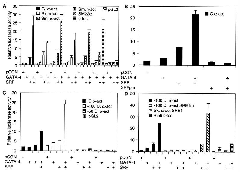
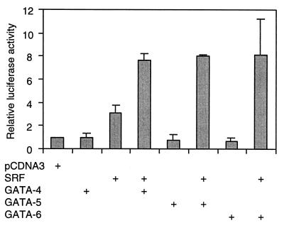
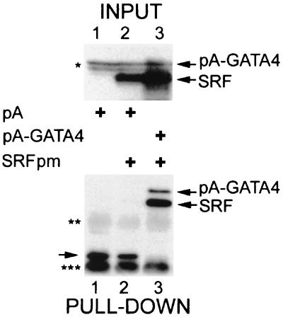
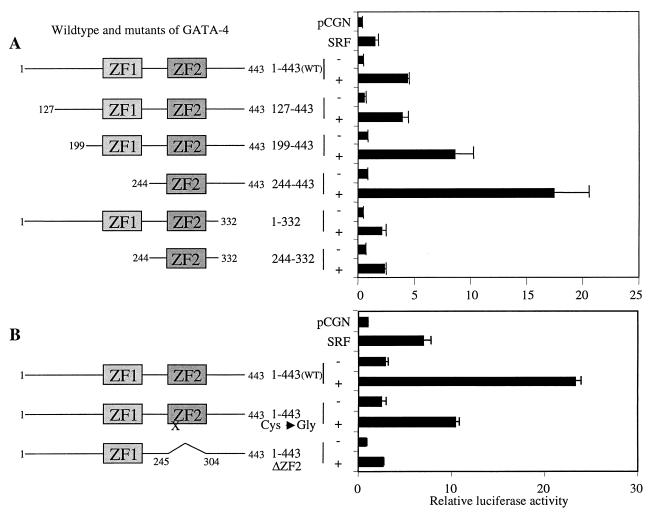
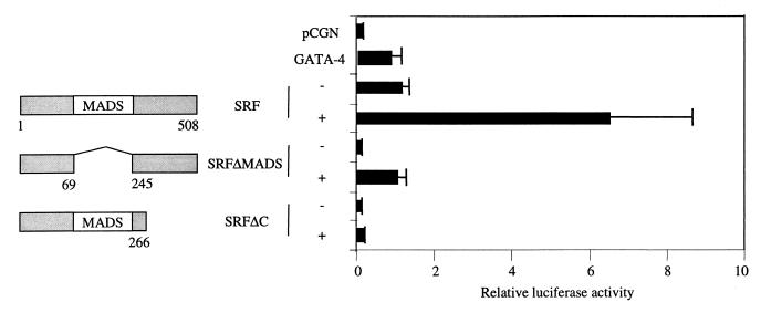
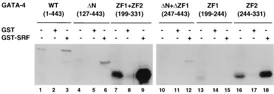
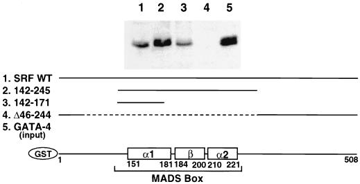
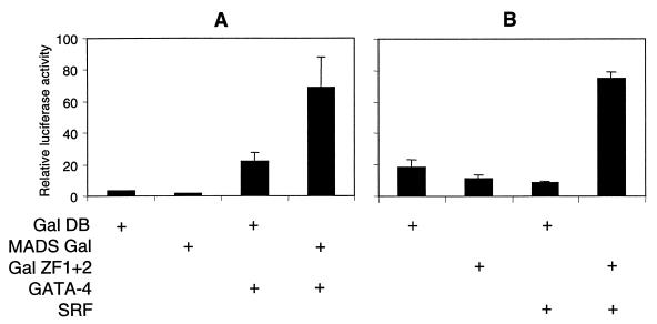
Combinatorial interaction among cardiac tissue-restricted enriched transcription factors may facilitate the expression of cardiac tissue-restricted genes. Here we show that the MADS box factor serum response factor (SRF) cooperates with the zinc finger protein GATA-4 to synergistically activate numerous myogenic and nonmyogenic serum response element (SRE)-dependent promoters in CV1 fibroblasts. In the absence of GATA binding sites, synergistic activation depends on binding of SRF to the proximal CArG box sequence in the cardiac and skeletal alpha-actin promoter. GATA-4's C-terminal activation domain is obligatory for synergistic coactivation with SRF, and its N-terminal domain and first zinc finger are inhibitory. SRF and GATA-4 physically associate both in vivo and in vitro through their MADS box and the second zinc finger domains as determined by protein A pullout assays and by in vivo one-hybrid transfection assays using Gal4 fusion proteins. Other cardiovascular tissue-restricted GATA factors, such as GATA-5 and GATA-6, were equivalent to GATA-4 in coactivating SRE-dependent targets. Thus, interaction between the MADS box and C4 zinc finger proteins, a novel regulatory paradigm, mediates activation of SRF-dependent gene expression.


Figures

FIG. 1
FIG. 1
SRF and GATA-4 synergistically activate the cardiac α-actin promoter. (A) Subconfluent CV1 cells were transfected with 1 μg of numerous myogenic and nonmyogenic promoter luciferase reporters (indicated), along with 150 ng of expression vector for SRF alone or in combination with 400 ng of GATA-4. (B) A DNA-binding mutant of SRF (SRFpm1) (150 ng) was used in addition to wild-type SRF and GATA-4. (C) The wild type and deletion mutants of the cardiac α-actin promoter and the control pGL2 basic luciferase reporters were used. (D) A deletion mutant of cardiac α-actin containing a single wild-type or mutated SRE1 and a truncated c-fos minimal promoter (Δ56 c-fos) with or without skeletal α-actin SRE1 cloned upstream was used in the cotransfection assay. The total amount of DNA was adjusted to 2 μg by balancing with the pCGN empty vector. Cells were harvested 48 h posttranscription, and the luciferase activity was measured. Results shown are means ± the standard errors of the means for three duplicate experiments (B and C) and two duplicate experiments (A and D).
FIG. 2
FIG. 2
GATA-5 and GATA-6 can substitute for GATA-4 in coactivation of the cardiac α-actin promoter. Subconfluent CV1 cells were transfected with 200 ng of wild-type cardiac α-actin luciferase reporter and 200 ng of an expression vector for SRF (pCGNSRF) either alone or in combination with 800 ng of pCDNA3GATA-4, -5, or -6. Cells were harvested 48 h posttranscription, and the luciferase activity was measured. Results shown are means ± standard errors of the means for two duplicate experiments.
FIG. 3
FIG. 3
GATA-4 and SRF associate in vivo. Bottom panel, CV1 cells transfected with HA-tagged SRFpm1 with either protein A (lane 2) or protein A–GATA-4 (lane 3) fusions. For lane 1, protein A vector was transfected alone. Cells were lysed, and the lysates were allowed to react with IgG-Sepharose beads. After extensive washing, proteins retained by protein A and protein A fusions were eluted by boiling in SDS sample buffer and analyzed by immunoblotting with anti-SRF antibody. The top panel shows the Western analysis of input proteins probed with HA antibody. Protein A and protein A fusions to SRF and GATA-4, as well as IgG heavy chains (double asterisk) and IgG light chains (triple asterisk), were visualized due to binding of the secondary antibody. Nonspecific anti-HA immunoreactive bands which migrate close to the pA-GATA4 band are indicated by a single asterisk.
FIG. 4
FIG. 4
The second zinc finger and the immediate C-terminal basic region of GATA-4 are essential for synergistic activation of the cardiac α-actin promoter. (A) Subconfluent CV1 cells were transfected with 1 μg of cardiac α-actin luciferase reporter and 400 ng of the wild type and various deletion mutants of GATA-4, either alone or in combination with 150 ng of SRF. The total amount of DNA was adjusted to 2 μg by balancing with the pCGN empty vector. (B) The −100 cardiac α-actin promoter containing the proximal SRE was used as the reporter. Cells were harvested 48 h posttranscription, and the luciferase activity was measured. Results shown are means ± standard errors of the means for three duplicate experiments (A) and two duplicate experiments (B). Domains of GATA-4 that are retained in each deletion mutant are diagrammatically represented on the left. ZF1 and ZF2 refer to the N- and C-terminal zinc fingers, respectively. The single amino acid mutation in ZF2 (cysteine 273 to glycine) that abolished DNA-binding activity of GATA-4 is indicated by an X.
FIG. 5
FIG. 5
Subconfluent CV1 cells were transfected with 1 μg of cardiac α-actin luciferase reporter along with 400 ng of GATA-4 and 150 ng of either the wild type or deletion mutants of SRF. The total amount of DNA was adjusted to 2 μg by balancing with the pCGN empty vector. Cells were harvested 48 h posttranscription, and the luciferase activity was measured. Results shown are means ± standard errors of the means for three duplicate experiments. Domains of SRF retained in each deletion mutant are diagrammatically represented on the left.
FIG. 6
FIG. 6
Physical interaction between GATA-4 and SRF is mediated by the second zinc finger and the immediate C-terminal basic region. In vitro-translated [35S]methionine-labeled wild-type (WT) GATA-4 (lanes 1 to 3), an N-terminally truncated GATA-4 (ΔN) (lanes 4 to 6), both zinc fingers of GATA-4 (ZF1 + ZF2) (lanes 7 to 9), N-terminal GATA-4 with a deletion of ZF1 (ΔN + ΔZF1) (lanes 10 to 12), the first zinc finger of GATA-4 (ZF1) (lanes 13 to 15), and the second zinc finger along with the immediate C-terminal basic region of GATA-4 (ZF2) (lanes 16 to 18) (7.5 μl each) were incubated with approximately 1 μg (lanes 2, 5, 8, 11, 14, and 17) of GST or GST-SRF fusion protein (lanes 3, 6, 9, 12, 15, and 18) immobilized on glutathione-agarose beads. The beads were washed extensively, and the bound proteins were resolved on an SDS–10% protein gel and visualized by autoradiography. For lanes 1, 4, 7, 10, and 16, 0.75-μl volumes of the lysates were run.
FIG. 7
FIG. 7
Physical interaction between SRF and GATA-4 is mediated by the N-terminal portion of the α-I helix of the MADS box of SRF. Approximately 1 μg of wild-type GST-SRF (lane 1), N- and C-terminally truncated SRF (lane 2), the N-terminal portion of the α-I helix of the MADS box of SRF (lane 3), and the SRF with a deletion of the MADS box (lane 4) were immobilized on glutathione-Sepharose beads and incubated with 100 μg of CV1 cell extract overexpressing GATA-4. The beads were washed extensively and resolved on an SDS–10% protein gel, and Western blot analysis was done with an anti-GATA-4 antibody. Ten micrograms of the lysate was run for lane 5. A schematic diagram at the bottom of the figure shows various regions of SRF retained in the deletion mutants.
FIG. 8
FIG. 8
Reciprocal recruitment of SRF and GATA-4 via one-hybrid assays. (A) Subconfluent CV1 cells were transfected with 200 ng of Gal4 luciferase reporter and 200 ng of Gal4 DNA-binding domain (Gal DB) or Gal DB fused to the MADS box (MADS Gal) in the presence or absence of 750 ng of GATA-4. (B) One microgram of Gal4 reporter and 1 μg of Gal DB or Gal DB fused to the first and the second zinc fingers of GATA-4 (Gal ZF1 + 2) were transfected in the presence or absence of 100 ng of SRF. Cells were harvested 48 h posttranscription, and the luciferase activity was measured. Results shown are means ± standard errors of the means for two duplicate experiments.

References

    1. Arsenian S, Weinhold B, Oelgeschlager M, Ruther U, Nordheim A. Serum response factor is essential for mesoderm formation during mouse embryogenesis. EMBO J. 1998;17:6289–6299. - PMC - PubMed
    1. Belaguli N S, Schildmeyer L A, Schwartz R J. Organization and myogenic restricted expression of the murine serum response factor gene. J Biol Chem. 1997;272:18222–18231. - PubMed
    1. Belaguli N S, Zhou W, Trinh T-H T, Majesky M W, Schwartz R J. Dominant negative murine serum response factor: alternative splicing within the activation domain inhibits transactivation of serum response factor binding targets. Mol Cell Biol. 1999;19:4582–4591. - PMC - PubMed
    1. Biesiada E, Hamamori Y, Kedes L, Sartorelli V. Myogenic basic helix-loop-helix proteins and Sp1 interact as components of a multiprotein transcriptional complex required for activity of the human cardiac α-actin promoter. Mol Cell Biol. 1999;19:2577–2584. - PMC - PubMed
    1. Black B L, Olson E N. Transcriptional control of muscle development by myocyte enhancer factor-2 (MEF2) proteins. Annu Rev Cell Dev Biol. 1998;14:167–196. - PubMed

Publication types

MeSH terms

LinkOut - more resources