. 2000 Oct;20(20):7662-72.
doi: 10.1128/MCB.20.20.7662-7672.2000.

Induction of human fetal globin gene expression by a novel erythroid factor, NF-E4

Affiliations

Induction of human fetal globin gene expression by a novel erythroid factor, NF-E4

W Zhou et al. Mol Cell Biol. 2000 Oct.

Abstract

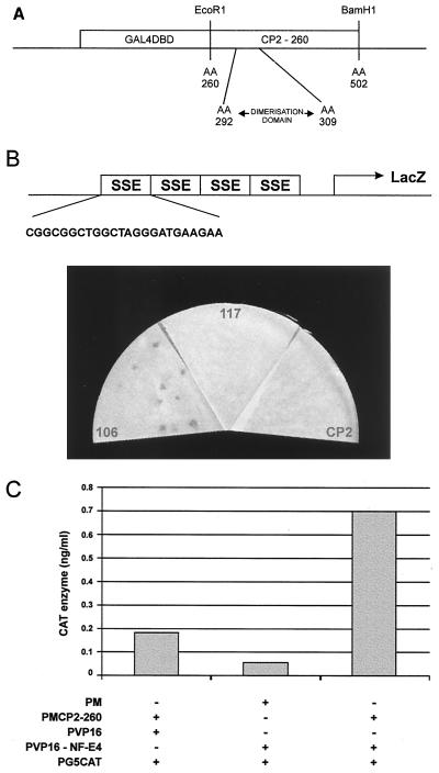
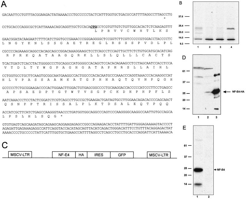
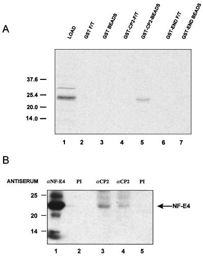
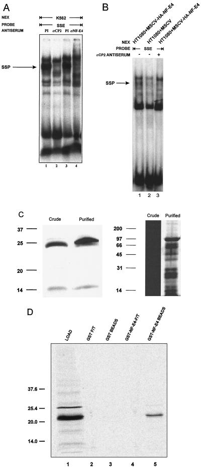
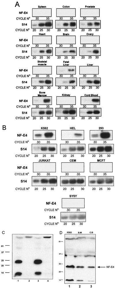
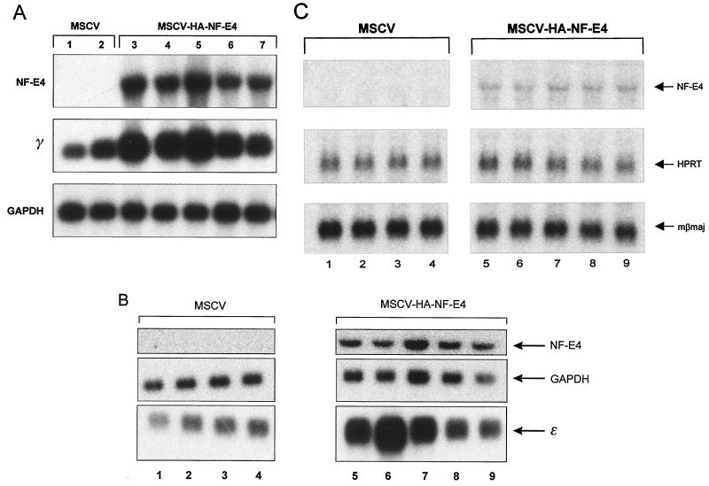
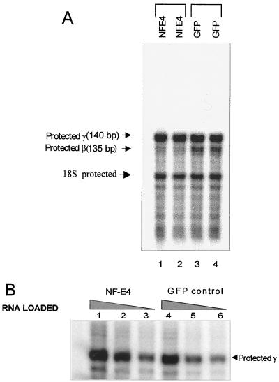
The stage selector protein (SSP) is a heteromeric complex involved in preferential expression of the human gamma-globin genes in fetal-erythroid cells. We have previously identified the ubiquitous transcription factor CP2 as a component of this complex. Using the protein dimerization domain of CP2 in a yeast two-hybrid screen, we have cloned a novel gene, NF-E4, encoding the tissue-restricted component of the SSP. NF-E4 and CP2 coimmunoprecipitate from extract derived from a fetal-erythroid cell line, and antiserum to NF-E4 ablates binding of the SSP to the gamma promoter. NF-E4 is expressed in fetal liver, cord blood, and bone marrow and in the K562 and HEL cell lines, which constitutively express the fetal globin genes. Enforced expression of NF-E4 in K562 cells and primary erythroid progenitors induces endogenous fetal globin gene expression, suggesting a possible strategy for therapeutic intervention in the hemoglobinopathies.


Figures

FIG. 1
FIG. 1
Isolation of CP2-interacting proteins. (A) Schematic of the bait construct used in the yeast two-hybrid screen of a K562 cDNA library. The coding sequence of CP2 from aa 260 to 502 was fused in frame with the GAL4DBD. The minimal dimerization domain (aa 292 to 309) is shown. (B) Yeast one-hybrid assay of CP2-interacting clones defined in the two-hybrid screen. Plasmids containing CP2 or c106 or c117 fused to the GAL4AD were transfected into a yeast reporter strain carrying four concatemerized SSE sites linked to a lacZ reporter gene. The sequence of a single SSE site is expanded. Transformants were selected on minimal medium plates lacking leucine and uracil, lifted onto filters, and assayed for β-galactosidase activity. Filters were photographed after 30 min, and equivalent transfection efficiencies were observed for all constructs. (C) Mammalian two-hybrid analysis of interaction between CP2 and NF-E4. 293 cells were transiently cotransfected with a reporter plasmid containing five GAL4DBD sites linked to the CAT gene (pG5CAT) and the indicated expression vectors. PM contains the GAL4DBD, PMCP2-260 contains the GAL4DBD fused to aa 260 to 502 of CP2, PVP16 has the VP16AD, and PVP16-NF-E4 has the NF-E4 sequence isolated from the two-hybrid clone fused to the VP16AD. Transfections were performed in triplicate, and cell lysates were assayed in triplicate for CAT activity.
FIG. 2
FIG. 2
NF-E4 is a 22-kDa protein which may initiate at a CUG codon. (A) Nucleotide and amino acid sequences of NF-E4. The potential initiator CUG is underlined and in boldface. The 5′ and 3′ termination codons are marked with asterisks. (B) In vitro transcription/translation of NF-E4. The NF-E4 cDNA with its native Kozak sequence was cloned into pSP72, and [35S]methionine-labeled protein was produced (lane 1). We also generated radiolabeled protein from NF-E4 constructs in which the CTG was mutated to a GGG or TTG but retained the native Kozak sequence (lanes 2 and 3) or was mutated to an ATG with a consensus Kozak sequence (lane 4); 2 μl of each sample was resolved by SDS-PAGE on a 12% gel. The gel was dried and subjected to autoradiography. The migration of molecular size standards is marked in kilodaltons. (C) Diagrammatic representation of the MSCV-NF-E4-HA vector. The vector consists of the MSCV backbone containing the NF-E4 coding sequence tagged at the COOH terminus with HA, followed by an encephalomyocarditis virus IRES linked to the GFP cDNA. (D) Western analysis of K562 cell pools expressing NF-E4-HA. Whole-cell lysates from K562 cells transduced with MSCV-NF-E4-HA (lane 2) or MSCV alone (lane 1) were resolved on a 12% polyacrylamide gel, transferred to a PVDF membrane, probed with polyclonal anti-HA antiserum, and developed with ECL. The specific NF-E4-HA band is arrowed. Recombinant NF-E4-HA served as a control (lane 3). The migration of molecular mass standards is indicated in kilodaltons. (E) Western analysis of native K562 cells. Nuclear extract from K562 cells was resolved on a 12% polyacrylamide gel, transferred to a PVDF membrane, and probed with polyclonal anti-NF-E4 (lane 1) or preimmune (lane 2) serum. Signal was developed with ECL. The migration of molecular mass standards is indicated in kilodaltons.
FIG. 3
FIG. 3
CP2 and NF-E4 interact in vitro and in vivo. (A) Direct interaction between CP2 and NF-E4. GST-CP2, GST-END, or GST alone was expressed in E. coli, and 1 μg of protein bound to glutathione-Sepharose beads. The beads were then incubated with 2 μl of 35S-labeled in vitro-translated NF-E4 in binding buffer for 1 h at room temperature (see Materials and Methods). After extensive washing, the beads were resuspended in SDS loading buffer and subjected to SDS-PAGE (lanes 1, 3, and 5). The supernatants (flowthrough [F/T]) of the binding reactions were also analyzed (lanes 2, 4, and 6). Positions of migration of the NF-E4 load and the molecular weight standards are labeled in kilodaltons. (B) Coimmunoprecipitation of CP2 and NF-E4 from native K562 cells. Nuclear extract from K562 cells was immunoprecipitated with polyclonal antiserum to NF-E4 (lane 1) or CP2 (lanes 3 [8 μl] and 4 [4 μl]) or the corresponding preimmune (PI) serum (lane 2 or 5, respectively). The immunoprecipitates were fractionated by SDS-PAGE and blotted with antisera to NF-E4. Positions of migration of NF-E4 and the molecular mass standards (in kilodaltons) are labeled.
FIG. 4
FIG. 4
NF-E4 is a component of the SSP. (A) NF-E4 antiserum disrupts the SSP-SSE complex. Crude K562 cell nuclear extract (NEX) was studied in EMSA with the SSE probe in the presence of anti-CP2 antiserum (lane 2), anti-NF-E4 antiserum (lane 4), or the corresponding preimmune (PI) serum (lane 1 or 3, respectively). Migration of the SSP-SSE complex is arrowed. (B) Expression of NF-E4 in a null cell line generates a de novo SSP complex. Nuclear extract was prepared from the human sarcoma cell line HT1080 transduced with MSCV-HA-NF-E4 (lane 1) or MSCV (lane 2) and analyzed by EMSA using an SSE probe. The effect of anti-CP2 antisera on the EMSA from MSCV-HA-NF-E4 was examined in lane 3. Migration of the SSP complex is arrowed. (C) Western analysis and silver stain of NF-E4 in purified SSP. Crude K562 cell nuclear extract or purified SSP was resolved on a 12% polyacrylamide gel, transferred to a PVDF membrane, probed with polyclonal anti-NF-E4 antiserum and de-veloped with ECL (left); the samples were also electrophoresed, and the gel was silver stained (right). Molecular size standards are indicated in kilodaltons. (D) NF-E4 forms homodimeric complexes. GST–NF-E4 or GST alone was expressed in E. coli, and 1 μg of protein was bound to glutathione-Sepharose beads. The beads were then incubated with 2 μl of 35S-labeled in vitro-translated NF-E4 in binding buffer for 1 h at room temperature (see Materials and Methods). After extensive washing, the beads were resuspended in SDS loading buffer and subjected to SDS-PAGE (lanes 1 and 3). The supernatants (flowthrough [F/T]) of the binding reactions were also analyzed (lanes 2 and 4). Positions of migration of NF-E4 and the molecular mass standards (in kilodaltons) are labeled.
FIG. 5
FIG. 5
Expression of NF-E4 in primary tissues and established cell lines. First-strand cDNA transcribed from poly(A)+ RNA from multiple primary tissues or cell lines was used as a template to PCR amplify a product using primers specific for S14. Samples were then diluted and reamplified to give comparable band intensities for the same number of amplification cycles of S14 RNA within the linear range of the assay; this represents 20, 25, and 30 cycles. (A) Expression of NF-E4 in primary human tissues. Based on the S14 quantitation, comparable amounts of cDNA from multiple primary human tissues were PCR amplified using primers specific for NF-E4. These primers span a 1.8-kb intron and thus discriminate between mRNA and genomic DNA-derived signal. Cycle numbers were chosen to represent the linear range of amplification; this represents 30 and 35 cycles. (B) Expression of NF-E4 in cell lines. Based on the S14 quantitation, comparable amounts of cDNA from multiple established cell lines were PCR amplified using primers specific for NF-E4. RNA was prepared from erythroid lines (K562 and HEL), T-cell lines (CEM and Jurkat), a human embryonic kidney cell line (293), a breast cell line (MCF7), and a brain cell line (SY5Y). All PCR products were electrophoresed on 1% agarose, transferred to nitrocellulose, and probed with an internal radiolabeled oligonucleotide specific for the predicted product. (C) Western analysis of NF-E4 in cell lines. Nuclear extract isolated from 293T (lane 1), COS (lane 2), K562 (lane 3), and HeLa (lane 4) cell lines was resolved on a 12% polyacrylamide gel, transferred to a PVDF membrane, probed with polyclonal anti-NF-E4 antiserum, and developed with ECL. Molecular size standards are indicated in kilodaltons. (D) NF-E4 protein in cord blood and bone marrow. CD34+ cells were isolated from fresh bone marrow (B.M; lane 2) or cord blood (C.B; lane 3) and cultured for 7 days (see Materials and Methods). Nuclear extracts were prepared from both samples and resolved by SDS-PAGE using a 12% gel. Crude K562 nuclear extract served as the control (lane 1). After transfer to PVDF, the samples were immunoblotted with anti-NF-E4 antiserum and developed with ECL. Positions of migration of NF-E4 and the molecular size standards (in kilodaltons) are indicated.
FIG. 6
FIG. 6
Enforced expression of NF-E4 induces γ- and ɛ-gene expression. (A) Northern analysis of K562 cell pools overexpressing NF-E4. K562 cells were transduced with either MSCV-HA-NF-E4 or MSCV alone, and GFP-positive cells were obtained by FACS. Cells were expanded, resorted, and cultured in oligoclonal pools; 10 μg of total RNA from five MSCV-HA-NF-E4 pools (lanes 3 to 7) or two MSCV pools (lanes 1 and 2) was analyzed with γ-gene and NF-E4 probes. GAPDH served as the control. (B) Enforced expression of NF-E4 induces ɛ-gene expression. K562 cells were transduced with either MSCV-HA-NF-E4 or MSCV, and GFP-positive cells were obtained by FACS. Cells were expanded, resorted, and cultured in oligoclonal pools; 10 μg of total RNA from four MSCV pools (lanes 1 to 4) or five MSCV-HA-NF-E4 pools (lanes 5 to 9) was analyzed with ɛ-gene and NF-E4 probes. GAPDH served as the control. (C) Northern analysis of MEL cell pools overexpressing NF-E4. MEL cells were transduced with either MSCV-HA-NF-E4 or MSCV alone, and GFP-positive cells were obtained by FACS. Cells were expanded, resorted, and cultured in oligoclonal pools; 10 μg of total RNA from four MSCV pools (lanes 1 to 4) or five MSCV-HA-NF-E4 pools (lanes 5 to 9) was analyzed with βmaj gene and NF-E4 probes. HPRT served as the control.
FIG. 7
FIG. 7
Enforced expression of NF-E4 in cord blood progenitors induces γ-gene and represses β-gene expression. (A) RNase protection assays on RNA from transduced cord blood CD34+ cells. CD34+ cells were isolated from fresh cord blood and transduced with either MSCV-HA-NF-E4 or the MSCV generated in the FLYRD18 packaging cell line (see Materials and Methods). Cells were expanded in in vitro differentiation culture (59) for 12 days, and then GFP- and glycophorin A-positive cells were obtained by FACS; 1 μg of total RNA was used in each lane, and the predicted protected fragments for human γ- and β-globin are shown on the left. (B) Quantitation of γ-gene expression by RNase protection assay. Total RNA (lanes 1 and 4, 0.8 μg; lanes 2 and 5, 0.4 μg; lanes 3 and 6, 0.1 μg) from MSCV-HA-NF-E4 (lanes 1 to 3) or MSCV (lanes 4 to 6) transduced cord blood CD34+ cells (as detailed above) was used; the predicted protected fragment for human γ-globin and 18S is shown on the right.

References

    1. Acland P, Dixon M, Peters G, Dickson C. The subcellular fate of the Int-2 oncoprotein is determined by choice of initiation codon. Nature. 1990;343:662–665. - PubMed
    1. Amrolia P J, Cunningham J M, Jane S M. Maximal activity of an erythroid-specific enhancer requires the presence of specific protein binding sites in linked promoters. J Biol Chem. 1998;273:13593–13598. - PubMed
    1. Anderson K P, Lloyd J A, Ponce E, Crable S C, Neumann J C, Lingrel J B. Regulated expression of the human β-globin gene in transgenic mice requires an upstream globin or nonglobin promoter. Mol Biol Cell. 1993;4:1077–1085. - PMC - PubMed
    1. Armstrong J A, Bieker J J, Emerson B M. A SWI/SNF-related chromatin remodelling complex, E-RC1, is required for tissue-specific transcriptional regulation by EKLF in vitro. Cell. 1998;95:93–104. - PubMed
    1. Asano H, Li X S, Stamatoyannopoulos G. FKLF, a novel Krüppel-like factor that activates human embryonic and fetal β-like globin genes. Mol Cell Biol. 1999;19:3571–3579. - PMC - PubMed

Publication types

MeSH terms