. 2000 Oct;20(20):7735-50.
doi: 10.1128/MCB.20.20.7735-7750.2000.

Inhibition of ErbB-2 mitogenic and transforming activity by RALT, a mitogen-induced signal transducer which binds to the ErbB-2 kinase domain

Affiliations

Inhibition of ErbB-2 mitogenic and transforming activity by RALT, a mitogen-induced signal transducer which binds to the ErbB-2 kinase domain

L Fiorentino et al. Mol Cell Biol. 2000 Oct.

Abstract

The product of rat gene 33 was identified as an ErbB-2-interacting protein in a two-hybrid screen employing the ErbB-2 juxtamembrane and kinase domains as bait. This interaction was reproduced in vitro with a glutathione S-transferase fusion protein spanning positions 282 to 395 of the 459-residue gene 33 protein. Activation of ErbB-2 catalytic function was required for ErbB-2-gene 33 physical interaction in living cells, whereas ErbB-2 autophosphorylation was dispensable. Expression of gene 33 protein was absent in growth-arrested NIH 3T3 fibroblasts but was induced within 60 to 90 min of serum stimulation or activation of the ErbB-2 kinase and decreased sharply upon entry into S phase. New differentiation factor stimulation of mitogen-deprived mammary epithelial cells also caused accumulation of gene 33 protein, which could be found in a complex with ErbB-2. Overexpression of gene 33 protein in mouse fibroblasts inhibited (i) cell proliferation driven by ErbB-2 but not by serum, (ii) cell transformation induced by ErbB-2 but not by Ras or Src, and (iii) sustained activation of ERK 1 and 2 by ErbB-2 but not by serum. The gene 33 protein may convey inhibitory signals downstream to ErbB-2 by virtue of its association with SH3-containing proteins, including GRB-2, which was found to associate with gene 33 protein in living cells. These data indicate that the gene 33 protein is a feedback inhibitor of ErbB-2 mitogenic function and a suppressor of ErbB-2 oncogenic activity. We propose that the gene 33 protein be renamed with the acronym RALT (receptor-associated late transducer).


Figures

FIG. 1
FIG. 1
Physical interaction of RALT with ErbB-2. (A) Physical interactions between the indicated LexA–ErbB-2 baits and VP16 fusions were detected in colonies of yeast transformants by assaying β-galactosidase activity on replica filters. Three independent colonies expressing VP16-clone 52 (cl. 52) and ErbB-2 baits were tested. The Ras-Raf interaction was used as positive control for β-galactosidase detection, whereas LexA-lamin was coexpressed with VP16-clone 52 as a negative control. (B) Lysates from NIH–ErbB-2, NIH–ErbB-2 5F, and NIH-EGFR/ErbB-2 transfectants were analyzed for receptor expression (top left blot) and receptor PTyr content (middle blot) by immunoblot analysis; EGFR–ErbB-2 receptors were activated by stimulation with 50 ng of EGF/ml for 5 min at 37°C. Solubilized ErbB-2 and EGFR–ErbB-2 receptors were tested for their ability to bind to the indicated GST fusion proteins immobilized onto glutathione-agarose beads (bottom left and top right blots). Proteins bound to agarose beads were immunoblotted with anti-ErbB-2 antibodies and detected with ECL reaction. WB, Western blot. (C) (Left) Bacterial lysates containing recombinant GST-SH2 Src, GST-clone 52 (GST-RALT), and GST were incubated with either glutathione-agarose or PTyr-agarose beads for 30 min at 4°C. After being washed, bound proteins were resolved by SDS-PAGE, transferred onto a nitrocellulose filter, and stained with Ponceau's red (blotting with anti-GST antibodies gave identical results; not shown). (Right) GST-clone 52 (GST-RALT) and GST-SH2 Src proteins were bound to glutathione-agarose beads and incubated for 30 min with either buffer alone (control) or the indicated phosphoamino acids. Resins were then assayed for the ability to bind to ErbB-2 solubilized from NIH–ErbB-2 transfectants, as described for panel B. P-Ser, phosphoserine (D) 293 cells were transiently transfected with the indicated expression vectors for RALT and/or various ErbB-2 alleles. Anti-ErbB-2 immunoprecipitates (IP) were analyzed by immunoblotting for receptor content (anti-ErbB-2 blot), receptor autophosphorylation (anti-PTyr blot), and the presence of coprecipitated RALT (S1 blot); expression of transfected RALT in each sample was assessed by Western blot analysis of 50 μg of total cell protein with affinity-purified S1 antibodies (bottom). Primary antibodies were detected with HRP-labeled secondary antibodies and the ECL reaction.
FIG. 2
FIG. 2
Structural features of RALT. (A) The 459-amino-acid sequence of RALT is shown. The sequence corresponding to that of the clone 52 insert is boxed; PEST sequences (as scored by the PEST-FIND program) are in boldface; candidate SH3 binding sequences are underlined, as is the nuclear localization signal (NLS) KRKH. Grey box, PLTP consensus sequence for phosphorylation by ERK 1 and ERK 2; asterisks, a putative binding sequence for 14-3-3 proteins. Note the presence of several consensus sequences for Thr or Ser phosphorylation by PKA (R/K2-X-S/T), PKC (S/T-X-R/K), and CKII (S/T-X2-D/E). (B) Alignment of the RALT homology region of human ACK (ack) with RALT (gene33) and Mig-6 (mig-6) homologous sequences, as defined by the CLUSTAL W program; asterisks, double dots, and single dots, identities and conserved and semiconserved substitutions, respectively. (C) Schematic representation of RALT as a multidomain protein.
FIG. 3
FIG. 3
Activation of the ErbB-2 kinase drives formation of RALT–ErbB-2 complexes and causes relocation of RALT to the membrane compartment. (A) 293 cells were transfected with expression vectors for RALT and/or EGFR–ErbB-2, as indicated. Lysates were prepared either before or after stimulation with EGF (50 ng/ml) for 5 min at 37°C. Antireceptor immunoprecipitates (IP) were analyzed for EGFR–ErbB-2 content (anti-ErbB-2 blot), EGFR–ErbB-2 autophosphorylation (anti-PTyr blot), and the presence of coprecipitating RALT (S1 blot, bottom). Expression of transfected RALT was assessed by Western blot (WB) analysis of 50 μg of total cell protein with S1 antibodies. (B) 293 cells expressing either EGFR–ErbB-2 or EGFR–ErbB-2 and RALT were stimulated for 2 or 15 min with 50 ng of EGF/ml at 37°C; lysates were subjected to immunoprecipitation with antibodies against RALT or SHC followed by Western blot analysis with anti-PTyr antibodies. The indicated bands correspond to tyrosine-phosphorylated EGFR–ErbB-2 molecules, as also proved by their reactivity with anti-ErbB-2 antibodies and their absence in IP prepared with lysates of 293 RALT cells (data not shown). (C) Expression of endogenous RALT protein was induced in quiescent NIH-EGFR/ErbB-2 cells by stimulation with 10 ng of EGF/ml or 10% serum for the indicated times. Antireceptor IP (prepared with a MAb against the extracellular domain of human EGFR) were analyzed by immunoblotting with anti-ErbB-2, anti-PTyr, and S1 antibodies. Expression of RALT was monitored by immunoblotting 50 μg of cell lysate with S1 antibodies. (D) NIH-EGFR/ErbB-2 transfectants infected with PINCO or PINCO-RALT retroviruses (see the legend for Fig. 8 for details) were serum starved for 24 h; cells were lysed either before (−) or after (+) stimulation with 10 ng of EGF/ml, 10% serum, or 50 ng of PMA/ml for the indicated times (minutes). Cytosolic (S100) and membrane fractions (P100) were prepared and analyzed by immunoblotting with S1 antibodies. Under these conditions the S1 antibody detects only ectopically expressed RALT; similar results were obtained with anti-RALT MAb 19C5/4 (not shown). (E) Expression of endogenous RALT protein was induced in quiescent NIH-EGFR/ErbB-2 cells by a 3-h stimulation with 10 ng of EGF/ml or 10% serum; cytosolic and membrane fractions were analyzed by immunoblotting with S1 antibodies. In all panels primary antibodies were detected by incubation with HRP-labeled secondary antibodies and the ECL reaction.
FIG. 4
FIG. 4
Expression of RALT is regulated during the cell cycle. Quiescent NIH-EGFR/ErbB-2 (A) and NIH 3T3 cells (B) were lysed either before (−) or after (+) stimulation for 3 h at 37°C with 10% serum or EGF at the indicated concentrations. Samples were normalized for protein content and analyzed by immunoblotting with S1 affinity-purified antibodies. A lysate of 293 and RALT transfectants was used as control for S1 reactivity in panel A. (C) Quiescent EGFR/ErbB-2 cells were stimulated at 37°C for the indicated times with 10 ng of EGF/ml. Lysates were normalized for protein content and analyzed by Western blotting with S1 antibodies. (D) NIH-EGFR/ErbB-2 cells were made quiescent and then challenged with EGF as in panel C. Total RNA from each sample was subjected to Northern hybridization with a RALT cDNA probe and exposed for autoradiography (top). The same filter was subsequently stripped of radioactivity and hybridized to a human β-actin probe (bottom). When indicated, actinomycin D (ACT-D) or cycloheximide (CHX) was added to medium along with EGF to inhibit de novo mRNA or protein synthesis, respectively. (E) Quiescent EGFR/ErbB-2 transfectants were lysed either before or after stimulation with 10 ng of EGF/ml for the indicated times; lysates were analyzed by blotting with the indicated antibodies. Detection of primary antibodies in Western blots shown in panels A to C and E was with the ECL reaction.
FIG. 5
FIG. 5
Specificity of regulation of RALT expression and RALT–ErbB-2 interaction. Subconfluent cultures of HC11 normal mammary epithelial cells (A) and T47D and MDA-MB 361 (B) and SK-Br3 (C) breast carcinoma cell lines were kept in MFM for 36 h and then either lysed or stimulated for the indicated lengths of time with NDF-β1 or TGF-α (each at 20 ng/ml). Equal amounts of total cell protein were analyzed by immunoblotting with anti-RALT antibodies. Filters were subsequently stripped and reprobed with anti-SHC antibodies. Note that the ECL reaction mixtures corresponding to the anti-RALT blots of T47D, MDA-MB 361, and SK-Br3 samples were exposed for the same times, thus allowing for a fair comparison of relative levels of expression of RALT. (D) T47D and MDA-MB 361 cultures were kept in MFM for 36 h and then stimulated for 3 h with the indicated ligands (each at 20 ng/ml). Lysates were subjected to immunoprecipitation (IP) with the anti-ErbB-2 MAb and immunoblotting with the anti-RALT antibody. The band below RALT in each lane corresponds to the MAb heavy chain. WB, Western blotting; IgG, immunoglobulin G. (E) Quiescent monolayers of NIH 3T3 transfectants expressing EGFR, TRK-A, or the chimeric EGFR-RET receptor were stimulated with either carrier or the indicated ligand for 5 min at 37°C. After lysis, equal amounts of cellular protein were assayed in binding reactions with the indicated GST fusion proteins immobilized onto glutathione-agarose beads. Precipitated proteins were analyzed by immunoblotting with the indicated antireceptor antibodies. Antireceptor immunoreactivity in 5% of the input lysate in corresponding binding reactions is shown.
FIG. 6
FIG. 6
RALT binds to SH3 domains. (A) Lysates of 293-RALT transfectants were incubated with the indicated recombinant GST fusion proteins immobilized onto glutathione-agarose beads. Proteins bound to resins were analyzed by immunoblotting with anti-RALT antibodies and the ECL reaction. Lysate in the left lane represents 5% of the input lysate in the binding reaction. (B) GST–GRB-2 fusions expressed from either wt GRB-2 cDNA or cDNA encoding the indicated GRB-2 mutant proteins were purified onto glutathione-agarose beads in similar amounts and used as affinity reagents for the capture of RALT solubilized from 293 transfectants; lysate in the left lane corresponds to 5% input in the binding reaction. Proteins bound to resin were subjected to immunoblot analysis with S1 anti-RALT antibodies. (C) Each of the indicated recombinant proteins (2 μg; purified by affinity chromatography on glutathione-agarose resin) was run on SDS-PAGE gel and transferred to nitrocellulose filters. After the proteins were stained with Ponceau's red to ensure that equal amounts of protein were present in each lane, independent filters were probed with biotin-labeled GST–RALT 1-262 or GST–RALT 263-459; detection was with HRP-conjugated streptavidin followed by the ECL reaction. No specific reactivity was observed with RALT 1-262, and therefore only the filter probed with GST–RALT 263-459 is shown. (D) Lysates from 293 cells transfected with RALT or RALT and GRB-2 expression vectors were analyzed for RALT and GRB-2 expression by immunoblotting (upper two panels). Anti-RALT immunoprecipitates from these lysates were blotted with anti-GRB-2 antibodies along with lysates representing 10% of the input protein in the immunoprecipitation reaction (lower two panels). ECL exposure of the third panel from top was 90 s, while that of the bottom panel was 45 s. WB, Western blotting; Tx, transfection.
FIG. 7
FIG. 7
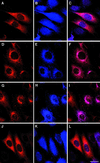
Immunofluorescence imaging of RALT in EGFR/ErbB-2 transfectants. Subconfluent monolayers of PINCO-RALT cells (see legend for Fig. 8A and B for details) were kept in MFM for 24 h. Dishes were processed for immunocytochemistry either before (A to C) or after (D to I) stimulation with EGF (10 ng/ml) for 15 (D to F) and 60 min (G to I). Samples shown in panels J to L were stimulated with 10% serum for 60 min. Samples shown in panels A, D, G, and J were stained with anti-RALT MAb 19C5/4; those shown in panels B, E, H, and K were stained with anti-ErbB-2 M6 antiserum. (C, F, I, L) Merged images. Bar (L), 20 μm.
FIG. 8
FIG. 8
RALT overexpression inhibits ErbB-2 mitogenic activity. (A) NIH-EGFR/ErbB-2 cells were infected with replication-defective PINCO and PINCO-RALT retroviruses; transduction efficiency was monitored by flow cytometry assessment of GFP-positive cells. Background fluorescence of parental cells is indicated by the left peak in each graph. (B) RALT expression in PINCO and PINCO-RALT cells was assessed by immunoblot analysis of lysates prepared either from cycling (left two lanes) or from quiescent cells stimulated for the indicated times (hours) with 5 ng of EGF/ml. WB, Western blotting. (C) NIH-EGFR/ErbB-2 cells were plated in 24-well plates, subjected to two rounds of infection with PINCO and PINCO-RALT viruses, and then switched to either MFM or MFM supplemented with serum or EGF. DNA synthesis was assessed after 40 h by pulse-labeling quadruplicate wells with [3H]thymidine. Data are expressed as fold increases of thymidine incorporation in mitogen-treated wells over that in mitogen-free wells. A typical dose-response analysis for EGF stimulation is shown. Open circles, PINCO; solid circles, PINCO-RALT. Bars at right, mitogenic response to 5% serum. Open bar, PINCO; solid bar, PINCO-RALT. (D) Open bars, average inhibition of ErbB-2-driven DNA synthesis in PINCO-RALT cells grown in EGF-supplemented MFM compared to that in PINCO cells; data were obtained from five experiments performed as detailed in the legend to panel C; solid bars, average inhibition of DNA synthesis in growth-arrested PINCO-RALT cells stimulated to enter S phase by EGF compared to that in PINCO cells. Assays were performed in quadruplicate, and data were obtained from four experiments. Error bars, standard deviations. (E) After infection, PINCO and PINCO-RALT cells were grown for 40 h either in MFM or MFM supplemented with 5 ng of EGF/ml or 5% serum. Distribution of cells in the cell cycle was assessed by staining with propidium iodide and analysis with an Epics flow cytometer. Percentages of cells in G1, S, and G2 phases are indicated.
FIG. 9
FIG. 9
RALT overexpression inhibits ErbB-2-driven cell transformation and late ERK activation. (A) NIH 3T3 fibroblasts were infected with either PINCO or PINCO-RALT retroviruses; >95% of target cells were transduced as indicated by GFP expression. PINCO or PINCO-RALT NIH 3T3 cells were superinfected with the indicated replication-defective recombinant retroviruses. Dishes were stained with methylene blue after 10 to 12 days. (B) NIH-EGFR/ErbB-2 fibroblasts transduced with PINCO or PINCO-RALT retroviruses were made quiescent and then challenged with either 10% serum or 5 ng of EGF/ml for the indicated times (hours). Lysates were analyzed by immunoblotting with anti-ERK 1 and 2 and anti-P-ERK antibodies, followed by the ECL reaction.

Similar articles

Cited by

References

    1. Alroy I, Yarden Y. The ErbB signaling network in embryogenesis and oncogenesis: signal diversification through combinatorial ligand-receptor interactions. FEBS Lett. 1997;410:83–86. - PubMed
    1. Anton E S, Marchionni M A, Lee K F, Rakic P. Role of GGF/neuregulin signaling in interactions between migrating neurons and radial glia in the developing cerebral cortex. Development. 1997;124:3501–3510. - PubMed
    1. Baulida J, Kraus M H, Alimandi M, Di Fiore P P, Carpenter G. All ErbB receptors other than the epidermal growth factor receptor are endocytosis impaired. J Biol Chem. 1996;271:5251–5257. - PubMed
    1. Beerli R R, Hynes N E. Epidermal growth factor-related peptides activate distinct subsets of ErbB receptors and differ in their biological activities. J Biol Chem. 1996;271:6071–6076. - PubMed
    1. Ben Levy R, Paterson H F, Marshall C J, Yarden Y. A single autophosphorylation site confers oncogenicity to the Neu/ErbB-2 receptor and enables coupling to the MAP kinase pathway. EMBO J. 1994;13:3302–3311. - PMC - PubMed

Publication types

MeSH terms