

Comparison of visual field progression in patients with normal pressure glaucoma between eyes with and without visual field loss that threatens fixation
- PMID: 11004102
- PMCID: PMC1723256
- DOI: 10.1136/bjo.84.10.1154
Comparison of visual field progression in patients with normal pressure glaucoma between eyes with and without visual field loss that threatens fixation
Abstract
Aim: To compare the frequency and site of visual field progression and changes in visual acuity in patients with normal pressure glaucoma (NPG) with and without pre-existing visual field loss.
Method: Patients with normal tension glaucoma were selected who had at least 10 visual fields over 5 or more years of follow up and no other condition that might influence the visual field or visual acuity. Alternate left and right eyes were selected from patients in random order. These eyes were then subdivided according to visual field defect threatening fixation, visual field defect not threatening fixation, and no visual field defect (fellow eyes). Eyes were defined as showing a threat to fixation according to the presence of a visual field defect involving one of more of four paracentral visual field locations. Pointwise linear regression analysis was applied to each visual field series using PROGRESSOR software. Progression of visual field loss was defined as the appearance of a regression slope 1 dB per year or more with a significance of p<0.01, which remained consistent with the addition of two of three successive visual fields to the series. The number of patients showing progression and the number where progression occurred in one of the four paracentral visual field locations was noted. The number of eyes losing two or more lines of Snellen visual acuity over the follow up period was also noted.
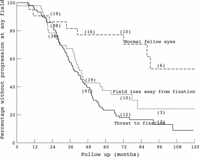
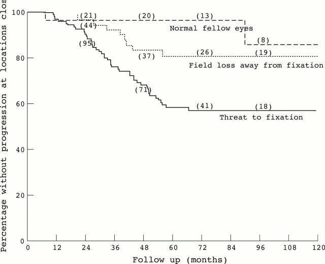
Results: 174 eyes of 174 patients were selected. 106 eyes had visual field loss threatening fixation, 46 eyes had visual field loss that did not threaten fixation, and 22 were fellow eyes with normal visual fields. The median follow up was 7.2 years. Eight eyes (36.4%) in the "normal visual fields" group, 31 eyes (67.4%) in the "visual field loss away from fixation" group, and 87 eyes (82.1%) in the "threat to fixation" group showed progression in any part of the visual field. Two eyes (9.1%) in the "normal visual fields" group, nine eyes (19.6%) in the "visual field loss away from fixation" group, and 45 eyes (42.5%) in the "threat to fixation" group showed progression at "threat to fixation". The Cox proportional hazards regression model showed an increased risk of progression at any part of the visual field for female sex and a decreased risk for eyes with normal visual fields. For progression at threat to fixation this model showed an increased risk with pre-existing threat to fixation. Eyes from older patients and those that went on to have progressive visual field loss at fixation were more likely to lose two lines of Snellen visual acuity over the follow up period.
Conclusion: Since 20-30% of previously field damaged eyes and over 60% without prior field loss fail to demonstrate progressive visual field damage over a long follow up it is recommended that normal pressure glaucoma patients be monitored for progression and that potentially harmful therapy be withheld until progression is demonstrated. Although the presence of visual field loss that threatens fixation does not constitute an increased risk of visual field progression it does indicate an increased risk of further loss of visual field close to fixation which is in turn associated with loss of central acuity. In the light of this finding, patients with visual field loss that threatens fixation should be managed more aggressively.
Figures
Similar articles
-
Threat to fixation at diagnosis and lifetime risk of visual impairment in open-angle glaucoma.Ophthalmology. 2015 May;122(5):1034-9. doi: 10.1016/j.ophtha.2014.12.004. Epub 2014 Dec 20. Ophthalmology. 2015. PMID: 25537196
-
Glaucoma surgery with or without adjunctive antiproliferatives in normal tension glaucoma: 2 Visual field progression.Br J Ophthalmol. 2001 Jun;85(6):696-701. doi: 10.1136/bjo.85.6.696. Br J Ophthalmol. 2001. PMID: 11371491 Free PMC article.
-
The effectiveness of intraocular pressure reduction in the treatment of normal-tension glaucoma. Collaborative Normal-Tension Glaucoma Study Group.Am J Ophthalmol. 1998 Oct;126(4):498-505. doi: 10.1016/s0002-9394(98)00272-4. Am J Ophthalmol. 1998. PMID: 9780094 Clinical Trial.
-
[Is trabeculectomy without danger in case of threatened fixation?].J Fr Ophtalmol. 1996;19(4):253-8. J Fr Ophtalmol. 1996. PMID: 8734217 Review. French.
-
[Visual field progression in glaucoma: cluster analysis].J Fr Ophtalmol. 2012 Nov;35(9):735-41. doi: 10.1016/j.jfo.2011.10.011. Epub 2012 Jul 6. J Fr Ophtalmol. 2012. PMID: 22771181 Review. French.
Cited by
-
Refinement of pointwise linear regression criteria for determining glaucoma progression.Invest Ophthalmol Vis Sci. 2013 Sep 19;54(9):6234-41. doi: 10.1167/iovs.13-11680. Invest Ophthalmol Vis Sci. 2013. PMID: 23908183 Free PMC article.
-
Defective angles of localized retinal nerve fiber layer reflect the severity of visual field defect- a cross-sectional analysis.BMC Ophthalmol. 2020 Apr 9;20(1):141. doi: 10.1186/s12886-020-01396-y. BMC Ophthalmol. 2020. PMID: 32272929 Free PMC article.
-
Rates of visual field progression in clinical glaucoma care.Acta Ophthalmol. 2013 Aug;91(5):406-12. doi: 10.1111/j.1755-3768.2012.02492.x. Epub 2012 Oct 16. Acta Ophthalmol. 2013. PMID: 23066646 Free PMC article.
-
Macular Structure-Function Relationships of All Retinal Layers in Primary Open-Angle Glaucoma Assessed by Microperimetry and 8 × 8 Posterior Pole Analysis of OCT.J Clin Med. 2021 Oct 28;10(21):5009. doi: 10.3390/jcm10215009. J Clin Med. 2021. PMID: 34768529 Free PMC article.
-
Detection of visual field progression in glaucoma with standard achromatic perimetry: a review and practical implications.Graefes Arch Clin Exp Ophthalmol. 2011 Nov;249(11):1593-616. doi: 10.1007/s00417-011-1787-5. Epub 2011 Aug 26. Graefes Arch Clin Exp Ophthalmol. 2011. PMID: 21870086 Review.
References
MeSH terms
LinkOut - more resources
Full Text Sources
Medical
Miscellaneous