

Ocular features in Rubinstein-Taybi syndrome: investigation of 24 patients and review of the literature
- PMID: 11004107
- PMCID: PMC1723270
- DOI: 10.1136/bjo.84.10.1177
Ocular features in Rubinstein-Taybi syndrome: investigation of 24 patients and review of the literature
Abstract
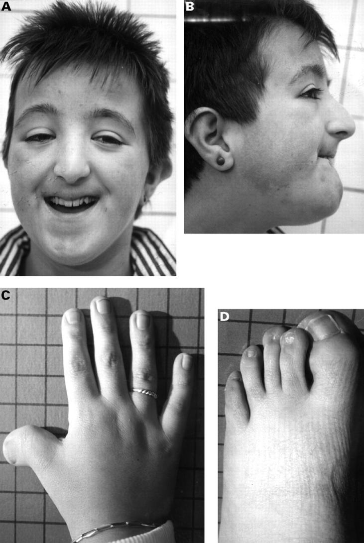
Aims: To delineate the nature and frequency of ocular pathology in Rubinstein-Taybi syndrome (RTs).
Methods: Literature was searched for reports describing ocular symptoms in patients with RTs. 24 RTs patients (out of a total of 73 Dutch known RTs individuals) were selected for ophthalmological and electrophysiological examination, selection being based only on the distance between a patient's residence and the place of investigation.
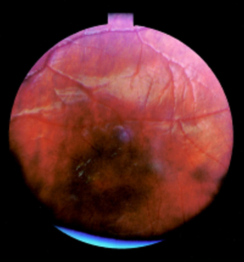
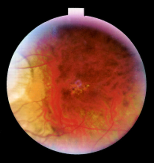
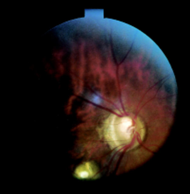
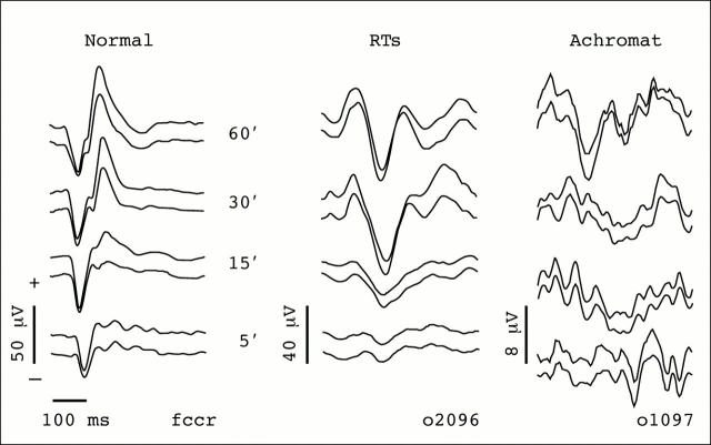
Results: Most frequently reported eye anomalies in the literature were lacrimal duct obstruction, corneal abnormalities, congenital glaucoma, congenital cataract, and colobomata. Abnormalities of almost any eye segment have been published in case reports. Ophthalmological examination of 24 Dutch RTs patients showed a visual acuity </=0.3 in five patients. The most frequently found eye anomalies were nasolacrimal duct problems (six patients), cataract (six patients, four congenital), and retinal abnormalities (18 patients). VEPs showed an abnormal waveform in 15 patients. It was possible to perform an ERG in 18 patients, of whom 14 were abnormal (eight showed cone dysfunction, six cone-rod dysfunction).
Conclusions: Ocular abnormalities occur in the majority of RTs patients and can be remarkably diverse. The high frequency of retinal dysfunction (78%) has not been described before. With age, retinal as well as electrophysiological abnormalities occur more frequently. In four patients no signs of retinal dysfunction were observed, indicating phenotypic heterogeneity. Further cytogenetic and molecular examination of the patients is needed before it becomes clear if this also represents genetic heterogeneity. Because of the high frequency of ocular abnormalities, visual function tests and electrophysiological investigations should be performed in every RTs patient at regular intervals.
Figures
References
Publication types
MeSH terms
LinkOut - more resources
Full Text Sources