. 2000 Oct;182(20):5872-9.
doi: 10.1128/JB.182.20.5872-5879.2000.

The last RNA-binding repeat of the Escherichia coli ribosomal protein S1 is specifically involved in autogenous control

Affiliations

The last RNA-binding repeat of the Escherichia coli ribosomal protein S1 is specifically involved in autogenous control

I V Boni et al. J Bacteriol. 2000 Oct.

Abstract

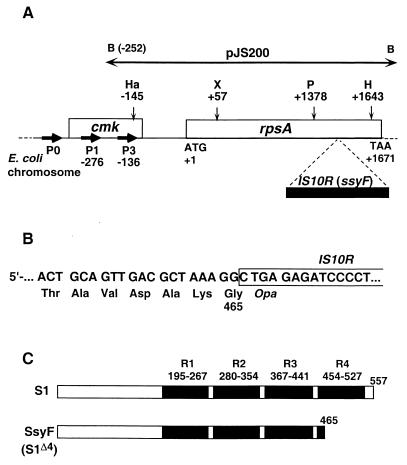
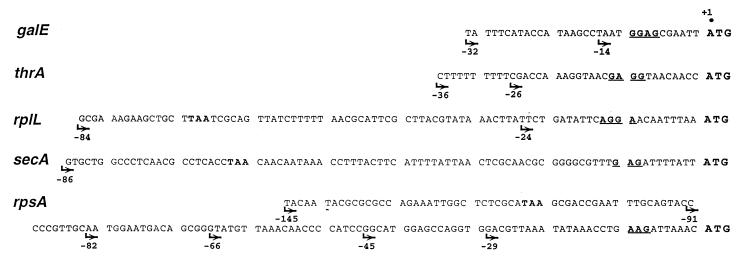
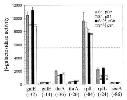
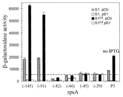
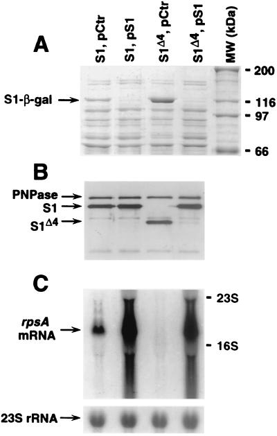
The ssyF29 mutation, originally selected as an extragenic suppressor of a protein export defect, has been mapped within the rpsA gene encoding ribosomal protein S1. Here, we examine the nature of this mutation and its effect on translation. Sequencing of the rpsA gene from the ssyF mutant has revealed that, due to an IS10R insertion, its product lacks the last 92 residues of the wild-type S1 protein corresponding to one of the four homologous repeats of the RNA-binding domain. To investigate how this truncation affects translation, we have created two series of Escherichia coli strains (rpsA(+) and ssyF) bearing various translation initiation regions (TIRs) fused to the chromosomal lacZ gene. Using a beta-galactosidase assay, we show that none of these TIRs differ in activity between ssyF and rpsA(+) cells, except for the rpsA TIR: the latter is stimulated threefold in ssyF cells, provided it retains at least ca. 90 nucleotides upstream of the start codon. Similarly, the activity of this TIR can be severely repressed in trans by excess S1, again provided it retains the same minimal upstream sequence. Thus, the ssyF stimulation requires the presence of the rpsA translational autogenous operator. As an interpretation, we propose that the ssyF mutation relieves the residual repression caused by normal supply of S1 (i.e., that it impairs autogenous control). Thus, the C-terminal repeat of the S1 RNA-binding domain appears to be required for autoregulation, but not for overall mRNA recognition.


Figures

FIG. 1
FIG. 1
Construction of E. coli strains in which TIRs originating from various genes are used to drive translation of the chromosomal lacZ gene. The DNA fragment carrying the TIR of interest (solid box) is first cloned in phase with the α-peptide gene (′lacZ′) of pEMBLΔ46, a pEMBL8+ derivative carrying a small deletion encompassing the lacZ RBS. The TIR is then transferred onto the chromosome of ENS0 by homologous recombination between lac sequences (lacI and lacZ) present on both the plasmid and chromosome. The chromosome of ENS0 (Lac) carries a slightly larger lac deletion than the plasmid, encompassing the lac promoter (Plac) and operator (op).
FIG. 2
FIG. 2
(A) General representation of the E. coli chromosome region encompassing genes cmk and rpsA, which encode cytidine monophosphate kinase and ribosomal protein S1, respectively (open boxes). The initiation (ATG) and termination (TAA) codons of rpsA are also indicated. The solid box indicates an IS10R insertion which has been found within the ssyF allele of rpsA (see text). B, Ha, X, P, and H designate restriction sites for the enzymes BamHI, HaeII, XmaI, PstI, and HindIII, respectively. The main promoters responsible for rpsA transcription are noted as P0, P1, and P3 (numbers below P1 and P3 refer to the positions of the corresponding transcription start points) (22). The horizontal, double-arrowed bar shows the rpsA region carried by plasmid pJS200 (29). (B) Sequence of the rpsA-IS10R junction in the ssyF allele showing premature interruption of translation within the inserted sequence. (C) General structure of the 557-residue-long S1 protein (S1) and of the truncated polypeptide encoded by the ssyF allele (SsyF, renamed here S1Δ4). Solid boxes indicate the four S1 motifs (R1 to R4), and numbers indicate the positions of the corresponding amino acids according to Subramanian (35).
FIG. 3
FIG. 3
Sequence of DNA fragments used as TIRs in this study. The name of the gene from which each fragment originated is indicated on the left (boldface, italic). Only sequences located upstream of the initiation codon (ATG) are shown in each case. SD sequences are underlined. Arrows indicate the exact 5′ boundaries of the different fragments that have actually been used as TIRs. Note that for rplL, secA, and rpsA, the longest fragments used retain the stop codon from the preceding gene (TAA in boldface).
FIG. 4
FIG. 4
Histograms showing the activities of the different TIRs listed in Fig. 3 (except rpsA TIR) as measured by the β-galactosidase activity resulting from their fusion to lacZ (Fig. 1). Each group of four vertical bars (from left to right; S1, pCtr; S1, pS1; S1Δ4, pCtr; S1Δ4, pS1) illustrates the activity of a given TIR when the chromosomal rpsA gene is either wild type or ssyF (i.e., encodes either the full-length [S1] or the truncated [S1Δ4] protein and the cell contains either plasmid pACYC184 [pCtr] or the same plasmid carrying the wild-type rpsA gene [pS1]). Below each group of four bars is indicated the gene from which the TIR originates and the 5′ boundary of the particular fragment used as TIR (Fig. 3). Given β-galactosidase activity (in nanomoles of ONPG hydrolyzed per minute per milligram of total protein) is the average of two to five experiments. n.d., not determined. The horizontal dotted line corresponds to the β-galactosidase expression observed with the genuine lacZ TIR in rpsA+ cells lacking any plasmid (5,600 U) (43).
FIG. 5
FIG. 5
Same as Fig. 4, except that the rpsA TIR has been used. Seven rpsA fragments differing in their 5′ end have been fused to lacZ, and the number below each group of four vertical bars corresponds to the 5′ boundary of the fragment used (Fig. 3). The 5′ boundary of fragment P3, which extends to nt −252, is not shown on Fig. 3. Since it carries the intact rpsAp3 (P3) promoter, it can drive β-galactosidase synthesis in the absence of IPTG. All symbols are defined as in Fig. 4.
FIG. 6
FIG. 6
(A) Total protein extracts of E. coli (10 μg of proteins per lane) separated on a 7.5% Laemmli gel and stained with Coomassie blue. In the experiment shown, the expression of the fusion β-galactosidase (arrowed) is driven by the rpsA TIR (−91). The exact extracts used are indicated above each lane (all symbols are defined as in Fig. 4). MW, standard proteins, with the corresponding molecular mass (kDa) given on the right. (B) Western blot analysis of samples from the same cultures as those described above. Samples (1 μg) were separated on a 10% Laemmli gel. The blot was revealed with polyclonal rabbit antibodies raised against purified S1 (3) mixed with the antibodies against PNPase to detect eventual lane-to-lane differences in total protein loading. Secondary antirabbit horseradish peroxidase-labelled antibodies (Promega) and ECL (enhanced chemiluminescence) reagent (Amersham) were used for detection. The positions of PNPase, S1, and S1Δ4 are marked with arrows. (C) Northern analysis of the rpsA mRNA in the same cultures as in panels A and B. Total RNA was separated on the 1% agarose–formaldehyde gel, blotted, and hybridized essentially as in reference . The 32P random-labelled rpsA-specific probe is described in Materials and Methods. The arrows show the positions of the main rpsA mRNA species and 16S and 23S rRNAs, as indicated. The strip below the main panel shows reprobing of the same membrane with a 23S rRNA-specific oligonucleotide probe.

Similar articles

Cited by

References

    1. Artamonova V S, Boni I V. The ssyF29 mutation in E. coli ribosomal protein S1 gene which suppresses the defect in protein export machinery results from an insertion of IS10R-element. Bioorg Khim. 1996;22:941–943. - PubMed
    1. Artamonova V S, Tsareva N V, Boni I V. Regulation of the ribosomal protein L7/12 synthesis: the role of the rplJL intercistronic region as a translational enhancer. Bioorg Khim. 1998;24:530–538. - PubMed
    1. Boni I V, Issaeva D M, Musychenko M L, Tzareva N V. Ribosome-messenger recognition: mRNA target sites for ribosomal protein S1. Nucleic Acids Res. 1991;19:155–162. - PMC - PubMed
    1. Boni I V, Zlatkin I V, Budowsky E I. Ribosomal protein S1 associates with Escherichia coli ribosomal 30S subunit by means of protein-protein interactions. Eur J Biochem. 1982;121:371–376. - PubMed
    1. Bycroft M, Hubbard T J P, Proctor M, Freund S M V, Murzin A G. The solution structure of the S1 RNA binding domain: a member of an ancient nucleic acid-binding fold. Cell. 1997;88:235–242. - PubMed

Publication types

MeSH terms

LinkOut - more resources