. 2000 Jul;5(3):196-206.
doi: 10.1379/1466-1268(2000)005<0196:cotmgf>2.0.co;2.

Characterization of the mouse gene for the heavy metal-responsive transcription factor MTF-1

Affiliations

Characterization of the mouse gene for the heavy metal-responsive transcription factor MTF-1

A Auf der Maur et al. Cell Stress Chaperones. 2000 Jul.

Abstract

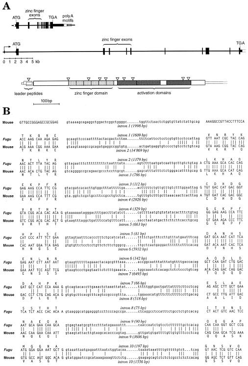
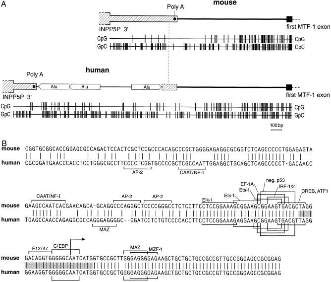
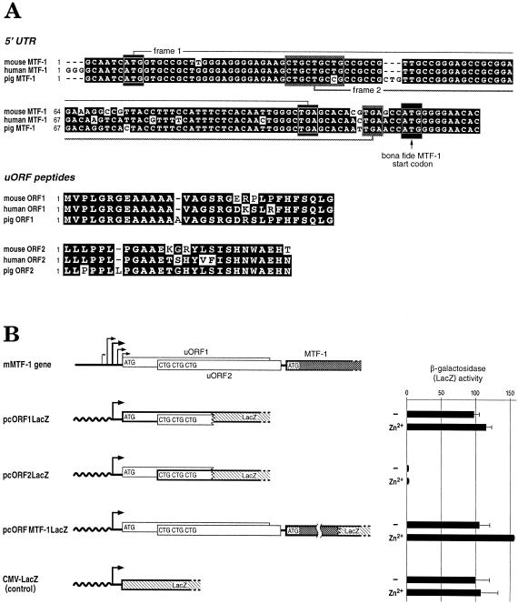
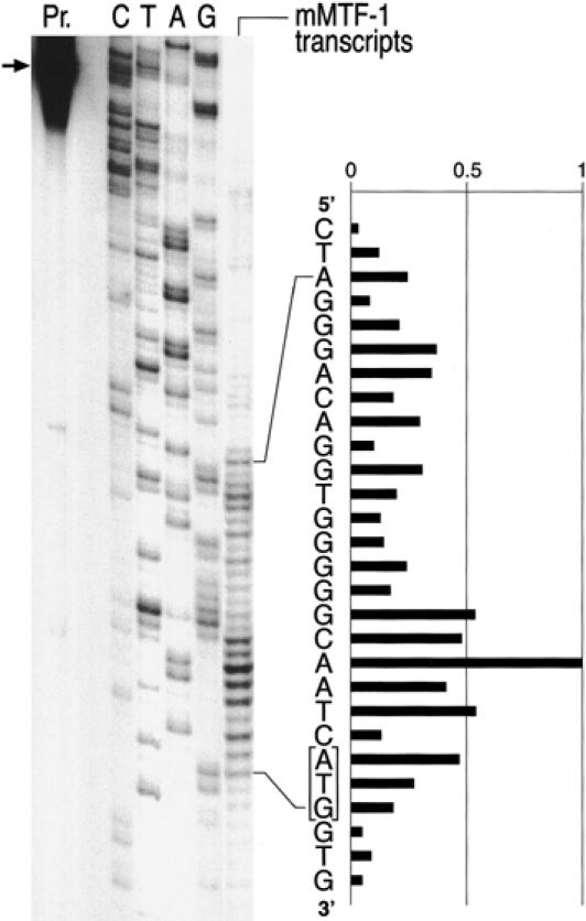
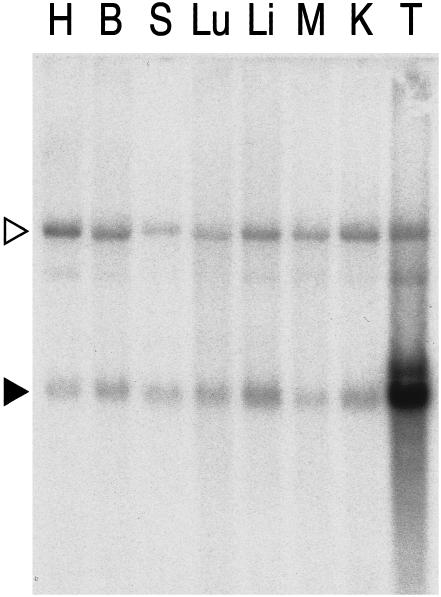
MTF-1 is a zinc finger transcription factor that mediates the cellular response to heavy metal stress; its targeted disruption in the mouse leads to liver decay and embryonic lethality at day E14. Recently, we have sequenced the entire MTF-1 gene in the compact genome of the pufferfish Fugu rubripes. Here we have defined the promoter sequences of human and mouse MTF-1 and the genomic structure of the mouse MTF-1 locus. The transcription unit of MTF-1 spans 42 kb (compared to 8.5 kb in Fugu) and is located downstream of the gene for a phosphatase (INPP5P) in mouse, human, and fish. In all of these species, the MTF promoter region has the features of a CpG island. In both mouse and human, the 5' untranslated region harbors conserved short reading frames of unknown function. RNA mapping experiments revealed that in these two species, MTF-1 mRNA is transcribed from a cluster of multiple initiation sites from a TATA-less promoter without metal-responsive elements. Transcription from endogenous and transfected MTF-1 promoters was not affected by heavy metal load or other stressors, in support of the notion that MTF-1 activity is regulated at the posttranscriptional level. Tissue Northern blots normalized for poly A+ RNA indicate that MTF-1 is expressed at similar levels in all tissues, except in the testes, that contain more than 10-fold higher mRNA levels.


Figures

Fig 1.
Fig 1.
Genomic structure of Fugu and mouse MTF-1 genes. (A) In the pufferfish (on top) and in the mouse, the transcription unit of MTF-1 spans 8.5 and 42 kb, respectively. The exons (black boxes), introns, and spacers (black lines) are drawn to approximate scale and clearly illustrate the compaction of the Fugu MTF-1 gene relative to MTF-1 of the mouse and probably most other vertebrates. Shown underneath the genomic structure is a schematic view of the mouse cDNA with protein reading frames indicated, including the 2 short overlapping uORFs (uORF1 and 2), followed by the MTF-1 ORF with its 6 zinc fingers for DNA binding and 3 activation domains. The position of introns is indicated by inverted open triangles. (B) Comparison of the Fugu and mouse MTF-1 splice junctions. Both genes contain 11 exons; 1 intron and flanking exon sequences are deleted in the mouse relative to the Fugu, while the mouse leader sequence contains an additional intron. Splice junction sequences are conserved to the nucleotide, even if located in a region of low similarity between the 2 genes (notably at intron 7 of Fugu vs intron 8 of the mouse). Accession numbers pufferfish: AJ131394; accession number mouse: AJ251880
Fig 2.
Fig 2.
Structure of the MTF-1 promoter region. (A) Genomic structure of the last exon of INPP5P, subsequent intergenic segment, and the promoter region of the MTF-1 gene in mouse (accession number AJ251880), and human (accession number AJ251881). In human, the polyadenylation site is shifted upstream relative to the mouse, probably as a consequence of the insertion of 3 Alu repeats. CpG-density and [G + C] content, as represented by the GpC-density, is also indicated by the bar graph below. In mammals (shown here) and pufferfish (Auf der Maur et al 1999), the MTF-1 promoter is embedded in a CpG island. In mouse and human, not only the CpG density but also the overall [G + C] content is increased, typical for warmblooded vertebrates, but not fish. Note that the CpG island features extend quite far upstream of the first MTF-1 exon in mouse and human, yet only about 60 bp of exon-proximal promoter sequences are conserved (Fig 2B). (B) Comparison of mouse and human MTF-1 promoters. While the typical CpG island properties extend further upstream (Fig 2A), the region of strong sequence similarity is confined to the immediate upstream region and the mRNA leader (5′ UTR). Putative transcription factor binding sites, as found using the TRANSFAC database (Heinemeyer et al 1998), are indicated by brackets if present in either 1 or both species. Putative binding sites correspond to the ones of previously characterized transcription factors but have not been verified experimentally for the mouse or human MTF-1 promoters. The hatched region denotes the region of multiple transcription starts, as shown in Figure 4, with the major start indicated by an arrow within the sequence CAATC
Fig 3.
Fig 3.
Sequence of the 5′ UTR region of the MTF-1 gene. (A) Alignment of mouse, human, and pig MTF-1 5′ UTR sequences and peptide ORFs. Two ORFs (uORF1 and uORF2) are located upstream of the actual MTF-1 ORF. For uORF1, AUG start and TGA stop are demonstrated by white rectangles, and uORF2 triple CUG and TGA by shadowed rectangles. The start codon of the bona fide MTF-1 ORF is marked with an arrow. Shown below is the alignment of uORF1 and 2 peptide sequences. Conserved nucleotides and amino acids are highlighted as white letters on a black background. The 5′ UTR of pig MTF-1 represents an EST (accession number Z84162). (B) Translation initiation at uORF start codons. To assess whether the uORFs are translated, recombinant plasmids were constructed wherein the start codon from either uORF1, uORF2, or hMTF-1 was fused in frame to the β-galactosidase gene ORF. Plasmids pcORF1lacZ, with β-Gal fused with the uORF1 AUG, pcORF2lacZ, with 3 CUGs from uORF2, and pcMTF1ORFlacZ, with AUG of the MTF-1 gene ORF, were separately transfected into HeLa cells. Zinc induction was performed with 100 μM ZnCl2 in the medium for 4 hours. The expression level of β-galactosidase from the start codon of each construct was quantified by the ONPG assay (see Materials and Methods). The expression level of the control CMV-lacZ plasmid is set as 100%. Note that the ORF fused to lacZ is indicated in bold. The standard deviation for the zinc-treated pcORFMTF-1lacZ is so small that the error bar is hardly visible. Shown on top for comparison is the genomic structure of the mouse MTF-1 gene with its multiple transcription starts, and the 2 uORFs (uORF1 and uORF2), followed by the bona fide start of the MTF-1 ORF
Fig 4.
Fig 4.
Precise mapping of the multiple transcription start sites in the MTF-1 gene. RNA was isolated from cells and subjected to S1 nuclease mapping with a labeled DNA oligonucleotide corresponding to the entire promoter region of MTF-1. While our β-globin reporter gene under these assay conditions yields a strong, narrowly defined site of transcription initiation (Westin et al 1987, and not shown), the MTF-1 promoter shows the scattered multiple starts characteristic of TATA-less, CpG-island type promoters. At least the longer transcripts are expected to allow for translation initiation from the AUG of uORF1 (framed) (see also Fig 3). Pr., position of input oligonucleotide probe (for details see Materials and Methods); C/T/A/G, sequencing reaction of the MTF-1 promoter region used as reference; “mMTF-1 transcripts” indicates the pattern of protection from S1 nuclease digest after hybridization of cellular mRNA to the probe oligonucleotide
Fig 5.
Fig 5.
Similar levels of MTF-1 mRNA in all tissues except testes. A tissue Northern blot containing a normalized amount of poly A+ RNA from various mouse organs was hybridized with a cDNA probe of mouse MTF-1. The autoradiograph reveals the presence of MTF-1 mRNA (black triangle) in all tissues: heart (H), brain (B), spleen (S), lung (Lu), liver (Li), skeletal muscle (M), kidney (K), and testes (T). An upper band (open triangle) that was not seen in a previous Northern blot from liver (not shown) was not further investigated. The extremely high representation of MTF transcripts in testicular tissue was confirmed by quantification of protein by electrophoretic mobility shift (not shown). Similar high testicular transcript levels have been previously reported for other components of the RNA Pol II transcriptional apparatus (Schmidt and Schibler 1995)

Similar articles

Cited by

References

    1. Auf der Maur A, Belser T, Elgar G, Georgiev O, Schaffner W. Characterization of the transcription factor MTF-1 from the Japanese pufferfish (Fugu rubripes) reveals evolutionary conservation of heavy metal stress response. Biol Chem. 1999;380:175–185. - PubMed
    1. Beg AA, Sha WC, Bronson RT, Ghosh S, Baltimore D. Embryonic lethality and liver degeneration in mice lacking the RelA component of NK-kappa B. Nature. 1995;376:167–170. - PubMed
    1. Bittel D, Dalton T, Samson SL, Gedamu L, Andrews GK. The DNA binding activity of metal response element-binding transcription factor-1 is activated in vivo and in vitro by zinc, but not by other transition metals. J Biol Chem. 1998;273:7127–7133. - PubMed
    1. Brown CY, Mize GJ, Pineda M, George DL, Morris DR. Role of two upstream open reading frames in the translational control of oncogene mdm2. Oncogene. 1999;18:5631–5637. - PubMed
    1. Cross SH, Bird AP. CpG islands and genes. Curr Opin Genet Dev. 1995;5:309–314. - PubMed

Publication types

MeSH terms

LinkOut - more resources