

Heat shock and arsenite induce expression of the nonclassical class I histocompatibility HLA-G gene in tumor cell lines
- PMID: 11005379
- PMCID: PMC312887
- DOI: 10.1379/1466-1268(2000)005<0207:hsaaie>2.0.co;2
Heat shock and arsenite induce expression of the nonclassical class I histocompatibility HLA-G gene in tumor cell lines
Abstract
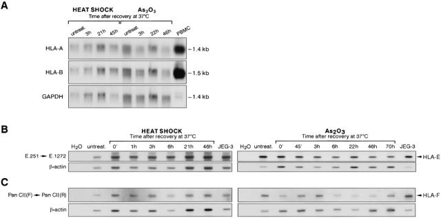
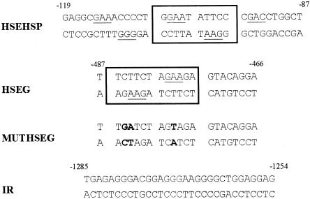
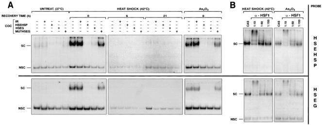
The nonclassical histocompatibility class I gene HLA-G has a tissue-restricted expression. To explore mechanisms involved in HLA-G transcriptional regulation, we have investigated the effect of stress, including heat shock and arsenite treatment, on HLA-G expression in tumor cell lines. We show that stress induces an increase of the level of the different HLA-G alternative transcripts without affecting other MHC class I HLA-A, -B, -E, and -F transcripts. A heat shock element (HSE) that binds to heat shock factor 1 (HSF1) on stress conditions was further identified within the HLA-G promoter. Considering the ability of HLA-G to modulate the function of immunocompetent cells, we hypothesize a new feature of HLA-G as a signal regulating the immune response to stress.
Figures

Similar articles
-
A specific interferon (IFN)-stimulated response element of the distal HLA-G promoter binds IFN-regulatory factor 1 and mediates enhancement of this nonclassical class I gene by IFN-beta.J Biol Chem. 2001 Mar 2;276(9):6133-9. doi: 10.1074/jbc.M008496200. Epub 2000 Nov 21. J Biol Chem. 2001. PMID: 11087747
-
Distinct stress-inducible and developmentally regulated heat shock transcription factors in Xenopus oocytes.Dev Biol. 1997 Jan 1;181(1):47-63. doi: 10.1006/dbio.1996.8441. Dev Biol. 1997. PMID: 9015264
-
Specific binding of nuclear factors to the HLA-G gene promoter correlates with a lack of HLA-G transcripts in first trimester human fetal liver.Hum Immunol. 1998 Dec;59(12):751-7. doi: 10.1016/s0198-8859(98)00081-0. Hum Immunol. 1998. PMID: 9831130
-
Differential in vivo and in vitro HLA-G expression in melanoma cells: potential mechanisms.Hum Immunol. 2003 Nov;64(11):1057-63. doi: 10.1016/j.humimm.2003.08.357. Hum Immunol. 2003. PMID: 14602236 Review.
-
Transcriptional regulation of the MHC class Ib genes HLA-E, HLA-F, and HLA-G.Hum Immunol. 2000 Nov;61(11):1102-7. doi: 10.1016/s0198-8859(00)00198-1. Hum Immunol. 2000. PMID: 11137213 Review.
Cited by
-
Role of HLA-G in Viral Infections.Front Immunol. 2022 Feb 14;13:826074. doi: 10.3389/fimmu.2022.826074. eCollection 2022. Front Immunol. 2022. PMID: 35237271 Free PMC article. Review.
-
Modulation of HLA-G expression in human neural cells after neurotropic viral infections.J Virol. 2005 Dec;79(24):15226-37. doi: 10.1128/JVI.79.24.15226-15237.2005. J Virol. 2005. PMID: 16306594 Free PMC article.
-
Implications of the polymorphism of HLA-G on its function, regulation, evolution and disease association.Cell Mol Life Sci. 2011 Feb;68(3):369-95. doi: 10.1007/s00018-010-0580-7. Epub 2010 Nov 24. Cell Mol Life Sci. 2011. PMID: 21107637 Free PMC article. Review.
-
Impact of HLA-G analysis in prevention, diagnosis and treatment of pathological conditions.World J Methodol. 2014 Mar 26;4(1):11-25. doi: 10.5662/wjm.v4.i1.11. eCollection 2014 Mar 26. World J Methodol. 2014. PMID: 25237627 Free PMC article. Review.
-
Non-classical transcriptional regulation of HLA-G: an update.J Cell Mol Med. 2009 Sep;13(9B):2973-89. doi: 10.1111/j.1582-4934.2009.00800.x. Epub 2009 Jun 5. J Cell Mol Med. 2009. PMID: 19508383 Free PMC article. Review.
References
-
- Amiot L, Onno M, Drénou B, Monvoisin C, Fauchet R. HLA-G class I gene expression in normal and malignant hematopoietic cells. Hum Immunol. 1998;59:524–528. - PubMed
-
- Amiot L, Onno M, Renard I, Drénou B, Guillaudeux T, Le Bouteiller P, Fauchet R. HLA-G transcription studies during the different stages of normal and malignant hematopoiesis. Tissue Antigens. 1996;47:408–413. - PubMed
-
- Blaschitz A, Lenfant F, Mallet V, Hartmann M, Bensussan A, Geraghty DE, Le Bouteiller P, Dohr G. Endothelial cells in chorionic fetal vessels of first trimester placenta express HLA-G. Eur J Immunol. 1997;27:3380–3388. - PubMed
-
- Botzler C, Ellwart J, Günther W, Eissner G, Multhoff G. Synergistic effects of heat and ET-18-OCH3 on membrane expression of hsp70 and lysis of leukemic K562 cells. Exp Haematol. 1999;27:470–478. - PubMed
Publication types
MeSH terms
Substances
LinkOut - more resources
Full Text Sources
Other Literature Sources
Research Materials