. 2000 Oct 10;97(21):11488-93.
doi: 10.1073/pnas.210319397.

Ribozyme rescue of photoreceptor cells in P23H transgenic rats: long-term survival and late-stage therapy

Affiliations

Ribozyme rescue of photoreceptor cells in P23H transgenic rats: long-term survival and late-stage therapy

M M LaVail et al. Proc Natl Acad Sci U S A. .

Abstract

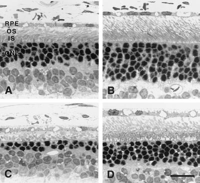
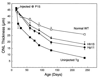
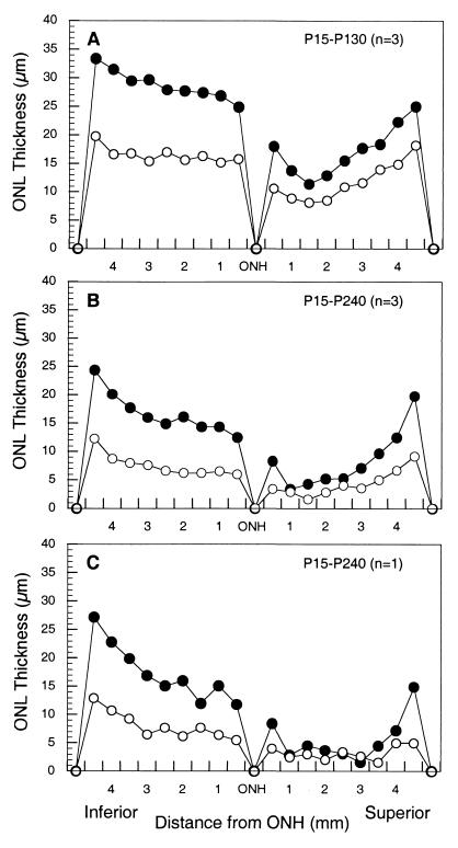
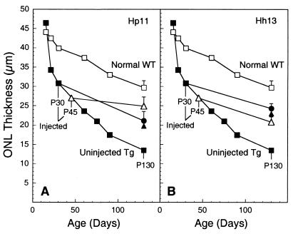
Ribozyme-directed cleavage of mutant mRNAs appears to be a potentially effective therapeutic measure for dominantly inherited diseases. We previously demonstrated that two ribozymes targeted to the P23H mutation in rhodopsin slow photoreceptor degeneration in transgenic rats for up to 3 months of age when injected before significant degeneration at postnatal day (P) 15. We now have explored whether ribozyme rescue persists at older ages, and whether ribozymes are effective when injected later in the degeneration after significant photoreceptor cell loss. Recombinant adeno-associated virus (rAAV) vectors incorporating a proximal bovine rod opsin promoter were used to transfer either hairpin or hammerhead ribozyme genes to photoreceptors. For the study of long-term survival, rAAV was administered by subretinal injection at P15, and the rats were allowed to live up to 8 months of age. For the study of late-stage gene transfer, rAAV was administered at P30 or P45, when 40-45% of the photoreceptors already had degenerated. Eyes were examined functionally by the electroretinogram and structurally by morphometric analysis. When injected at P15, expression of either ribozyme markedly slowed the rate of photoreceptor degeneration for at least 8 months and resulted in significantly greater electroretinogram amplitudes at least up to P180. When injected at P30 or P45, virtually the same number of photoreceptors survived at P130 as when injected at P15. Ribozyme rescue appears to be a potentially effective, long-term therapy for autosomal dominant retinal degeneration and is highly effective even when the gene transfer is done after significant photoreceptor cell loss.


Figures

Figure 1
Figure 1
Light micrographs of plastic-embedded P23H transgenic rat retinas taken at P130 (A and B) or P240 (C and D) from the posterior inferior region along the vertical meridian of the eye. (A) Uninjected eye from a rat at P130, which shows 3–4 rows of photoreceptor nuclei in the ONL, reduced from the normal 8–9 at this age. Photoreceptor inner segments (IS) and outer segments (OS) are shorter than normal, although still intact. RPE, retinal pigment epithelium. (B) Retina from the opposite eye from the same rat as in A, which was injected subretinally injected with Hh13 ribozyme at P15. About 6–7 rows of photoreceptor nuclei are present, and the photoreceptor inner and outer segments are about 70–80% of normal length. (C) Uninjected eye from a rat at P240, which shows only 1–2 rows of photoreceptor nuclei surviving, and only remnants of inner and outer segments present. (D) Retina from the opposite eye from the same rat in C, which was injected subretinally injected with Hh13 ribozyme at P15. About 4–5 rows of photoreceptor nuclei are still present and the inner and outer segments are almost as long as at P130 (B). Toluidine blue stain. (Bar = 20 μm.)
Figure 2
Figure 2
Measurements of ONL thickness in rats killed at different ages. □ denote normal wild-type (WT), nontransgenic animals. P23H-3 transgenic (Tg) rats were either uninjected (■) or injected subretinally with rAAV vectors carrying either Hp11 (●) or Hh13 (⧫) ribozymes. All injections were done at P15. The symbols represent mean ± SD, and error bars were omitted if they fell within the symbol. The number of animals in each group is shown in Table 1. The data points at P60, P75, and P90 are taken from our earlier study (25).
Figure 3
Figure 3
Measurement of the ONL thickness along the vertical meridian of the eye from the optic nerve head (ONH) to the ora serrata (anterior margin of the retina) of P23H-3 mutant rhodopsin transgenic rats. The rats were either uninjected (○) or injected (●) with Hh13 ribozymes at P15 and the eyes taken either at P130 (A) or P240 (B and C). Means of three representative retinas are shown in A and B, and the plot of a single rat of those plotted in B is shown in C. For each rat, three measurements were made in each of nine 440-μm fields in both the superior and inferior hemispheres.
Figure 4
Figure 4
Measurements of ONL thickness in rats killed at P130 after late-stage ribozyme injection at P30 and P45. Data for the normal wild-type (WT) nontransgenic rats (□) and the uninjected P23H-3 transgenic (Tg) rats (■) are the same as in Fig. 2. P23H-3 rats were injected subretinally with rAAV vectors carrying either Hp11 (A) or Hh13 (B) ribozymes at either P30 (●) or P45 (▵). For comparison, the data point from injection at P15 and killed at P130 is shown (▴). The values represent mean ± SD, and error bars were omitted if they fell within the symbol. The number of animals in each group is shown in Table 1.

References

    1. Humphries P, Farrar G J, Kenna P, McWilliam P. Clin Genet. 1990;38:1–13. - PubMed
    1. Berson E L. Invest Ophthalmol Visual Sci. 1993;34:1659–1676. - PubMed
    1. Gorin M B, Breitner J C S, De Jong P T V M, Hageman G S, Klaver C C W, Kuehn M H, Seddon J M. Mol Vis. 1999;5:29. - PubMed
    1. Zack D J, Dean M, Molday R S, Nathans J, Redmond T M, Stone E M, Swaroop A, Valle D, Weber B H. Mol Vis. 1999;5:30. - PubMed
    1. Wong F. Arch Ophthalmol. 1995;113:1245–1247. - PubMed

Publication types

LinkOut - more resources