

Molecular mechanisms underlying differential odor responses of a mouse olfactory receptor
- PMID: 11005853
- PMCID: PMC27088
- DOI: 10.1073/pnas.97.20.10712
Molecular mechanisms underlying differential odor responses of a mouse olfactory receptor
Abstract
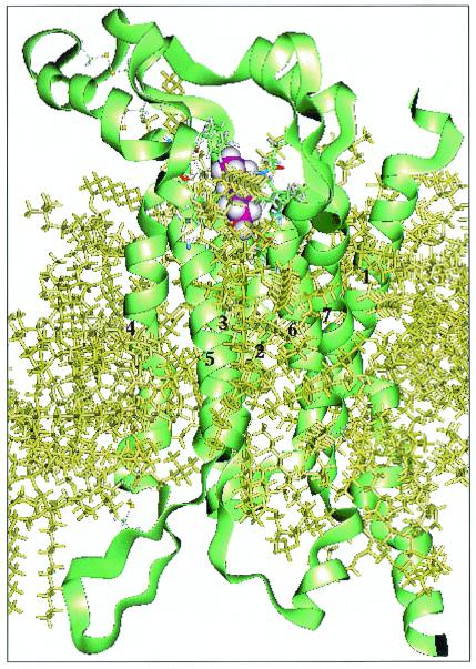
The prevailing paradigm for G protein-coupled receptors is that each receptor is narrowly tuned to its ligand and closely related agonists. An outstanding problem is whether this paradigm applies to olfactory receptor (ORs), which is the largest gene family in the genome, in which each of 1,000 different G protein-coupled receptors is believed to interact with a range of different odor molecules from the many thousands that comprise "odor space." Insights into how these interactions occur are essential for understanding the sense of smell. Key questions are: (i) Is there a binding pocket? (ii) Which amino acid residues in the binding pocket contribute to peak affinities? (iii) How do affinities change with changes in agonist structure? To approach these questions, we have combined single-cell PCR results [Malnic, B., Hirono, J., Sato, T. & Buck, L. B. (1999) Cell 96, 713-723] and well-established molecular dynamics methods to model the structure of a specific OR (OR S25) and its interactions with 24 odor compounds. This receptor structure not only points to a likely odor-binding site but also independently predicts the two compounds that experimentally best activate OR S25. The results provide a mechanistic model for olfactory transduction at the molecular level and show how the basic G protein-coupled receptor template is adapted for encoding the enormous odor space. This combined approach can significantly enhance the identification of ligands for the many members of the OR family and also may shed light on other protein families that exhibit broad specificities, such as chemokine receptors and P450 oxidases.
Figures
References
-
- Malnic B, Hirono J, Sato T, Buck L B. Cell. 1999;96:713–723. - PubMed
-
- Donnelly D. Biochem Soc Trans. 1993;21:36–39. - PubMed
-
- Ding H Q, Karasawa N, Goddard W A., III J Chem Phys. 1992;97:4309–4315.
-
- Ding H Q, Karasawa N, Goddard W A., III Chem Phys Lett. 1992;196:6–10.
-
- Jain A, Vaidehi N, Rodriguez G. J Comp Physiol. 1993;106:258–268.
Publication types
MeSH terms
Substances
LinkOut - more resources
Full Text Sources
