

Enrichment for murine keratinocyte stem cells based on cell surface phenotype
- PMID: 11005869
- PMCID: PMC27131
- DOI: 10.1073/pnas.97.20.10960
Enrichment for murine keratinocyte stem cells based on cell surface phenotype
Abstract
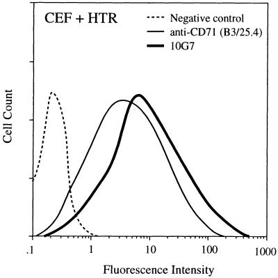
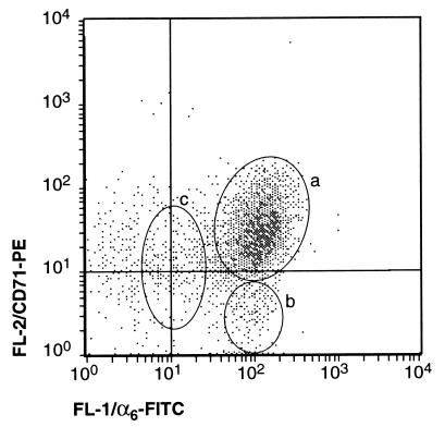
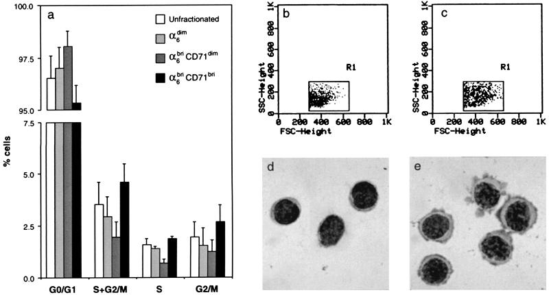
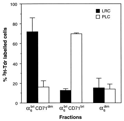
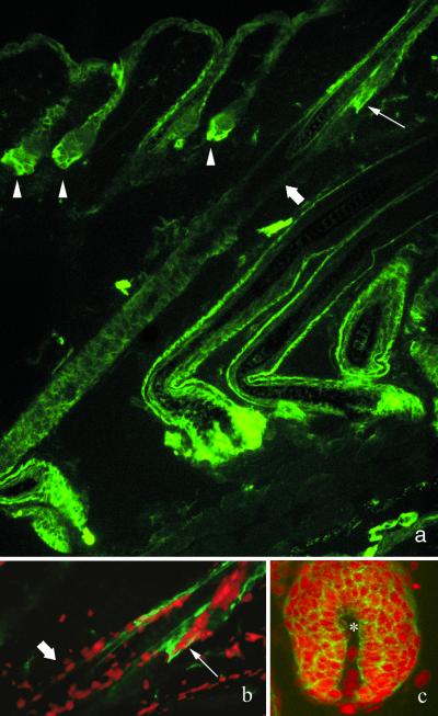
The identification and physical isolation of epithelial stem cells is critical to our understanding of their growth regulation during homeostasis, wound healing, and carcinogenesis. These stem cells remain poorly characterized because of the absence of specific molecular markers that permit us to distinguish them from their progeny, the transit amplifying (TA) cells, which have a more restricted proliferative potential. Cell kinetic analyses have permitted the identification of murine keratinocyte stem cells (KSCs) as slowly cycling cells that retain [(3)H]thymidine ([(3)H]Tdr) label, termed label-retaining cells (LRCs), whereas TA cells are visualized as rapidly cycling cells after a single pulse of [(3)H]Tdr, termed pulse-labeled cells (PLCs). Here, we report on the successful separation of KSCs from TA cells through the combined use of in vivo cell kinetic analysis and fluorescence-activated cell sorting. Specifically, we demonstrate that murine dorsal keratinocytes characterized by their high levels of alpha(6) integrin and low to undetectable expression of the transferrin receptor (CD71) termed alpha(6)(bri)CD71(dim) cells, are enriched for epithelial stem cells because they represent a minor ( approximately 8%) and quiescent subpopulation of small blast-like cells, with a high nuclear:cytoplasmic ratio, containing approximately 70% of label-retaining cells, the latter being a well documented characteristic of stem cells. Conversely, TA cells could be enriched in a phenotypically distinct subpopulation termed alpha(6)(bri)CD71(bri), representing the majority ( approximately 60%) of basal keratinocytes that are actively cycling, and importantly contain approximately 70% of [(3)H]Tdr pulse-labeled cells. Importantly, immunostaining of dorsal skin revealed the presence of CD71(dim) cells in the hair follicle bulge region, a well documented location for KSCs.
Figures
Comment in
-
Epidermal stem cells: properties, markers, and location.Proc Natl Acad Sci U S A. 2000 Dec 5;97(25):13473-5. doi: 10.1073/pnas.250380097. Proc Natl Acad Sci U S A. 2000. PMID: 11087834 Free PMC article. No abstract available.
References
-
- Bickenbach J R. J Dent Res. 1981;60:1611–1620. - PubMed
-
- Potten C S, editor. Stem Cells: Their Identification and Characterization. London: Churchill Livingstone; 1983. pp. 200–232.
-
- Morris R J, Fischer S M, Slaga T J. J Invest Dermatol. 1985;84:277–281. - PubMed
-
- MacKenzie I C, Bickenbach J R. Cell Tissue Res. 1985;242:551–556. - PubMed
-
- Potten C S. Int J Radiat Biol. 1986;49:257–278. - PubMed
Publication types
MeSH terms
Substances
Grants and funding
LinkOut - more resources
Full Text Sources
Other Literature Sources
Medical
Research Materials
