. 2000 Oct;41(11):3365-73.

Corneal opacity in lumican-null mice: defects in collagen fibril structure and packing in the posterior stroma

Affiliations

Corneal opacity in lumican-null mice: defects in collagen fibril structure and packing in the posterior stroma

S Chakravarti et al. Invest Ophthalmol Vis Sci. 2000 Oct.

Abstract

Purpose: Gene targeted lumican-null mutants (lum(tm1sc)/lum(tm1sc)) have cloudy corneas with abnormally thick collagen fibrils. The purpose of the present study was to analyze the loss of transparency quantitatively and to define the associated corneal collagen fibril and stromal defects.

Methods: Backscattering of light, a function of corneal haze and opacification, was determined regionally using in vivo confocal microscopy in lumican-deficient and wild-type control mice. Fibril organization and structure were analyzed using transmission electron microscopy. Biochemical approaches were used to quantify glycosaminoglycan contents. Lumican distribution in the cornea was elucidated immunohistochemically. RESULTS; Compared with control stromas, lumican-deficient stromas displayed a threefold increase in backscattered light with maximal increase confined to the posterior stroma. Confocal microscopy through-focusing (CMTF) measurement profiles also indicated a 40% reduction in stromal thickness in the lumican-null mice. Transmission electron microscopy indicated significant collagen fibril abnormalities in the posterior stroma, with the anterior stroma remaining relatively unremarkable. The lumican-deficient posterior stroma displayed a pronounced increase in fibril diameter, large fibril aggregates, altered fibril packing, and poor lamellar organization. Immunostaining of wild-type corneas demonstrated high concentrations of lumican in the posterior stroma. Biochemical assessment of keratan sulfate (KS) content of whole eyes revealed a 25% reduction in KS content in the lumican-deficient mice.

Conclusions: The structural defects and maximum backscattering of light clearly localized to the posterior stroma of lumican-deficient mice. In normal mice, an enrichment of lumican was observed in the posterior stroma compared with that in the anterior stroma. Taken together, these observations indicate a key role for lumican in the posterior stroma in maintaining normal fibril architecture, most likely by regulating fibril assembly and maintaining optimal KS content required for transparency.


Figures

Figure 1
Figure 1
In vivo CMTF three-dimensional image and profile of lum+/lum+ and lumtm1sc/lumtm1sc. Three-dimensional CMTF images of typical corneas from 5-month-old wild-type (A) and lumican-null mutants (B) show the stroma (b and b′) spanned by an epithelium (a and a′) and the endothelium (c and c′). The backscatter in the mutant corneal stroma increased maximally near the endothelium. A traced profile of the scanned images (C) shows a marked increase in the intensity of backscattered light in the mutant stroma (b′) with a sharp increase approaching saturation at the endothelium (c′).
Figure 2
Figure 2
Fibril defects are localized to the posterior corneal stroma in corneas from 7.5-month-old lumtm1sc/lumtm1sc mice. Transmission electron micrographs comparing collagen fibril structure in the anterior (A, B) and posterior (C, D) stroma of lumican-deficient (B, D) and wild-type (A, C) corneas. Fibril structure and packing are comparable in the anterior stroma of wild-type (A) and null mice (B). In contrast, the fibrils in the posterior stroma of lumican-deficient mice (D) contain abnormally large-diameter fibrils. Numerous examples of fibrils with irregular contours or laterally associated fibrils are present (arrows). These structures are indicative of abnormal lateral growth. Bar, 100 nm.
Figure 3
Figure 3
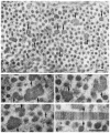
Transmission electron micrographs of corneal sections illustrate structural defects in collagen fibrils indicative of abnormal lateral fusion in the posterior stroma. An overview of a region that contained numerous large-diameter collagen fibrils, many with irregular contours, indicative of abnormal lateral growth (A). The mean diameter of fibrils indicated by the arrows was 87.5 ± 17.7 nm (SD) compared with a normal diameter of 35 nm. A gallery of higher magnification micrographs provides structural details of the abnormal fibrils (B, C, D). Diameters of the fibrils indicated by the arrows were: (B) 135, 91, and 79 nm; (C) 93 and 101 nm; (D) 109 and 63 nm; and (E) 56 nm. (B, C) Especially obvious images indicative of abnormal lateral association and fusion. Corneas were from 7.5-month-old lumican-deficient mice. Bars, 100 nm.
Figure 4
Figure 4
Mean fibril diameter and fibril diameter distribution in anterior versus posterior stroma. The fibril diameter distributions were analyzed in the anterior (A) and posterior stromas (B) in corneas of 7.5-month-old wild-type (lum+/lum+) and lumican-deficient (lumtm1sc/lumtm1sc) mice. Masked samples selected randomly from the different regions were analyzed. (A) The anterior stroma of the wild-type and lumican-deficient corneas were nearly identical in mean fibril diameter and distribution, although with a small but reproducible increase in fibril diameter. The fibril diameter range in the anterior stroma was 32 nm and 31 nm for wild-type (minimum, 14 nm; maximum, 46 nm) and mutant (minimum, 17 nm; maximum, 48 nm), respectively. (B) The posterior stroma showed a significant (P < 0.005) increase in mean fibril diameter as well as a shift in the distribution toward larger diameter fibrils. The diameter range was 41 nm (minimum, 22 nm; maximum, 63 nm) and 79 nm (minimum, 21 nm; maximum, 100 nm) for wild-type and mutant posterior stromas, respectively. A population of larger diameter fibrils was observed in the mutant stromas, as seen in the electron micrographs (arrows).
Figure 5
Figure 5
Fibril packing and lamellar organization disrupted in the posterior stroma of lumican-deficient cornea. Transmission electron micrographs taken from approximately 10 μm of the posteriormost stroma from 7.5-month-old wild-type (+/+, A) and lumican-deficient (−/−, B, C) corneas. (A) The lamellar organization of the posterior stroma in lum+/lum+ is regular (bold arrows), with uniformly packed fibrils. (B, C) In contrast, the lamellar architecture of the posteriormost stroma is disrupted in lumtm1sc/lumtm1sc (−/−) corneas. The fibrils also demonstrate irregular packing and disorganization (*). Even at this magnification, the large-diameter fibril present in the posterior stroma of the lumican-deficient mice can be seen (B, arrows). Bar, 1 μm.
Figure 6
Figure 6
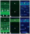
Increased immunostaining for lumican in the posterior stroma of wild-type control corneas. Corneas were stained with anti-lumican antisera (A, D, E) or secondary antibody only, omitting anti-lumican (negative control, B), or stained with Hoescht to visualize cells (C, F). Three-month-old wild-type cornea showed strong lumican immunostaining in the posterior stroma (S; A, arrows). In 7.5-month-old wild-type stronger lumican expression throughout the stroma reduced the anterior-to-posterior gradient in lumican expression somewhat, although highest lumican expression was still seen in the 20- to 30-μm zone of the posteriormost region (D). Within this zone Descemet’s layer is a thin posteriormost region adjacent to the endothelium (arrows). With the exception of the epithelium, which showed some nonspecific background staining in all samples, lumican-deficient corneas were negative for specific lumican staining, as expected (E). Exposure (integration) times were varied for different samples (A and B: 2 seconds, D: 0.5 seconds, and E: 4 seconds). The 3-month corneas (A, B) were exposed longer than the older corneas (D) to demonstrate the weaker reactivity in the anterior stroma of the younger corneas. The lumican-deficient cornea (E) was exposed eight times longer than the wild-type cornea (D) to demonstrate that the reactivity in the stroma was entirely specific. Bar, 20 μm.

References

    1. Maurice DM. The structure and transparency of the cornea. J Physiol. 1957;136:268–286. - PMC - PubMed
    1. McKusick VA. Mendelian Inheritance in Man. Baltimore, MD: The Johns Hopkins University Press; 1992.
    1. Alroy J, Haskins M, Birk D. Altered corneal stromal matrix organization is associated with mucopolysaccharidosis I, III and VI. Exp Eye Res. 1999;68:523–530. - PubMed
    1. Quantock AJ, Meek KM, Fullwood NJ, Zabel RW. Scheie’s syndrome: the architecture of corneal collagen and distribution of corneal proteoglycans. Can J Ophthalmol. 1993;28:266–272. - PubMed
    1. Hassell JR, Newsome DA, Krachmer JH, Rodrigues MM. Macular corneal dystrophy: failure to synthesize a mature keratan sulfate proteoglycan. Proc Natl Acad Sci USA. 1980;77:3705–3709. - PMC - PubMed

Publication types