. 2000 Sep;12(9):1619-32.
doi: 10.1105/tpc.12.9.1619.

anthocyanin1 of petunia encodes a basic helix-loop-helix protein that directly activates transcription of structural anthocyanin genes

Affiliations

anthocyanin1 of petunia encodes a basic helix-loop-helix protein that directly activates transcription of structural anthocyanin genes

C Spelt et al. Plant Cell. 2000 Sep.

Abstract

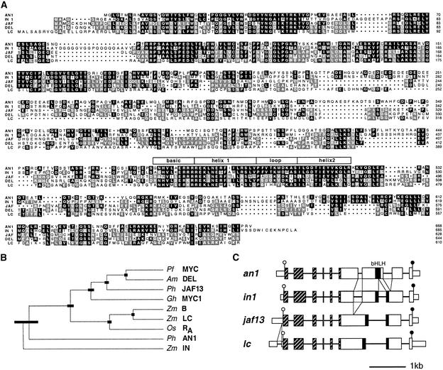
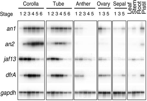
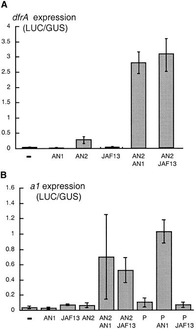
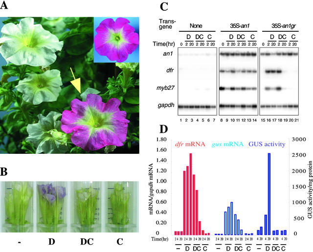
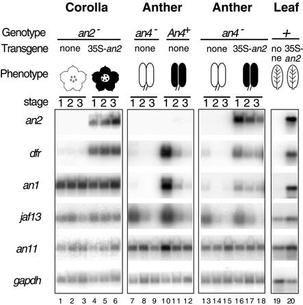
The petunia loci anthocyanin1 (an1), an2, an4, and an11 are required for the transcription of anthocyanin biosynthetic genes in floral organs. The an2 and an11 loci were recently cloned and shown to encode a MYB-domain transcriptional activator and a cytosolic WD40 protein, respectively. Here, we report the isolation of an1 by transposon tagging. an1 encodes a new member of the basic helix-loop-helix family of transcription factors that is functionally and evolutionarily distinct from JAF13, the apparent petunia ortholog of maize RED1 and snapdragon DELILA. We provide genetic evidence that the transcription factors encoded by an1, an2, and an4 operate in an unexpectedly complex regulatory hierarchy. In leaves, ectopic expression of AN2 induces an1 expression, whereas in anthers, an1 expression depends on an4, encoding (or controlling) a MYB protein that is paralogous to AN2. Experiments with transgenic plants expressing a post-translationally controlled AN1-GLUCOCORTICOID RECEPTOR fusion protein indicated that independent of protein synthesis, AN1 directly activates the expression of the dfrA gene encoding the enzyme dihydroflavonol 4-reductase and of Pmyb27 encoding a MYB-domain protein of unknown function.


Figures

Figure 1.
Figure 1.
Phenotypes of Flowers Harboring Different an1 Alleles. (A) Flower of line W138, homozygous for the allele an1-W138. (B) Flower of a plant heterozygous for an1-W138 and a full revertant An1+ allele. (C) Flower of a plant heterozygous for an1-W138 (evident from the red spots) and an excision allele with partial activity (evident from the pink background color). (D) Flower of a plant homozygous for a stable recessive null allele derived from an1-W138. (E) Flower of a homozygote for an1-A2255, a stable allele with low activity. (F) Flower of a homozygote for the unstable an1-X2191 allele.
Figure 2.
Figure 2.
Isolation of the an1 Gene. (A) Map of the an1 gene. Boxes represent exon sequences, separated by thin lines representing introns. Protein coding sequences are indicated by boxes of double height. The dTph1 insertion in the an1-W138 allele is represented by a triangle. (B) Ethidium bromide–stained PCR products amplified from plants harboring the parental An1-R27 allele (+), the mutable an1-W138 allele (m), 16 independently isolated full-revertant alleles (r1 to r16), or three independently isolated stable recessive (−) an1 alleles (W2406, X2076, and X2211). The genotypes indicated above the lanes were determined by phenotypic analysis of progeny obtained by self-pollination. The size of the PCR products is indicated at left. (C) Sequences of the parental An1-R27 allele, the unstable an1-W138 allele, and derived excision alleles. The flower diagrams indicate the petal color specified by each group of alleles. The 8-bp target site duplication caused by the dTph1 insertion in an1-W138 is underlined. Nucleotides that were lost during the attempted dTph1 excision are indicated by asterisks. The open and closed triangles in An1-R27 and an1-X2198 indicate confirmed acceptor splice sites. Triangles in other alleles indicate putative acceptor splice sites that were inferred by using a consensus sequence (Brown et al., 1996). Closed triangles indicate (putative) splice sites that generate an an1 transcript with an intact reading frame, whereas open triangles indicate (putative) splice sites that generate an1 transcripts with a frameshift.
Figure 3.
Figure 3.
Molecular Analysis of the an1 Gene. (A) Alignment of the proteins encoded by an1 and jaf13 (JAF) from petunia, in1 and lc1 (LC) from maize, and del from snapdragon. Sequence identity between AN1 and any one of the other proteins is indicated by black shading. Identical amino acids that are not conserved in AN1 are indicated by gray shading. Numbering of the protein sequences is shown at right, and the position of the bHLH domain is indicated by the box overlying this region. Dots represent gaps introduced to improve the alignment. (B) Phylogenetic tree of bHLH proteins implicated in pigmentation constructed by the unweighted pair method using arithmetic averages (UPGMA) algorithm (Sokal and Michener, 1958). The sequences are from the following sources: snapdragon (Am DEL, Goodrich et al., 1992; GenBank accession number M84913), Perilla frutescens (Pf MYC, Gong et al., 1999; accession number AB0204051), Gerbera hybrida (Gh MYC1, Elomaa et al., 1998; accession number AJ007709), rice (Os RA,, Hu et al., 1996; accession number U39860), maize (Zm B, Radicella et al., 1991; accession number X57276; Zm LC, Ludwig et al., 1989; accession number M26227; Zm IN, Burr et al., 1996; accession number U57899), and petunia (Ph JAF13, Quattrocchio et al., 1998; accession number AF020543; and Ph AN1, this paper, accession numbers AF260918 and AF260919). The thick bars indicate the standard error in the positions of the branch point. (C) Intron–exon structures of an1, in1, jaf13, and lc1 (LC). Exons are drawn to scale, and coding sequences are indicated by double height. Introns are not drawn to scale but are presented in such a way that the conserved positions of 3′ acceptor splice sites are aligned. The positions of additional introns that are not conserved in all genes are indicated by the triangles between the diagrams. The regions encoding the conserved N-terminal domains and the bHLH domain are indicated by striped and black boxes, respectively. The open and closed circles denote the start and stop codons, respectively, of the protein coding region.
Figure 4.
Figure 4.
Spatiotemporal Expression Pattern of an1. Transcripts of an1, an2, jaf13, dfrA, and gapdh were detected by quantitative RT-PCR in the corolla and tube, anthers, ovaries, and sepals from flowers at various developmental stages and from leaves, stems, roots, and pistils of the wild-type line V30, as indicated above the lanes.
Figure 5.
Figure 5.
Activation of dfr and a1 by Ectopically Expressed AN1, AN2, JAF13, and P. (A) Activation of dfr-luc by AN1, AN2 and JAF13. (B) Activation of a1-luc by AN1, AN2, JAF13 and P. The columns and error bars in (A) and (B) denote the mean and standard error of the activity of the dfr-luc or a1-luc reporter gene after bombardment with 35S-an1, 35S-an2, 35S-jaf13, or 35S-p alone or in combination. Reporter gene activity, measured as luciferase (LUC) enzyme activity, is expressed in arbitrary units and was normalized to GUS enzyme activity expressed from a cobombarded reference gene, 35S-gus, which consisted of the β-glucuronidase gene driven by the 35S promoter.
Figure 6.
Figure 6.
Direct Activation of dfr and myb27 by an AN1GR Fusion Protein. (A) A plant of the an1 line W242 harboring 35S-an1, 20 hr after one floral bud (arrow) had been dipped in 10 μM DEX. For comparison, the inset in the top right corner shows a flower of a transgenic W242 plant complemented with the 35S-an1 transgene. (B) Phenotype of a transgenic W242 flower harboring 35S-an1 gr after 20 hr of incubation in a test tube containing water (−), 10 μM DEX (D), 10 μM DEX plus 100 μM CHX (DC), or 100 μM CHX (C). (C) Detection of an1, dfr, myb27, and gapdh transcripts in petal limbs of W242 flowers containing 35S-an1 gr, 35S-an1, or the empty vector after incubation in 10 μM DEX (D), 10 μM DEX plus 100 μM CHX (DC), or 100 μM CHX (C) for 2 or 20 hr. The numbering of the lanes is indicated at the bottom. (D) dfr-GUS expression in an1 mutant plants harboring 35S-an1 gr. Flowers treated for various periods with DEX or CHX or both, as indicated in (B) and (C), were assayed for gus mRNA, dfr mRNA, and GUS enzyme activity. gus and dfr mRNA amounts, expressed in arbitrary units, were determined by hybridization of RT-PCR products, quantification of the radioactivity by PhosphorImaging, and normalization to the amount of gapdh mRNA.
Figure 7.
Figure 7.
Genetic Control of an1 Expression. Transcripts of an2, dfr, an1, jaf13, an11, and gapdh were detected by RT-PCR in different developmental stages of an2 corollas from the hybrid W115/W59 and complementants harboring 35S-an2 (lanes 1 to 6), in anthers from selected an4 and An4+ progeny of the backcross (W138 × V30) × W138 (lanes 7 to 12), in an4 anthers from the hybrid W115/W59 and complementants harboring 35S-an2 (lanes 13 to 18), and in leaves of W115/W59 and W115/W59 plants harboring 35S-an2 (lanes 19 and 20). The presence of anthocyanin pigments in the various tissues is indicated by cartoons. The numbering of the lanes is indicated at bottom.

Similar articles

Cited by

References

    1. Alfenito, M.R., Souer, E., Goodman, C.D., Buell, R., Mol, J., Koes, R., and Walbot, V. (1998). Functional complementation of anthocyanin sequestration in the vacuole by widely divergent glutathione S-transferases. Plant Cell 10, 1135–1149. - PMC - PubMed
    1. Brown, J.W., Smith, P., and Simpson, C.G. (1996). Arabidopsis consensus intron sequences. Plant Mol. Biol. 32, 531–535. - PubMed
    1. Bruce, W., Folkerts, O., Garnaat, C., Crasta, O., Roth, B., and Bowen, B. (2000). Expression profiling of the maize flavonoid pathway genes controlled by estradiol-inducible transcription factors CRC and P. Plant Cell 12, 65–80. - PMC - PubMed
    1. Burr, F.A., Burr, B., Scheffler, B.E., Blewitt, M., Wienand, U., and Matz, E.C. (1996). The maize repressor-like gene intensifier1 shares homology with the r1/b1 multigene family of transcription factors and exhibits missplicing. Plant Cell 8, 1249–1259. - PMC - PubMed
    1. de Vetten, N., Quattrocchio, F., Mol, J., and Koes, R. (1997). The an11 locus controlling flower pigmentation in petunia encodes a novel WD-repeat protein conserved in yeast, plants and animals. Genes Dev. 11, 1422–1434. - PubMed

Publication types

MeSH terms

Associated data

LinkOut - more resources