. 2000 Sep;12(9):1719-36.
doi: 10.1105/tpc.12.9.1719.

Identification and characterization of a novel microtubule-based motor associated with membranous organelles in tobacco pollen tubes

Affiliations

Identification and characterization of a novel microtubule-based motor associated with membranous organelles in tobacco pollen tubes

G Cai et al. Plant Cell. 2000 Sep.

Abstract

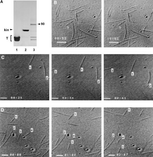
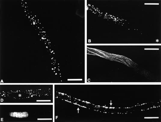
Pollen tube growth depends on the differential distribution of organelles and vesicles along the tube. The role of microtubules in organelle movement is uncertain, mainly because information at the molecular level is limited. In an effort to understand the molecular basis of microtubule-based movement, we isolated from tobacco pollen tubes polypeptides that cosediment with microtubules in an ATP-dependent manner. Major polypeptides released from microtubules by ATP (ATP-MAPs) had molecular masses of 90, 80, and 41 kD. Several findings indicate that the 90-kD ATP-MAP is a kinesin-related motor: binding of the polypeptide to microtubules was enhanced by the nonhydrolyzable ATP analog AMP-PNP; the 90-kD polypeptide reacted specifically with a peptide antibody directed against a highly conserved region in the motor domain of the kinesin superfamily; purified 90-kD ATP-MAP induced microtubules to glide in motility assays in vitro; and the 90-kD ATP-MAP cofractionated with microtubule-activated ATPase activity. Immunolocalization studies indicated that the 90-kD ATP-MAP binds to organelles associated with microtubules in the cortical region of the pollen tube. These findings suggest that the 90-kD ATP-MAP is a kinesin-related microtubule motor that moves organelles in the cortex of growing pollen tubes.


Figures

Figure 1.
Figure 1.
Silver-Stained SDS Gel Showing Intermediate Fractions Obtained during Preparation of ATP-MAPs from a High-Speed Supernatant of Tobacco Pollen Tubes. Lane 1, Mr standards (indicated at left in kilodaltons); lanes 2 and 3, the LSS and the high-speed supernatant (HSS), respectively; lane 4, the supernatant after hexokinase treatment; lane 5, the taxol supernatant; lane 6, the corresponding pellet; lane 7, the pellet obtained after ATP elution; and lane 8, the ATP-released polypeptides (ATP-MAPs). Polypeptides of 90, 80, and 41 kD are indicated. Protein loading was 3 μg in lanes 2 to 5 and 8. Volumes identical to that in lane 8 were loaded in lanes 6 and 7. T, tubulin.
Figure 2.
Figure 2.
Differential Binding of Pollen Tube Proteins to Microtubules. (A) Addition of 10 mM ATP instead of 10 mM AMP-PNP during the binding step prevents recovery of polypeptides in the final ATP supernatants (only the tubulin doublet is present; the lane is overloaded). (B) Silver-stained SDS gel showing ATP-dependent release of pollen ATP-MAPs from microtubules. In this case, the taxol pellet was first washed with ATP and then centrifuged to yield the ATP supernatant (lane 1). The corresponding pellet was then washed with ATP/KCl to obtain a second supernatant (lane 2). Polypeptides of 90, 80, and 41 kD are indicated by the arrowheads. Identical volumes were loaded in each lane. (C) AMP-PNP–sensitive binding of pollen ATP-MAPs to microtubules. Before addition of bovine brain microtubules, ATP-depleted HSS was divided into two parts, one of which was not supplemented with AMP-PNP. Both taxol pellets were then processed as reported. The silver-stained SDS gel (identical volumes loaded in each lane) shows the final ATP-MAPs fractions, obtained without AMP-PNP (lane 1) and with AMP-PNP (lane 2). T, tubulin.
Figure 3.
Figure 3.
Gel Filtration Chromatography of the Pollen Tube ATP-MAPs and Quantitation of Mg-ATP Activity. (A) The ATP-MAPs fraction was fractionated on a Superdex 200 HR 10/30 chromatography column. The column was eluted at 0.75 mL min−1, and 0.5-mL fractions were collected. The absorbance of the eluate was monitored at a wavelength of 280 nm and is expressed as absorbance units (mAU). Arrows indicate the position of protein standards: from left to right, catalase, IgG, BSA, and ovalbumin. Numbers in the graph indicate the main peaks. (B) Mg-ATPase activity with and without microtubules was analyzed for the same fractions. Activity is expressed as nmol of Pi min−1 mL−1.
Figure 4.
Figure 4.
Silver-Stained SDS Gels of the Gel Filtration Fractions from the Preparation Shown in Figure 3 and Fractionation of Pollen ATP-MAPs by Sucrose Gradient Centrifugation. (A) Superdex 200 HR 10/30 gel filtration of pollen ATP-MAPs. The numbers and arrowheads at left indicate Mr standards (M) in kilodaltons. The numbers at top are fraction numbers. The numbers and arrowheads at right indicate 90-, 80-, and 41-kD ATP-MAPs, which peaked in fractions 27 to 28, 24, and 31, respectively. The column (1 × 30 cm) was eluted with HEMD buffer. Proteins eluted between fractions 15 and 38, but only fractions 17 to 34 are shown. L, ATP-eluted MAPs loaded onto the column. (B) Fractionation of pollen ATP-MAPs by sucrose gradient centrifugation. The silver-stained SDS gel shows that the 90- and 41-kD polypeptides cosedimented in a single peak centered around fraction 13, whereas the 80-kD polypeptide eluted around fraction 14. In this experiment, 4.8 mL of 5 to 25% sucrose in HEEM buffer plus 2 mM DTT was overlaid with 200 μL of ATP-MAPs and centrifuged at 83,700g for 15 hr at 4°C in a Sorvall AH-650 swinging-bucket rotor. Arrows at bottom indicate positions of standard proteins: from left to right, thyroglobulin, catalase, and BSA. L, loaded sample; T, tubulin.
Figure 5.
Figure 5.
Nucleotide-Sensitive Binding of Gel Filtration–Purified Pollen ATP-MAPs to Bovine Brain Microtubules by Sedimentation Assay. (A) Control experiment showing that a known nonmotor microtubule binding protein (MAP2) binds to taxol-stabilized microtubules (pellet in lane 2), whereas BSA does not (pellet in lane 4). Lanes 5 and 6 show the pattern of microtubules. MAP2 alone did not sediment under the binding condition (supernatant and pellet in lanes 7 and 8). BSA alone did not pellet again (supernatant and pellet in lanes 9 and 10). (B) Control experiment showing that bovine brain kinesin alone does not pellet (lanes 1 and 2). Lanes 3 and 4 show kinesin plus microtubules (kinesin pellets in the presence of microtubules). Lanes 5 and 6 show kinesin plus microtubules plus AMP-PNP (kinesin binds more efficiently to microtubules). Lanes 7 and 8 show kinesin plus microtubules plus ATP (now kinesin does not pellet). KHC, kinesin heavy chain; KLC, kinesin light chain. (C) Binding assay of the 90-kD polypeptide. . The numbers and arrowheads at left indicate Mr standards (M) in kilodaltons. Lanes 1 and 2 show the 90-kD ATP-MAP plus microtubules (it binds to microtubules and pellets). Lanes 3 and 4 show that the binding affinity of the 90-kD polypeptide increases in the presence of AMP-PNP. Lanes 5 and 6 show the 90-kD ATP-MAP plus microtubules plus ATP (no pellet forms). Lanes 7 and 8 show that the 90-kD ATP-MAP alone does not pellet. (D) Binding assay of the 80-kD polypeptide. Conditions are as described for the 90-kD polypeptide in (C); lanes 1 and 2 are 80-kD polypeptide plus microtubules; lanes 3 and 4 are 80-kD polypeptide plus microtubules plus AMP-PNP; lanes 5 and 6 are 80-kD polypeptide plus microtubules plus ATP; and lanes 7 and 8 show that the 80-kD polypeptide alone does not pellet. (E) Binding experiment with the 41-kD polypeptide. Conditions are as described in (C). Lanes 1 and 2 are in the absence of nucleotides; lanes 3 and 4 are with AMP-PNP; lanes 5 and 6 are with ATP; and lanes 7 and 8 show that the 41-kD polypeptide alone does not pellet. P, pellet; S, supernatant; T, tubulin.
Figure 6.
Figure 6.
Microtubules Gliding on Recombinant Kinesin and the 90-kD ATP-MAPs. (A) Polypeptides used in the assay. Lane 1, tubulin (T); lane 2, commercial recombinant kinesin (kin); and lane 3, purified 90-kD ATP-MAP. (B) Time-lapse sequences showing that microtubules alone do not move. Numbers above scale bars denote time (min:sec). (C) and (D) Time-lapse sequences of microtubules gliding on recombinant kinesin (C) and purified 90-kD ATP-MAP bound to a cover slip (D). The time (min:sec) is indicated at bottom left and was generated automatically by Argus-20. In (C), microtubules are gliding on commercially available recombinant kinesin. Frames were captured every 9 sec. Three single microtubules are numbered. In (D), microtubules are moving on purified 90-kD ATP-MAP bound to a cover slip. Frames were captured every 59 sec. Numbers indicate some moving microtubules. Bars in (B) to (D) = 2 μm, computed automatically by Argus-20.
Figure 7.
Figure 7.
Anti-Kinesin Immunoblot Analysis. (A) Silver-stained SDS gel of bovine brain kinesin (lane 2, 2 μg); purified bovine brain tubulin (lane 3, 20 μg); gel filtration–purified 80-kD ATP-MAPs (lane 4, 40 μL), 90-kD ATP-MAPs (lane 5, 40 μL), and 41-kD ATP-MAPs (lane 6, 40 μL); LSS from tobacco pollen tubes (lane 7, 40 μg); and cytosolic fraction (lane 8, 40 μg) and membrane fraction (lane 9, 40 μg) of tobacco pollen tubes. Lane 1 contains Mr standards, with masses indicated at left in kilodaltons. T, tubulin. (B) MMR44 antibody recognized the kinesin heavy chain (KHC, lane 11) but not bovine brain tubulin (lane 12). The antibody did not recognize the 80-kD (lane 13) or the 41-kD polypeptide (lane 15), but it did label purified 90-kD ATP-MAP (lane 14). Note the band at ∼102 kD in LSS (lane 16) and in cytosolic fraction (lane 17). The MMR44 antibody labeled 90-kD ATP-MAP in the LSS (lane 16) and in the membrane fraction (lane 18). A faint band at the same molecular mass was also detected in the cytosolic fraction (lane 17). Lane 10 contains biotinylated marker proteins, with sizes indicated at left in kilodaltons. Lanes 7, 8, 9, 16, 17, and 18 were overloaded to evaluate antibody specificity.
Figure 8.
Figure 8.
Immunofluorescence Localization of MMR44-Labeled Polypeptide Associated with Organelles from Tobacco Pollen Tubes. (A) DIC micrograph of organelle fraction from tobacco. (B) Fluorescence microscopy of the field shown in (A). Only a small percentage of organelles were labeled. (C) A detail of organelles immobilized on a poly-l-lysine–coated slide. Organelles labeled by the MMR44 antibody (arrows) were not uniform in size. (D) Fluorescence microscopy of the field shown in (C). Bars in (A) to (D) = 4 μm.
Figure 9.
Figure 9.
Immunolocalization of MMR44 Antibody in Tobacco Pollen Tubes. (A) The MMR44 antibody labeled in a punctate fashion throughout the vegetative cytoplasm, indicating that the antigen it recognized is associated with organelles. (B) The punctate pattern was not observed in the apical region of the pollen tube, where only a few, faint dots could be seen. The asterisk indicates the pollen tube apex. (C) Typical immunostaining of microtubules in the pollen tube reveals that the distribution of MMR44-labeled organelles coincides with regions in which microtubules are abundant. (This is not the same pollen tube shown in [B].) (D) and (E) In (D), the MMR44 antibody did not label the generative cell (asterisk) as compared by DNA staining in (E). Some fluorescent dots can be seen at the edge of the generative cell. (F) A confocal section through the middle of a pollen tube. MMR44 staining is concentrated in the cortical region (arrows), and only faint spots can be seen in the central regions. Bars in (A) to (F) = 20 μm.
Figure 10.
Figure 10.
Double Immunolocalization of MMR44 and Anti-Tubulin Antibodies in Tobacco Pollen Tubes. (A) Colocalization of organelles labeled with the MMR44 antibody along microtubules. Bundles of microtubules (green) extend along the longitudinal axis of the pollen tube. Rows of punctate dots stained by MMR44 (red) are aligned with microtubule bundles (arrow). Bar = 20 μm. (B) Colocalization of organelles labeled with the MMR44 antibody (red) along microtubules (green). Organelles labeled with MMR44 are aligned in rows where microtubules are located (arrows). Bar = 10 μm. (C) Magnification of the enclosed region in (B) showing organelles precisely aligned with microtubules (arrows). Bar = 2 μm. (D) A confocal section through pollen tube cortex showing organelles (red, arrows) that colocalize with a couple of microtubule bundles (green). Bars in (A) to (D) = 5 μm.

References

    1. Asada, T., and Collings, D. (1997). Molecular motors in higher plants. Trends Plant Sci. 2, 29–37.
    1. Asada, T., and Shibaoka, H. (1994). Isolation of polypeptides with microtubule-translocating activity from phragmoplasts of tobacco BY-2 cells. J. Cell Sci. 107, 2249–2257. - PubMed
    1. Asada, T., Kuriyama, R., and Shibaoka, H. (1997). TKRP125, a kinesin-related protein involved in the centrosome-independent organization of the cytokinetic apparatus in tobacco BY-2 cells. J. Cell Sci. 110, 179–189. - PubMed
    1. Åström, H., Sorri, O., and Raudaskoski, M. (1995). Role of microtubules in the movement of the vegetative nucleus and generative cell in tobacco pollen tubes. Sex. Plant Reprod. 8, 61–69.
    1. Baxter-Gabbard, K.L. (1972). A simple method for the large-scale preparation of sucrose gradients. FEBS Lett. 20, 117–119. - PubMed

Publication types

LinkOut - more resources