. 2000 Sep;12(9):1769-82.
doi: 10.1105/tpc.12.9.1769.

Biogenesis of the chloroplast-encoded D1 protein: regulation of translation elongation, insertion, and assembly into photosystem II

Affiliations

Biogenesis of the chloroplast-encoded D1 protein: regulation of translation elongation, insertion, and assembly into photosystem II

L Zhang et al. Plant Cell. 2000 Sep.

Abstract

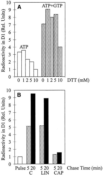
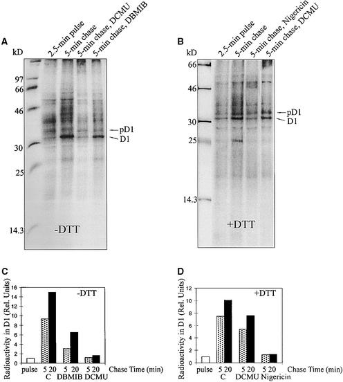
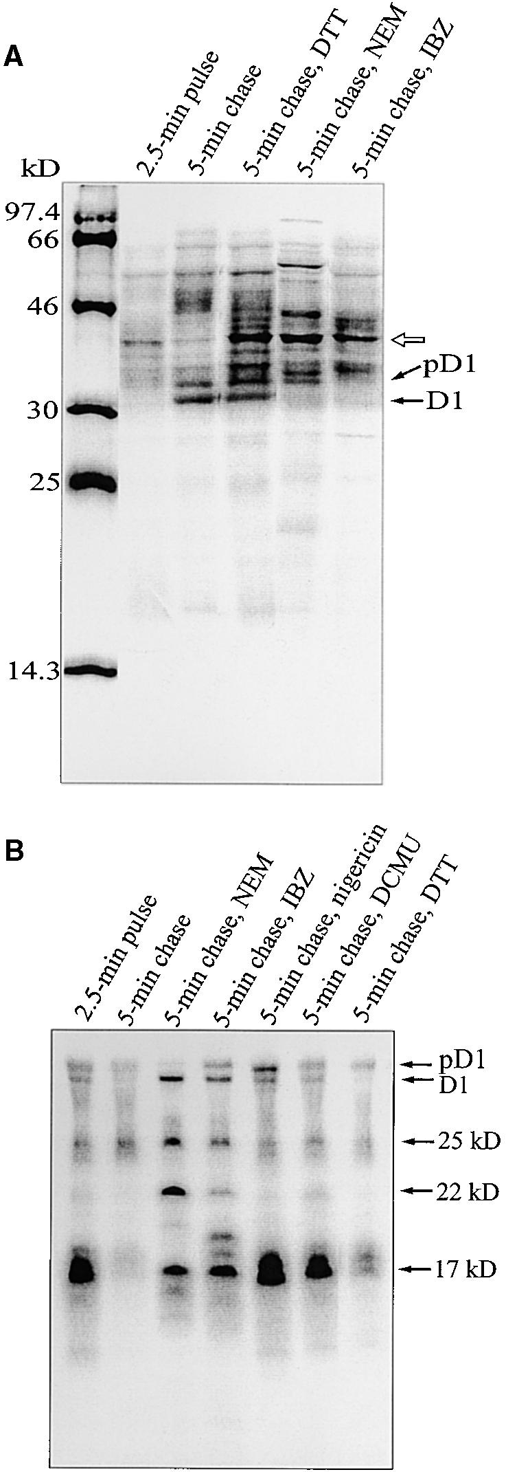
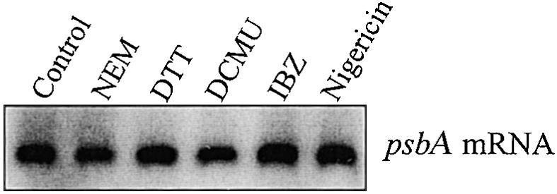
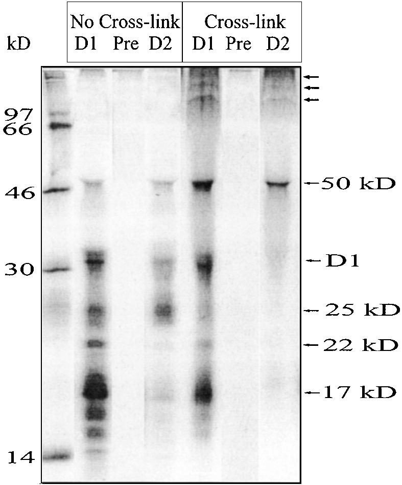
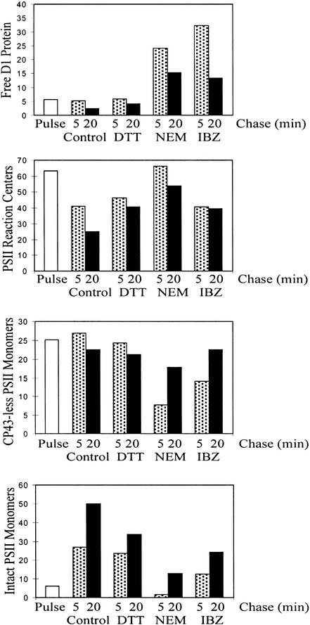
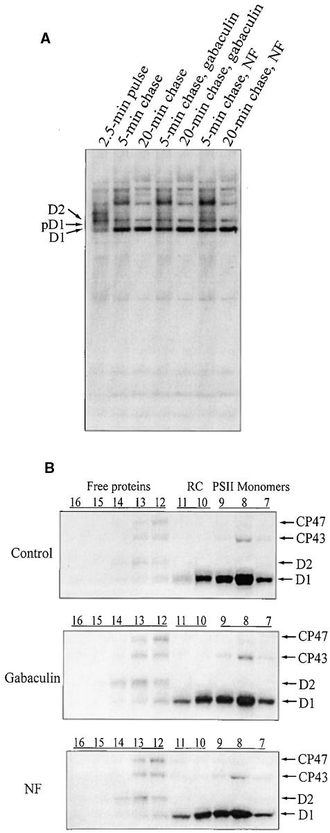
Regulation of translation elongation, membrane insertion, and assembly of the chloroplast-encoded D1 protein of photosystem II (PSII) was studied using a chloroplast translation system in organello. Translation elongation of D1 protein was found to be regulated by (1) a redox component that can be activated not only by photosynthetic electron transfer but also by reduction with DTT; (2) the trans-thylakoid proton gradient, which is absolutely required for elongation of D1 nascent chains on the thylakoid membrane; and (3) the thiol reactants N-ethylmaleimide (NEM) and iodosobenzoic acid (IBZ), which inhibit translation elongation with concomitant accumulation of distinct D1 pausing intermediates. These results demonstrate that D1 translation elongation and membrane insertion are tightly coupled and highly regulated processes in that proper insertion is a prerequisite for translation elongation of D1. Cotranslational and post-translational assembly steps of D1 into PSII reaction center and core complexes occurred independently of photosynthetic electron transfer or trans-thylakoid proton gradient but were strongly affected by the thiol reactants DTT, NEM, and IBZ. These compounds reduced the stability of the early PSII assembly intermediates, hampered the C-terminal processing of the precursor of D1, and prevented the post-translational reassociation of CP43, indicating a strong dependence of the D1 assembly steps on proper redox conditions and the formation of disulfide bonds.


Figures

Figure 1.
Figure 1.
D1 Protein Translation in Intact Chloroplasts. (A) Optimization of D1 synthesis. Intact chloroplasts were pulse labeled in the light for 10 min. Reactions were performed in the presence of 10 mM ATP or 10 mM ATP and 2 mM GTP and several concentrations of DTTred (DTT; 0, 1, 2, 5, and 10 mM). The relative amount (Rel. Units) of radioactivity incorporated into the precursor and mature D1 protein after 10 min of pulse labeling in the presence of 10 mM DTTred and 10 mM ATP, which are the conditions usually used for translation in vitro in intact chloroplasts (Mullet et al., 1986), was set as 1. (B) Translation elongation of the D1 protein during the chase. Intact chloroplasts were pulse labeled for 2.5 min and chased for 5 or 20 min in the presence of lincomycin (100 μg mL−1) or chloramphenicol (100 μg mL−1), which were added to the chase buffer immediately after the pulse. The relative amount of radioactivity incorporated into the precursor and mature D1 protein after 2.5 min of pulse labeling was set as 1. C, control; CAP, chloramphenicol; LIN, lincomycin.
Figure 2.
Figure 2.
Effects of Photosynthetic Electron Transfer Inhibitors and Uncouplers on D1 Translation Elongation. Intact chloroplasts were incubated for 5 min at 23°C and 50 μE m−2 sec−1 in a translation mixture in the absence (−DTT) ([A] and [C]) or the presence (+DTT) ([B] and [D]) of 5 mM DTTred. After 2.5 min of pulse labeling in intact chloroplasts, nigericin (1 μM), DCMU (10 μM), or DBMIB (2 μM) together with unlabeled methionine (10 mM) was added to the chase buffer. (A) and (B) Autoradiography of 35S-labeled translation products in the thylakoid membrane. The molecular mass markers in kilodaltons are indicated at left. (C) and (D) Quantification of 35S-methionine incorporation into the precursor and mature D1 protein. The relative amount (Rel. Units) of radioactivity incorporated into the precursor and mature D1 protein after 2.5 min of pulse labeling was set as 1. C, control (i.e., only 10 mM unlabeled methionine was added in the beginning of the chase).
Figure 3.
Figure 3.
Regulation of Translation Elongation. (A) Autoradiography of labeled thylakoid membrane proteins. After 2.5 min of pulse labeling in intact chloroplasts, the accumulation of label in the D1 protein was chased in the light for 5 min in the presence of 10 mM unlabeled methionine and NEM (5 mM), IBZ (10 mM), or DTTred (DTT; 20 mM). The molecular mass markers in kilodaltons are indicated at left. The open arrow indicates a possible tRNA–D1 nascent chain complex. (B) Immunoprecipitation of labeled D1 intermediates from RNCs. After 2.5 min of pulse labeling in intact chloroplasts, followed by 5 min of chase in the presence of NEM (5 mM), IBZ (10 mM), DTTred (DTT; 20 mM), DCMU (10 μM), or nigericin (1 μM), the thylakoid-bound polysomes were isolated and solubilized with SDS, and immunoprecipitation was performed by adding an excess of N-terminal D1 antiserum. The precipitated products were separated by SDS-PAGE and visualized by autoradiography. The D1 intermediates of 17, 22, and 25 kD are indicated at right. Minor amounts of D1 and pD1 were always found in RNCs.
Figure 4.
Figure 4.
RNA Gel Blot Analysis of psbA mRNA Associated with Thylakoid-Bound Polysomes. Thylakoid-bound polysomes were isolated after 2.5 min of pulse labeling followed by a 5-min chase in intact chloroplasts in the presence of the following reagents: NEM (5 mM), IBZ (10 mM), DTTred (DTT; 20 mM), DCMU (10 μM), or nigericin (1 μM). The polysome-associated RNA was isolated by extraction with phenol/chloroform and then probed with a random prime-labeled Synechocystis sp 6803 psbA2 gene.
Figure 5.
Figure 5.
Interaction of D1 Nascent Chains with the D2 Protein. After 2.5 min of pulse labeling in intact chloroplasts, thylakoid-bound ribosomes were isolated and incubated with the homobifunctional sulfhydryl cross-linker BMH. After cross-linking, the samples were denatured with SDS and immunoprecipitated with antisera raised against the N-terminal residues (58 to 86) of the D1 protein (lanes labeled D1) or against the residues (230 to 245) of the D2 protein (lanes labeled D2). Lanes labeled Pre indicate immunoprecipitation with preserum (similar results were obtained with both the D1 and D2 presera). The precipitated products were separated by SDS-PAGE and visualized by autoradiography. The molecular mass markers in kilodaltons are indicated at left.
Figure 6.
Figure 6.
Thiol Reactants Hamper Incorporation of Newly Synthesized D1 Protein into PSII Subcomplexes. Intact chloroplasts were radiolabeled for 2.5 min and subsequently chased for 5 and 20 min in the light with no additions or in the presence (+) of 5 mM NEM or 20 mM DTTred (DTT). After translation, thylakoids were solubilized, and different PSII subcomplexes were separated in the sucrose gradients. The gradients were fractionated into 20 equal fractions, and the proteins in each fraction were separated by SDS-PAGE and visualized by autoradiography. According to earlier assignments (Zhang et al., 1999), unassembled PSII proteins were found in fractions 12 to 16, PSII reaction centers (RC) in fractions 10 and 11, PSII core complexes lacking CP43 in fraction 9, and intact PSII cores (including CP43) in fractions 7 and 8.
Figure 7.
Figure 7.
Quantification of Incorporation of Newly Synthesized D1 Protein into PSII Subcomplexes. Accumulation of radioactivity in the precursor and mature D1 protein in different PSII subpopulations was quantified after 5- and 20-min chases. Each chase was conducted with no additional chemicals (Control) or in the presence of DTTred (DTT), NEM, or IBZ, and PSII subcomplexes were separated, as given in the legend to Figure 6. Relative amounts of radioactivity in unassembled D1 protein (free D1 protein), PSII reaction centers, CP43-less PSII monomers, and intact PSII monomers (including CP43) are indicated. The data are expressed as the percentage of accumulated total labeled precursor and mature D1 protein.
Figure 8.
Figure 8.
Effect of Pigment Synthesis Inhibitors on D1 Translation Elongation and Assembly into PSII during the Repair Process. (A) Autoradiography of labeled thylakoid membrane proteins. After 2.5 min of pulse labeling in intact chloroplasts, gabaculin (1 mM) or norflurazon (NF; 0.1 mM) was added, and the accumulation of label in the precursor and mature D1 protein was chased for 5 and 20 min in the light. After translation, the thylakoids were isolated and subjected to SDS-PAGE and autoradiography. (B) Incorporation of 35S-methionine into PSII subcomplexes. To analyze the assembly process of PSII, the thylakoids were isolated after translations in vitro as in (A) and solubilized, and PSII subcomplexes were separated in the sucrose gradients. The gradients were fractionated into 20 equal fractions, and the proteins of each fraction were separated by SDS-PAGE and visualized by autoradiography. For more details, see the legend to Figure 6.

Similar articles

Cited by

References

    1. Adir, N., Shochat, S., and Ohad, I. (1990). Light-dependent D1 protein synthesis and translocation is regulated by reaction center II: Reaction center II serves as an acceptor for the D1 precursor. J. Biol. Chem. 265, 12563–12568. - PubMed
    1. Aro, E.-M., Virgin, I., and Andersson, B. (1993). Photoinhibition of photosystem II. Inactivation, protein damage and turnover. Biochim. Biophys. Acta 1143, 113–143. - PubMed
    1. Baena-Gonzalez, E., Barbato, R., and Aro, E.-M. (1999). Role of phosphorylation in the repair cycle and oligomeric structure of photosystem II. Planta 208, 196–204.
    1. Barbato, R., Friso, G., Rigoni, F., Dalla-Vecchia, F., and Giacometti, G.M. (1992). Structural changes and lateral redistribution of photosystem II during donor side photoinhibition of thylakoids. J. Cell Biol. 119, 325–335. - PMC - PubMed
    1. Bowyer, J.R., Packer, J.C.L., McCormack, B.A., Whitelegge, J.P., Robinson, C., and Tayor, M. (1992). Carboxyl-terminal processing of the D1 protein and photoactivation of water splitting in photosystem II. J. Biol. Chem. 267, 5424–5433. - PubMed

Publication types

MeSH terms

LinkOut - more resources