. 2000 Oct 1;20(19):7258-67.
doi: 10.1523/JNEUROSCI.20-19-07258.2000.

Phosphorylation of the AMPA receptor subunit GluR2 differentially regulates its interaction with PDZ domain-containing proteins

Affiliations

Phosphorylation of the AMPA receptor subunit GluR2 differentially regulates its interaction with PDZ domain-containing proteins

H J Chung et al. J Neurosci. .

Abstract

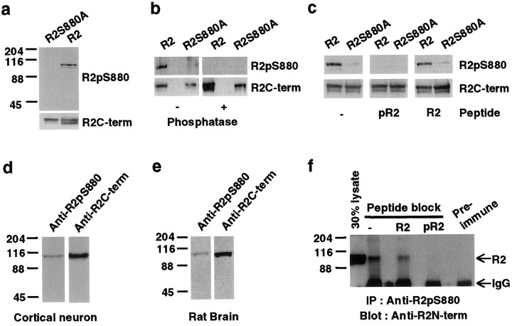
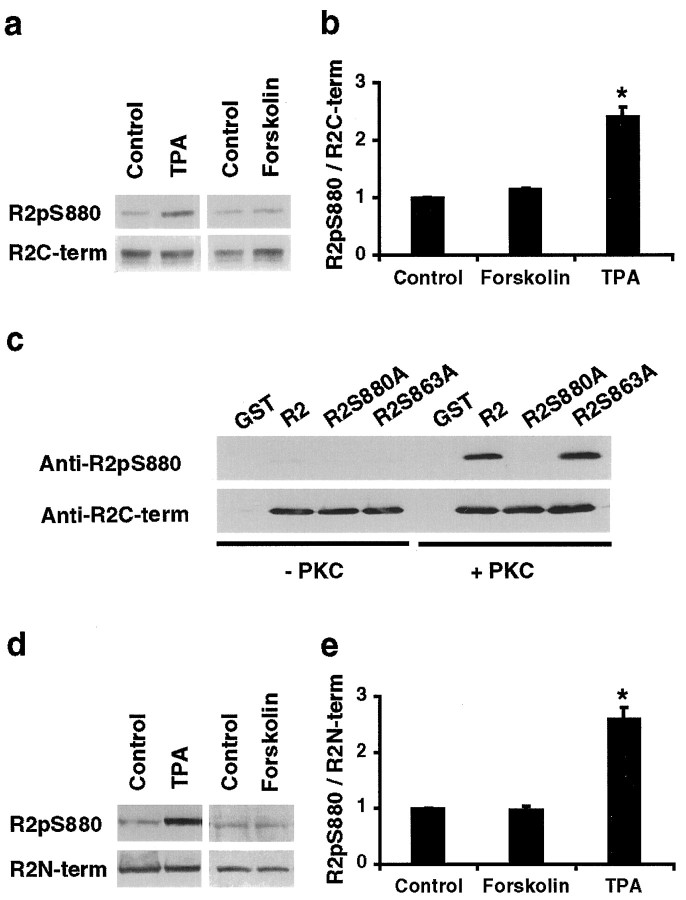
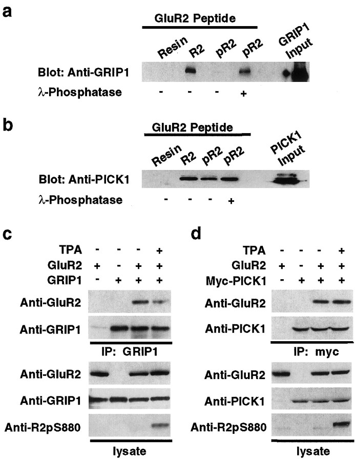
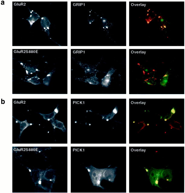
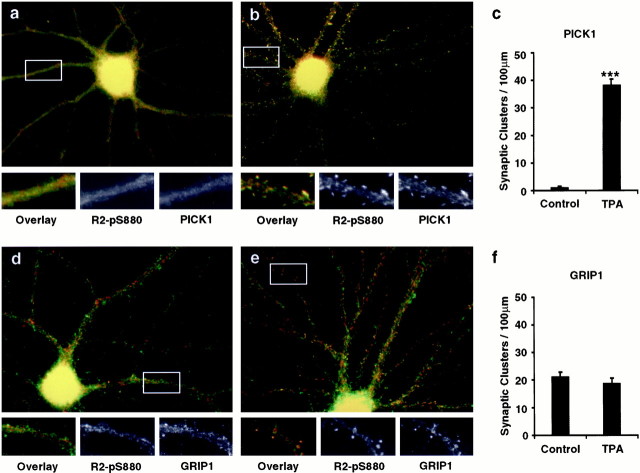
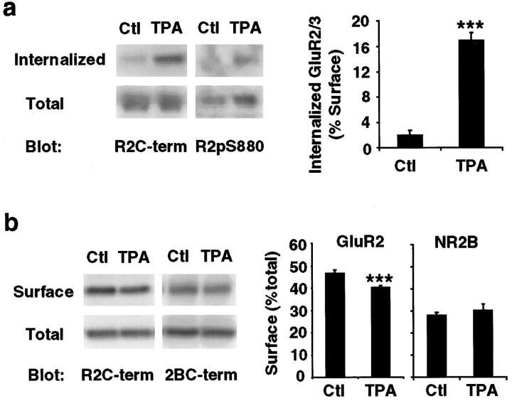
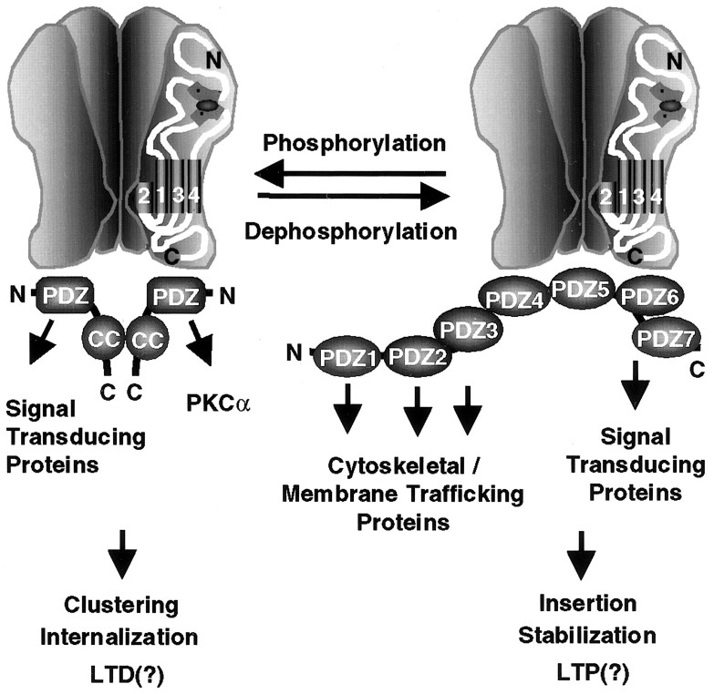
PSD-95, DLG, ZO-1 (PDZ) domain-mediated protein interactions have been shown to play important roles in the regulation of glutamate receptor function at excitatory synapses. Recent studies demonstrating the rapid regulation of AMPA receptor function during synaptic plasticity have suggested that AMPA receptor interaction with PDZ domain-containing proteins may be dynamically modulated. Here we show that PKC phosphorylation of the AMPA receptor GluR2 subunit differentially modulates its interaction with the PDZ domain-containing proteins GRIP1 and PICK1. The serine residue [serine-880 (Ser880)] in the GluR2 C-terminal sequence (IESVKI) critical for PDZ domain binding is a substrate of PKC and is phosphorylated in vivo. In vitro binding and coimmunoprecipitation studies show that phosphorylation of serine-880 within the GluR2 PDZ ligand significantly decreases GluR2 binding to GRIP1 but not to PICK1. Immunostaining of cultured hippocampal neurons demonstrates that the Ser880-phosphorylated GluR2 subunits are enriched and colocalized with PICK1 in the dendrites, with very little staining observed at excitatory synapses. Interestingly, PKC activation in neurons increases the Ser880 phosphorylation of GluR2 subunits and recruits PICK1 to excitatory synapses. Moreover, PKC stimulation in neurons results in rapid internalization of surface GluR2 subunits. These results suggest that GluR2 phosphorylation of serine-880 may be important in the regulation of the AMPA receptor internalization during synaptic plasticity.


Figures

Fig. 1.
Fig. 1.
The GluR2 subunit C-terminal PDZ ligand is phosphorylated on Ser880. a–c, The phosphorylation site-specific anti-GluR2-pS880 antibody was characterized on immunoblots of HEK293T cells expressing GluR2. The expression of GluR2 was analyzed by using a phosphorylation-independent anti-GluR2-C-terminal antibody (Anti-R2C-term). a, Anti-GluR2-pS880 antibody recognized wild-type GluR2 but not the mutant GluR2 in which serine-880 was mutated to alanine.b, Anti-GluR2-pS880 no longer recognized GluR2 when the PVDF membrane was treated with λ-phosphatase. c, Preabsorption of the anti-GluR2-pS880 antibody with Ser880-phosphorylated R2 peptide (pR2) but not unphosphorylated peptide (R2) blocked GluR2 recognition by anti-GluR2-pS880. d, e, Western Blot for GluR2-pS880 in homogenates of cortical culture neurons (d) and rat brain (e).f, Preabsorption of the anti-GluR2-pS880 antibody with Ser880-phosphorylated R2 peptide (pR2) but not unphosphorylated peptide (R2) blocked immunoprecipitation of GluR2 with the anti-GluR2-pS880.
Fig. 2.
Fig. 2.
PKC phosphorylates serine-880 of GluR2 in vitro and in vivo. a, Western blot for GluR2-pS880 in transfected HEK293T cells that were treated with control solution (Control), 1 μm phorbol 12-myristate 13-acetate (TPA), or 20 μm forskolin for 15 min. Total amount of GluR2 was detected by anti-GluR2-C-terminal antibody. b, The ratios of intensity of the signal (intensity of anti-GluR2-pS880 antibody labeling/intensity of anti-GluR2 C-terminal antibody labeling) were calculated and normalized to the control cells. PKC stimulation by TPA significantly increases Ser880 phosphorylation of GluR2.c, Immunoblot analysis of the in vitrophosphorylation reaction of purified GST fusion proteins corresponding to the C termini of GluR2, GluR2S880A, and GluR2S863A with purified PKC. PKC directly phosphorylates GluR2 but not GluR2S880A mutant fusion proteins. d, Western blot for GluR2-pS880 in 3-week-old, high-density cortical culture neurons, which were treated with control solution (Control), 200 nm TPA, or 20 μm forskolin for 15 min. Total amount of GluR2 was detected by anti-GluR2-N-terminal antibody (Chemicon).e, The ratios of intensity of the signals (intensity of anti-GluR2-pS880 antibody labeling/intensity of anti-GluR2 N-terminal antibody labeling) were calculated and normalized to the control neurons. PKC stimulation in neurons significantly increases Ser880 phosphorylation of GluR2.
Fig. 3.
Fig. 3.
Ser880 phosphorylation of GluR2 C terminus differentially regulates the interaction of GluR2 with GRIP1 and PICK1.a, b, In vitro binding of GRIP1 (a) or PICK1 (b) with Ser880-phosphorylated GluR2 peptides. Extracts of HEK293T cells expressing GRIP1 or PICK1 were incubated with Ser880-phosphorylated R2 peptides (pR2) or unphosphorylated peptides (R2) immobilized on Affigel resins, and bound GRIP1 or PICK1 was detected by immunoblotting. a, GRIP1 did not interact with pR2 peptide, whereas phosphatase treatment of pR2 peptide recovered GRIP1 binding. b, PICK1 binds to both pR2 and R2 peptide. c, d, Coimmunoprecipitation of GluR2 with GRIP1 (c) or PICK1 (d) from transfected HEK293T cells, with or without 1 μm TPA treatment. Increase in Ser880 phosphorylation of GluR2 C terminus by PKC activation attenuated GRIP1 interaction with GluR2 (c) but had no effect on the binding affinity of PICK1 to GluR2 (d).
Fig. 4.
Fig. 4.
S880E mutation of GluR2 disrupts GRIP1 interaction with GluR2 but not PICK1. a, Immunostaining of HEK293T cells that were transfected with Narp, GRIP1, and GluR2 or GluR2S880E mutant. The transfected cells were double-labeled with the mouse anti-GluR2-Nterm (red) and anti-GRIP1 antibodies (green). Narp induces clustering of GluR2, and GRIP1 coclusters with GluR2; however, S880E mutation disrupts GRIP1 coclustering with GluR2. b, Immunostaining of HEK293T cells that were transfected with Narp, PICK1, and GluR2 or GluR2S880E mutant. The transfected cells were double-labeled with the mouse anti-GluR2-Nterm (red) and anti-PICK1 antibodies (green). S880E mutation did not disrupt coclustering of PICK1 and GluR2.
Fig. 5.
Fig. 5.
Localization of GluR2-pS880 in cultured hippocampal neurons. a, b, Three-week-old low-density hippocampal culture neurons were double-labeled with the rabbit anti-GluR2-pS880 (red) and the mouse anti-GluR2-Nterm antibodies (green).a, In control neurons, GluR2-pS880 colocalizes with N-terminal GluR2 staining in dendrites but not with synaptic GluR2 at synapses. b, In the neurons treated with 200 nm TPA for 15 min, PKC activation dramatically increased the staining of Ser880-phosphorylated GluR2 in the spine heads as well as in the dendrites. c, d, Neurons were double-labeled with the rabbit anti-GluR2-pS880 (red) and the mouse anti-synaptophysin antibodies (green). c, GluR2-pS880 does not colocalize with synaptophysin. d, In the neurons treated with 200 nm TPA for 15 min, PKC activation increases the colocalization of Ser880-phosphorylated GluR2 with synaptophysin at synapses. e, f, Neurons were double-labeled with the rabbit anti-GluR2-pS880 (red) and the mouse anti-NR1 N-terminal antibodies (green). e, GluR2-pS880 does not colocalize with NR1. f, In the neurons treated with 200 nm TPA for 15 min, PKC activation increases the colocalization of Ser880-phosphorylated GluR2 with NR1 at synapses.g, Statistical analysis of the effect of PKC activation on the number of synaptic GluR2-pS880 clusters per 100 μmdendrite. TPA treatment significantly increased synaptic GluR2-pS880 clusters (n = 36 for control andn = 40 for TPA, t test,p < 0.001). However, there was no change in the total number of synaptic GluR2 clusters (n = 15 for control and n = 20 for TPA, t test,p > 0.05) and synaptic NR1 clusters (n = 10 for control and n = 10 for TPA, t test, p > 0.05).
Fig. 6.
Fig. 6.
PKC activation dramatically increases the synaptic GluR2-pS880 and PICK1 in cultured hippocampal neurons.a–c, Three-week-old low-density hippocampal culture neurons were double-labeled with anti-PICK1 antibodies (green) and the anti-GluR2-pS880 antibodies (red). a, In control neurons, PICK1 colocalizes with GluR2-pS880 in dendrites. b, In the neurons treated with 200 nm TPA for 15 min, PKC activation increases synaptic PICK1 staining. c, Statistical analysis of the effect of PKC activation on number of synaptic PICK1 clusters per 100 μm dendrite. PKC activation significantly increased the synaptic PICK1 clusters (n = 16 for control and n = 15 for TPA, t test, p < 0.001).d–f, Neurons were double-labeled with anti-GRIP1 antibodies (green) and the anti-GluR2-pS880 antibodies (red). d, In control neurons, GRIP1 does not colocalize well with GluR2-pS880 in both dendrites and spines. e, In the neurons treated with 200 nm TPA for 15 min, no significant change in the synaptic GRIP1 distribution was observed. f, Statistical analysis of the effect of PKC activation on number of synaptic GRIP1 clusters per 100 μm dendrite. PKC activation moderately decreased GRIP1 clusters at synapse (n = 7 for control andn = 8 for TPA, t test,p > 0.05).
Fig. 7.
Fig. 7.
PKC activation induces internalization of GluR2 in cultured cortical neurons. a, Internalization assay was performed in 3-week-old high-density cultured cortical neurons to examine the internalized GluR2 subunits after TPA treatment. The biotinylated, internalized GluR2 subunits were analyzed by immunoblotting with anti-GluR2-C-terminal and anti-GluR2-pS880 antibodies. The internalized GluR2 subunits were quantified as the percentage of total surface GluR2 subunits (n = 8 for both control and TPA, t test; p< 0.001). PKC activation induces rapid internalization of surface GluR2 subunits. b, Surface biotinylation was performed in cortical neurons after TPA treatment to examine the steady-state level of total surface GluR2 subunits. The biotinylated GluR2 subunits were analyzed by immunoblotting with anti-GluR2-C-terminal antibody. The steady-state level of surface GluR2 subunits was quantified as the percentage of total GluR2 subunits (n = 8 for both control and TPA, t test; p < 0.001). PKC stimulation results in a decrease in total level of surface GluR2 subunits but not surface NR2B subunits of NMDA receptors (n = 8 for both control and TPA, ttest; p > 0.05).
Fig. 8.
Fig. 8.
Model for modulation of the macromolecular AMPA receptor protein complex by phosphorylation of GluR2 PDZ ligand. PKC phosphorylation of Ser880 in GluR2 C-terminal PDZ ligand differentially regulates its interaction with PDZ domains of GRIP and PICK1. This differential regulation of PDZ domain-mediated interaction may modulate surface expression of AMPA receptors as well as the composition of the AMPA receptor complex and the subsequent downstream signaling cascades at excitatory synapses.

References

    1. Bliss TV, Collingridge GL. A synaptic model of memory: long-term potentiation in the hippocampus. Nature. 1993;361:31–39. - PubMed
    1. Bolshakov VY, Siegelbaum SA. Postsynaptic induction and presynaptic expression of hippocampal long-term depression. Science. 1994;264:1148–1152. - PubMed
    1. Brenman JE, Chao DS, Gee SH, McGee AW, Craven SE, Santillano DR, Wu Z, Huang F, Xia H, Peters MF, Froehner SC, Bredt DS. Interaction of nitric oxide synthase with the postsynaptic density protein PSD-95 and alpha1-syntrophin mediated by PDZ domains. Cell. 1996;84:757–767. - PubMed
    1. Carroll RC, Nicoll RA, Malenka RC. Effects of PKA and PKC on miniature excitatory postsynaptic currents in CA1 pyramidal cells. J Neurophysiol. 1998;80:2797–2800. - PubMed
    1. Carroll RC, Lissin DV, von Zastrow M, Nicoll RA, Malenka RC. Rapid redistribution of glutamate receptors contributes to long-term depression in hippocampal cultures. Nat Neurosci. 1999;2:454–460. - PubMed

Publication types

MeSH terms