. 2000 Oct 1;20(19):7325-33.
doi: 10.1523/JNEUROSCI.20-19-07325.2000.

NGF signals through TrkA to increase clathrin at the plasma membrane and enhance clathrin-mediated membrane trafficking

Affiliations

NGF signals through TrkA to increase clathrin at the plasma membrane and enhance clathrin-mediated membrane trafficking

E C Beattie et al. J Neurosci. .

Abstract

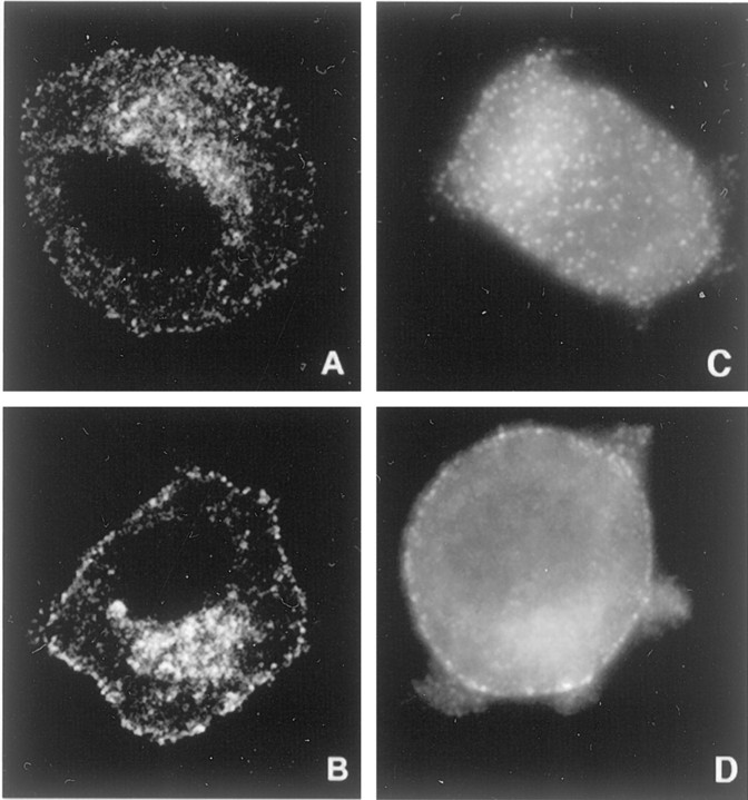
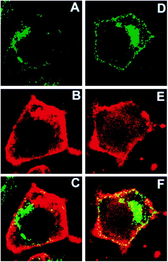
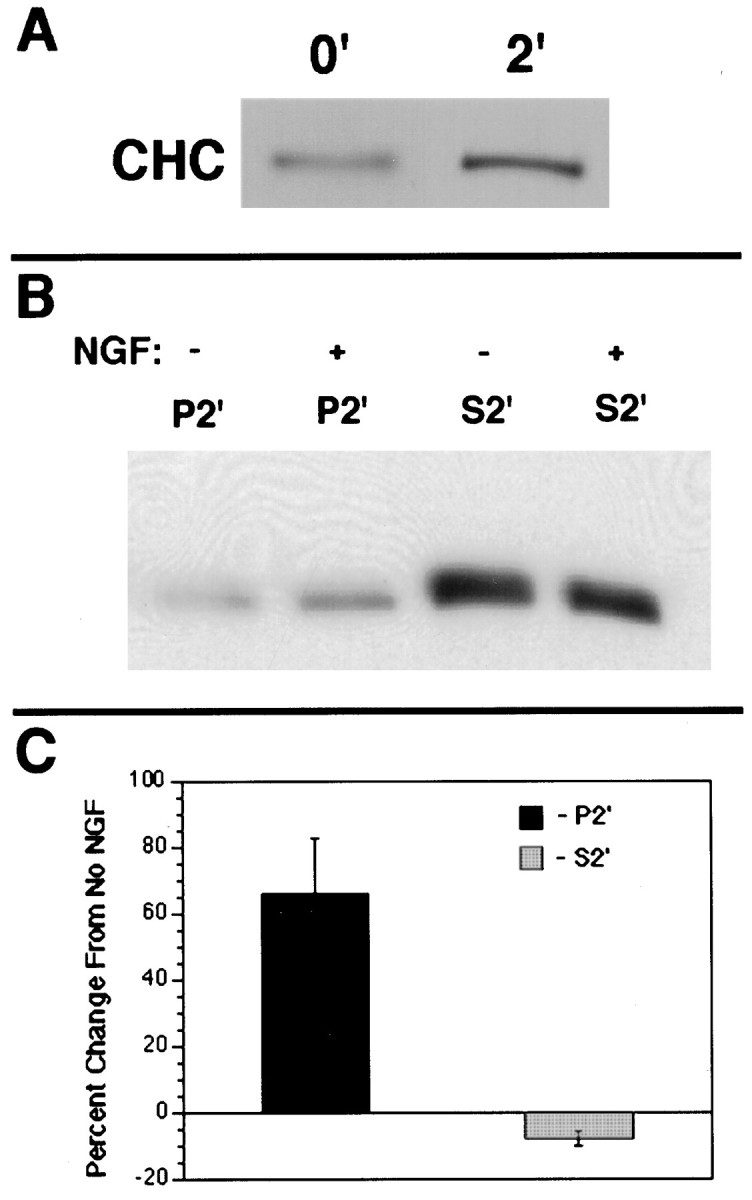
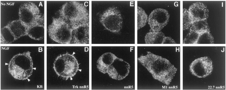
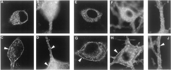
Neurotrophin (NT) signals may be moved from axon terminals to neuron cell bodies via signaling endosomes-organelles in which NTs continue to be bound to their activated receptors. Suggesting that clathrin-coated membranes serve as one source of signaling endosomes, in earlier studies we showed that nerve growth factor (NGF) treatment increased clathrin at the plasma membrane and resulted in colocalization of clathrin with TrkA, the receptor tyrosine kinase for NGF. Strikingly, however, we also noted that most clathrin puncta at the surface of NGF-treated cells did not colocalize with TrkA, raising the possibility that NGF induces a general increase in clathrin-coated membrane formation. To explore this possibility further, we examined the distribution of clathrin in NGF- and BDNF-treated cells. NGF signaling in PC12 cells robustly redistributed the adaptor protein AP2 and the clathrin heavy chain (CHC) to surface membranes. Using confocal and epifluorescence microscopy, as well as biochemical assays, we showed the redistribution of clathrin to be attributable to the activation of TrkA. Significantly, NGF signaled through TrkA to induce an increase in clathrin-mediated membrane trafficking, as revealed in the increased endocytosis of transferrin. In that BDNF treatment increased AP2 and clathrin at the surface membranes of hippocampal neurons, these findings may represent a physiologically significant response to NTs. We conclude that NT signaling increases clathrin-coated membrane formation and clathrin-mediated membrane trafficking and speculate that this effect contributes to their trophic actions via the increased internalization of receptors and other proteins that are present in clathrin-coated membranes.


Figures

Fig. 1.
Fig. 1.
NGF treatment caused clathrin and AP2 redistribution in PC12 cells. A, B, PC12 cells were cultured (i.e., primed) in the presence of NGF (2 nm) for 7 d. After being washed three times with fresh serum-free medium without NGF, the cells were chilled to 4°C and incubated for 1 hr in either the absence (A; i.e., with vehicle alone) or presence (B) of 2 nm NGF in serum-free medium. Then the cells were warmed at 37°C for 2 min, quickly chilled (4°C), fixed, and processed for CHC immunostaining with X22. The panels show confocal micrographs. The width of each panel is 45 μm. C, D, The localization of the adaptor protein AP2 was examined by epifluorescence microscopy. Unprimed PC12 cells were treated with vehicle (C) or NGF (2 nm; D) at 37°C for 2 min. Then they were chilled, fixed, and processed for immunostaining for AP2 with AP.6. NGF increased AP2 near the plasma membrane. The width of each panel is 45 μm.
Fig. 2.
Fig. 2.
NGF treatment resulted in increased movement of clathrin to the plasma membrane. Confocal microscopy was used to show that CHC immunostaining colocalized with that for the membrane marker DiI. PC12 cells were chilled to 4°C and incubated with the vehicle (A–C) or with NGF (2 nm;d–f). Then the cells were warmed to 37°C for 2 min, chilled, fixed, prepared for CHC immunostaining with X22 (A, D), and stained with DiI (B, E). The merged images for a control (C) and an NGF-treated cell (F) show that CHC colocalized with DiI at the surface of both cells and that the extent of colocalization was much greater in the NGF-treated cell. The width of each panel is 55 μm.
Fig. 3.
Fig. 3.
NGF induced an increase in membrane-associated clathrin. To quantify the amount of clathrin that was associated with membranes, we examined CHC in membrane and cytosolic fractions via two methods. A, In the first method equal numbers of PC12 cells were treated with either 2 nm NGF or with the vehicle for 2 min at 37°C; the ghost of gently disrupted cells was separated from the cytosol by pelleting at 8000 × g. CHC was immunoprecipitated with X22 and submitted to SDS-PAGE, followed by transfer to nitrocellulose and immunoblotting with TD.1. NGF treatment caused a 158 ± 9% increase (n = 4;p < 0.01) in membrane-associated clathrin.B, In the second method the cells were disrupted more thoroughly by three cycles of free/thaw. Using samples normalized for protein from the P2′ (membrane-associated) fraction or from the S2′ (cytosolic) fraction, we immunoprecipitated CHC with X22, submitted it to SDS-PAGE, transferred the CHC to nitrocellulose, and immunoblotted it with TD.1. NGF treatment caused a significant increase in CHC in P2′ (166 ± 18% of the vehicle-treated control; n= 3; p < 0.05). There was a concomitant small decrease in CHC in S2′ (92 ± 3.5% of the vehicle-treated control; n = 3; p = 0.06).C, The bands developed in B were quantified by National Institutes of Health Image program; the data from three separate experiments are shown. Error bars represent SEM.
Fig. 4.
Fig. 4.
NGF induced redistribution of clathrin to the surface of 3T3 cells expressing TrkA, but not p75NTR. National Institutes of Health 3T3 fibroblasts were examined. Parental cells (i.e., cells without Trk or p75NTR) are shown (E, F), as are cells transfected with p75NTR (C, D) or with TrkA (A, B). Cells were treated with NGF (2 nm; B, D, F) or vehicle (A, C, E) for 2 min at 37°C. Then they were chilled at 4°C, fixed, and processed to show the distribution of clathrin by CHC immunostaining. The panels shown are confocal micrographs, and their width is 65 μm. Only the TrkA-expressing cell line displayed an increase in clathrin at the plasma membrane (B).
Fig. 5.
Fig. 5.
NGF induced clathrin redistribution in PC12 cells expressing wild-type TrkA. PC12 cells, PC12 nnr5 cells, and nnr5 variants were chilled (4°C) and then incubated with vehicle or 2 nm NGF for 1 hr before being warmed at 37°C for 2 min. After treatment the cells were chilled quickly, fixed, and processed to determine the distribution of clathrin by immunostaining for CHC with X22. The cell lines that were examined were KB PC12 cells expressing endogenous wild-type TrkA (KB; A, B), nnr5 cells transfected with wild-type TrkA (TrkA nnr5;C, D), nnr5 parental cells (nnr5;E, F), nnr5 cells transfected with kinase-inactivated TrkA (M1 nnr5; G, H), and nnr5 cells transfected with activation loop-mutated TrkA (22.7 nnr5; I, J). Confocal microscopy was used to assess the distribution of clathrin in the vehicle-treated (top row) and NGF-treated (bottom row) conditions. Only cells with wild-type TrkA (B, D) responded to NGF with an increase in clathrin near the plasma membrane (arrowheads). The width of each panel is 55 μm.
Fig. 6.
Fig. 6.
BDNF induced an increase in AP2 and clathrin at the surface of hippocampal neurons. To show whether NT treatment induced an increase in AP2 and clathrin associated with surface membranes in primary neurons, we used BDNF to treat rat hippocampal neurons. The distribution of AP2 was assessed by confocal (A, C) and epifluorescence (B, D) microscopy of neurons immunostained with AP.6. Clathrin distribution was assessed with confocal (E, G) and epifluorescence (F, H–J) microscopy of neurons immunostained for CHC with X22. BDNF (2 nm;C, D, G, H, J) or vehicle (A, B, E, F, I) was applied to cultured neurons for 2 min at 37°C before they were chilled, fixed, and processed for immunostaining. BDNF increased staining for AP2 (C, D) and CHC (G, H) at the plasma membrane (arrowheads). I and J show sections of neuronal processes and indicate that the BDNF effect also was registered here (J). The width of all panels is 55 μm.
Fig. 7.
Fig. 7.
NGF treatment increased the phosphorylation of CHC. PC12 cells were treated with NGF or vehicle for 2 min at 37°C. The cells were chilled quickly (4°C) and lysed in lysis buffer. Samples equalized for protein were immunoprecipitated with X22 and subjected to SDS-PAGE, transferred to nitrocellulose, and immunoblotted (IB) with the anti-phosphotyrosine antibody 4G10. Thep-tyrrow shows that CHC phosphorylation was increased by NGF. The CHC row confirms that equal amounts of CHC were present in the NGF and in vehicle-treated samples.
Fig. 8.
Fig. 8.
NGF enhanced the uptake of FITC-dextran and of125I-Tfn in PC12 cells. A, PC12 cells were incubated with FITC-dextran for 0–10 min at 37°C in the absence or presence of 2 nm NGF. The amount of internalized FITC-dextran was determined by measuring the absorbance at 490 nm of the lysates of the washed cell pellets. The values are expressed as a percentage of the vehicle-treated samples that were warmed for 10 min. NGF increased the uptake of FITC-dextran by 20 ± 3% (n = 3; p = 0.02) at 5 min and by 60 ± 6% (n = 3; p = 0.01) at 10 min. The increase at 10 min resulted in a value that was 157% of the vehicle-treated control. The error bars represent SEM.B, PC12 cells were incubated with125I-Tfn in the absence or presence of 2 nm NGF for 2, 5, 15, or 30 min at 37°C. Then they were chilled (4°C) and quickly pelleted before acid stripping of the surface-bound Tfn. Cell-associated counts represent internalized125I-Tfn. The values are expressed as a percentage of the vehicle-treated samples at 30 min. NGF treatment increased the uptake of 125I-Tfn by 35 ± 10% (n = 3;p = 0.001) at 5 min to a value that was approximately twice that of the control. By 15 min the increase was 13 ± 1% (n = 3; p = 0.001). Error bars represent SEM. C, TrkA activation was required for the NGF effect on increased endocytosis of125I-Tfn. Cells were incubated with 125I-Tfn in the absence or presence of 2 nm NGF for 5 min at 37°C. Although NGF induced an increased endocytosis of 125I-Tfn in KB PC12 cells (177 ± 6% of the vehicle-treated;n = 3; p = 0.001), it had no significant effect in KB PC12 cells pretreated with 200 nmK252a (110 ± 3%; n = 3;p = 0.08) or in PC12 nnr5 cells (96 ± 3%;n = 3; p = 0.38).

Similar articles

Cited by

References

    1. Beattie EC, Zhou J, Grimes ML, Bunnett NW, Howe CL, Mobley WC. A signaling endosome hypothesis to explain NGF actions: potential implications for neurodegeneration. Cold Spring Harb Symp Quant Biol. 1996;61:389–406. - PubMed
    1. Beck KA, Chang M, Brodsky FM, Keen JH. Clathrin assembly protein AP-2 induces aggregation of membrane vesicles: a possible role for AP-2 in endosome formation. J Cell Biol. 1992;119:787–796. - PMC - PubMed
    1. Benmerah A, Gagnon J, Bègue B, Mégarbané B, Dautry-Varsat A, Cerf-Bensussan N. The tyrosine kinase substrate Eps15 is constitutively associated with the plasma membrane adaptor AP-2. J Cell Biol. 1995;131:1831–1838. - PMC - PubMed
    1. Berninger B, Schinder AF, Poo MM. Synaptic reliability correlates with reduced susceptibility to synaptic potentiation by brain-derived neurotrophic factor. Learn Mem. 1999;6:232–242. - PMC - PubMed
    1. Bhattacharyya A, Watson FL, Bradlee TA, Pomeroy SL, Stiles CD, Segal RA. Trk receptors function as rapid retrograde signal carriers in the adult nervous system. J Neurosci. 1997;17:7007–7016. - PMC - PubMed

Publication types

MeSH terms