. 2000 Oct 1;20(19):7417-23.
doi: 10.1523/JNEUROSCI.20-19-07417.2000.

Activity-dependent release of endogenous brain-derived neurotrophic factor from primary sensory neurons detected by ELISA in situ

Affiliations

Activity-dependent release of endogenous brain-derived neurotrophic factor from primary sensory neurons detected by ELISA in situ

A Balkowiec et al. J Neurosci. .

Abstract

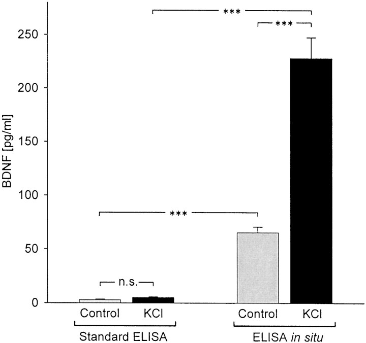
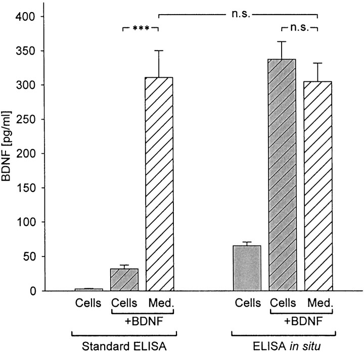
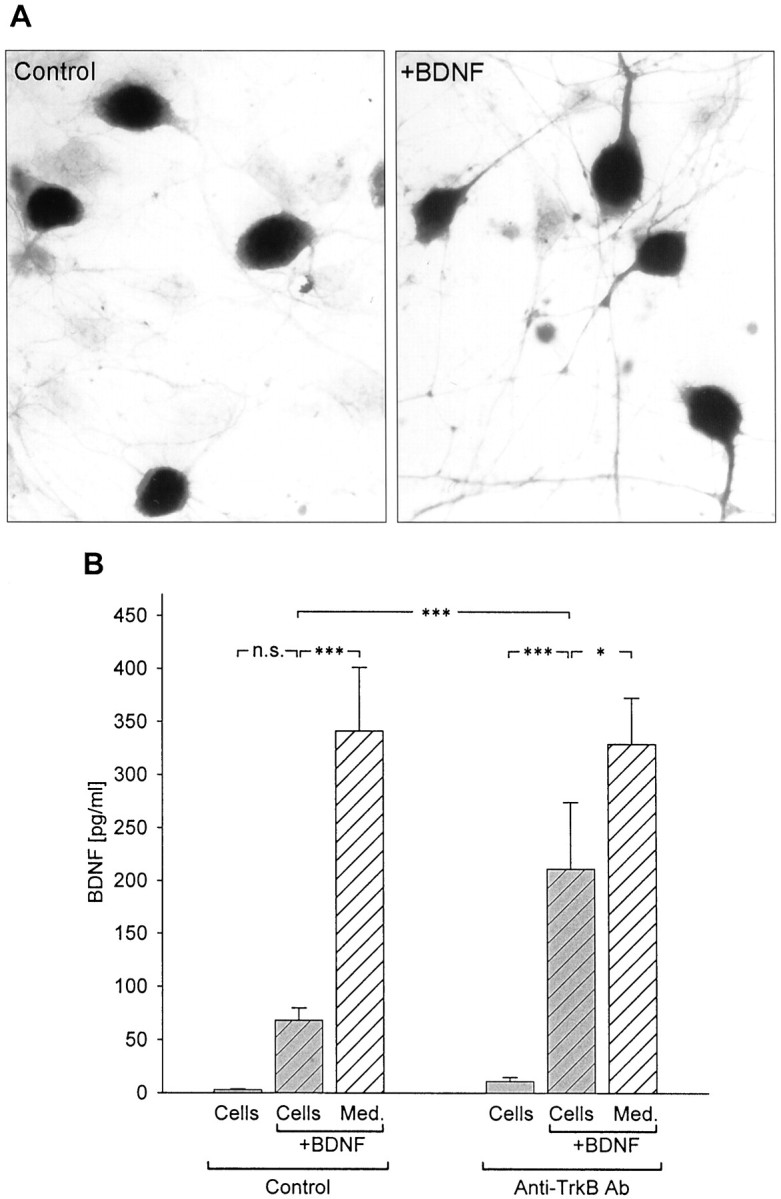
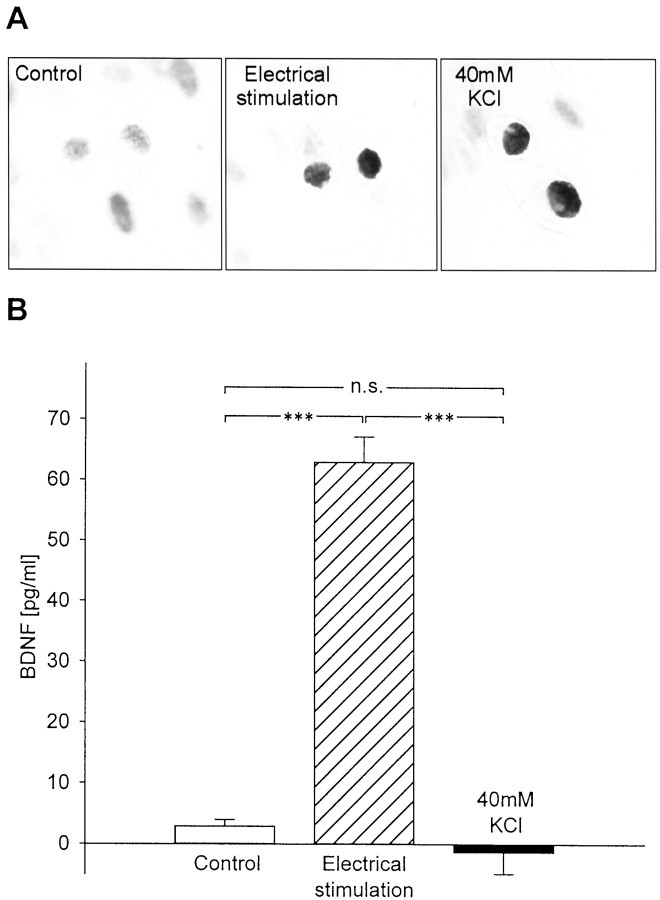
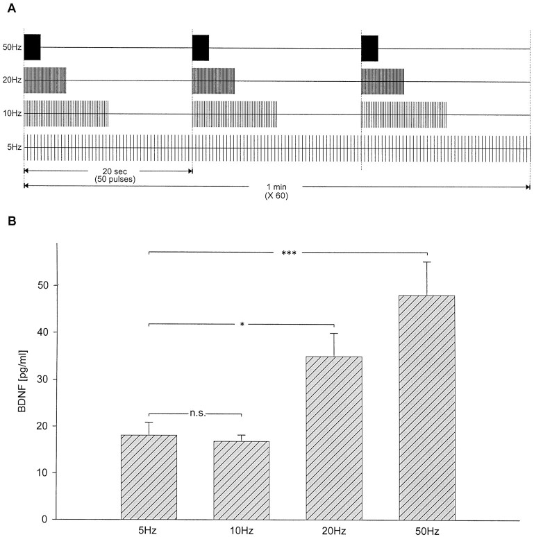
To define activity-dependent release of endogenous brain-derived neurotrophic factor (BDNF), we developed an in vitro model using primary sensory neurons and a modified ELISA, termed ELISA in situ. Dissociate cultures of nodose-petrosal ganglion cells from newborn rats were grown in wells precoated with anti-BDNF antibody to capture released BDNF, which was subsequently detected using conventional ELISA. Conventional ELISA alone was unable to detect any increase in BDNF concentration above control values following chronic depolarization with 40 mM KCl for 72 hr. However, ELISA in situ demonstrated a highly significant increase in BDNF release, from 65 pg/ml in control to 228 pg/ml in KCl-treated cultures. The efficacy of the in situ assay appears to be related primarily to rapid capture of released BDNF that prevents BDNF binding to the cultured cells. We therefore used this approach to compare BDNF release from cultures exposed for 30 min to either continuous depolarization with elevated KCl or patterned electrical field stimulation (50 biphasic rectangular pulses of 25 msec, at 20 Hz, every 5 sec). Short-term KCl depolarization was completely ineffective at evoking any detectable release of BDNF, whereas patterned electrical stimulation increased extracellular BDNF levels by 20-fold. In addition, the magnitude of BDNF release was dependent on stimulus pattern, with high-frequency bursts being most effective. These data indicate that the optimal stimulus profile for BDNF release resembles that of other neuroactive peptides. Moreover, our findings demonstrate that BDNF release can encode temporal features of presynaptic neuronal activity.


Figures

Fig. 1.
Fig. 1.
Long-term exposure to elevated extracellular potassium induces BDNF release from newborn NPG neurons in culture. Mean BDNF levels were measured with standard ELISA and ELISA in situ in sister cultures grown for 72 hr in the absence (Control, gray bars) or presence of 40 mmpotassium (KCl, black bars). n = 21; ***p < 0.001; n.s., not significant.
Fig. 2.
Fig. 2.
Detectability of exogenous BDNF by standard ELISAversus ELISA in situ. BDNF (500 pg/ml) was added at plating to newborn NPG cultures (hatched gray bars) and to culture medium (Med.) alone (hatched white bars) and incubated for 72 hr in the absence (Standard ELISA) or presence of anti-BDNF monoclonal capture antibody (ELISA in situ). BDNF levels were also measured in control cultures (solid gray bars) to which exogenous BDNF was not added. ***p < 0.001; n.s., not significant.
Fig. 3.
Fig. 3.
Inhibition of BDNF binding to cells increases BDNF detectability by standard ELISA. A, Immunostaining with an antibody against the extracellular domain of TrkB (Chemicon) in newborn NPG cultures grown for 3 d in the absence (Control) or presence of exogenous BDNF (+BDNF, 500 pg/ml). B, Mean levels of BDNF, detected with standard ELISA, in the absence (Control) or presence of anti-TrkB antibody (Anti-TrkB Ab, 10 μg/ml) in NPG cultures (hatched gray bars) and culture medium alone (hatched white bars) 48 hr after addition of 500 pg/ml BDNF. BDNF levels were also measured in control cultures (solid gray bars) to which exogenous BDNF was not added. ***p < 0.001;*p < 0.05;n.s., not significant.
Fig. 4.
Fig. 4.
Patterned electrical stimulation is markedly more effective at releasing BDNF from newborn NPG neurons than KCl-induced continuous depolarization. A, P-CREB immunostaining of newborn NPG cultures after 30 min electrical field stimulation (20 Hz;Electrical stimulation) or 30 min continuous depolarization (40mm KCl) compared with unstimulated controls. B, Mean levels of BDNF released in sister cultures of newborn NPG neurons during 30 min of control conditions (no stimulation), electrical field stimulation (50 biphasic rectangular pulses of 25 msec, at 20 Hz, every 5 sec), or continuous depolarization with 40 mm KCl. Each value represents the difference between the BDNF level measured after stimulation and the level measured in sister cultures at the beginning of the stimulus period. ***p < 0.001;n.s., not significant.
Fig. 5.
Fig. 5.
Activity-dependent release of BDNF is regulated by the pattern of stimulation. A, Schematic representation of the stimulation patterns applied to each group of cultures.B, Mean levels of BDNF released from newborn NPG neurons during 60 min of electrical field stimulation with 50 biphasic rectangular pulses of 10 msec, delivered at 5, 10, 20, and 50 Hz, with interburst intervals, respectively, of 0, 10, 15, and 18 sec as shown in A. ***p < 0.001; *p < 0.05; n.s., not significant.

References

    1. Acheson A, Lindsay RM. Non target-derived roles of the neurotrophins. Philos Trans R Soc Lond B Biol Sci. 1996;351:417–422. - PubMed
    1. Agoston DV, Conlon JM, Whittaker VP. Selective depletion of the acetylcholine and vasoactive intestinal polypeptide of the guinea-pig myenteric plexus by differential mobilization of distinct transmitter pools. Exp Brain Res. 1988;72:535–542. - PubMed
    1. Androutsellis-Theotokis A, McCormack WJ, Bradford HF, Stern GM, Pliego-Rivero FB. The depolarisation-induced release of [125I]BDNF from brain tissue. Brain Res. 1996;743:40–48. - PubMed
    1. Apfel SC, Wright DE, Wiideman AM, Dormia C, Snider WD, Kessler JA. Nerve growth factor regulates the expression of brain-derived neurotrophic factor mRNA in the peripheral nervous system. Mol Cell Neurosci. 1996;7:134–142. - PubMed
    1. Balkowiec A, Kunze DL, Katz DM. Brain-derived neurotrophic factor acutely inhibits AMPA-mediated currents in developing sensory relay neurons. J Neurosci. 2000;20:1904–1911. - PMC - PubMed

Publication types

Substances

LinkOut - more resources