. 2000 Oct 1;20(19):7446-54.
doi: 10.1523/JNEUROSCI.20-19-07446.2000.

Definition of neuronal circuitry controlling the activity of phrenic and abdominal motoneurons in the ferret using recombinant strains of pseudorabies virus

Affiliations

Definition of neuronal circuitry controlling the activity of phrenic and abdominal motoneurons in the ferret using recombinant strains of pseudorabies virus

I Billig et al. J Neurosci. .

Abstract

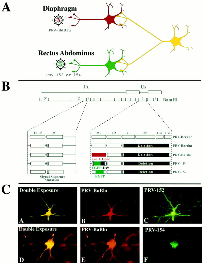
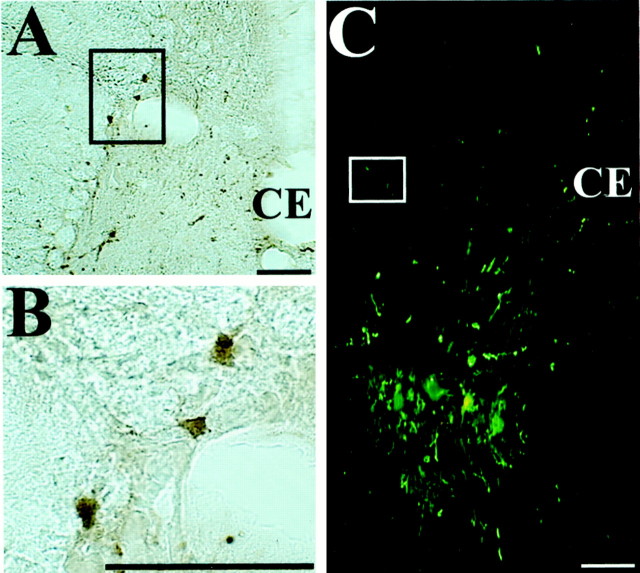
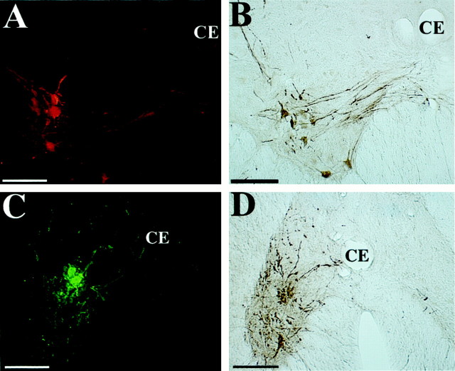
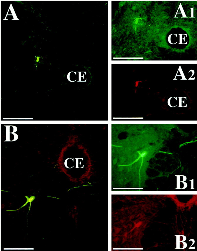
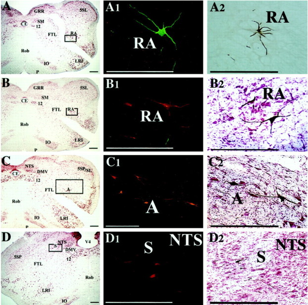
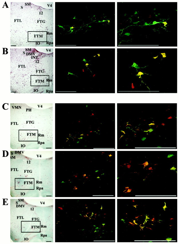
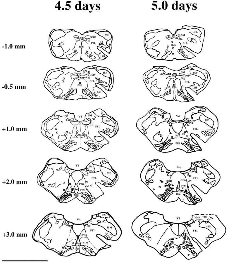
During a number of behaviors, including vomiting and some postural adjustments, activity of both the diaphragm and abdominal muscles increases. Previous transneuronal tracing studies using injection of pseudorabies virus (PRV) into either the diaphragm or rectus abdominis (RA) of the ferret demonstrated that motoneurons innervating these muscles receive inputs from neurons in circumscribed regions of the spinal cord and brainstem, some of which have an overlapping distribution in the magnocellular part of the medullary reticular formation (MRF). This observation raises two possibilities: that two populations of MRF neurons provide independent inputs to inspiratory and expiratory motoneurons or that single MRF neurons have collateralized projections to both groups of motoneurons. The present study sought to distinguish between these prospects. For this purpose, recombinant isogenic strains of PRV were injected into these respiratory muscles in nine ferrets; the strain injected into the diaphragm expressed beta-galactosidase, whereas that injected into RA expressed green fluorescent protein. Immunofluorescence localization of the unique reporters of each virus revealed three populations of infected premotor neurons, two of which expressed only one virus and a third group that contained both viruses. Dual-infected neurons were predominantly located in the magnocellular part of the MRF, but were absent from both the dorsal and ventral respiratory cell groups. These data suggest that coactivation of inspiratory and expiratory muscles during behaviors such as emesis and some postural adjustments can be elicited through collateralized projections from a single group of brainstem neurons located in the MRF.


Figures

Fig. 1.
Fig. 1.
A, The experimental strategy used in this study. Two recombinant strains of pseudorabies virus were used to produce retrograde transynaptic infection of neurons synaptically linked to motoneurons innervating the diaphragm and rectus abdominis muscles. Each viral strain expressed a unique reporter that could be detected with the use of monospecific antisera. B, The genomic organization of the recombinant viruses used in this study. The genome of PRV contains unique long (UL) and unique short (US) regions. The parental strain used to produce the recombinants used in this study was an attenuated vaccine strain known as PRV-Bartha. PRV-Bartha has several well characterized mutations and deletions that distinguish it from the wild-type virus (PRV-Becker). These alterations are mapped on the diagram. All recombinants involved insertion of the transgene at the gG locus of the viral genome. In PRV-BaBlu, gG was replaced with the gene encoding β-galactosidase. In PRV-152 and PRV-154, the gene encoding green fluorescent protein was inserted into the gG gene, either alone (PRV-152) or as part of a fusion protein (PRV-154). C, The distribution of the reporters within two infected neurons that were both double-labeled. β-galactosidase expression in neurons infected with PRV-Bablu provided staining of neuronal perikarya and dendrites (red fluorescence, panels B and E). Two EGFP-expressing viruses (PRV-152 and PRV-154) labeled cells differently. PRV-152 produced staining of perikarya and dendrites comparable to that produced by PRV-Bablu (green fluorescence, C). In contrast, the fusion protein produced by PRV-154 (EGFP+Us9) was differentially concentrated in the Golgi and rough endoplasmic reticulum of infected cells (green fluorescence,F). The majority of experiments reported here used PRV-152. Neurons containing both PRV-Bablu and PRV-152 or -154 appeared as having yellow fluorescence(A and D).
Fig. 2.
Fig. 2.
Photomicrographs illustrating restricted infection of sympathetic preganglionic neurons after injection of PRV into respiratory muscles. A, B, Infected neurons in the intermediolateral cell column of the T8 spinal segment after injection of PRV-152 into rectus abdominis; the section was immunoprocessed using immunoperoxidase. B is a higher magnification view of the region in A indicated by abox. Note that the infection was confined to the nucleus, cell body, and proximal dendrites of the labeled sympathetic preganglionic neurons. C, Infected neurons in the T11 spinal cord segment after the combined injection of PRV-Bablu into the diaphragm and PRV-152 into rectus abdominis. The section was immunoprocessed using immunofluorescence and photographed under illumination that excited both the CY2 and CY3 fluorophors. Note that the labeling of sympathetic preganglionic neurons in the intermediolateral cell column, which is indicated by abox, is much weaker than that of motoneurons located in the ventral horn. Scale bars, 100 μm.
Fig. 3.
Fig. 3.
Photomicrographs of presumed motoneurons infected 4 and 4.5 d after injection of PRV-Bablu into the diaphragm and PRV-152 into rectus abdominis. A, B, Large presumed motoneurons immunostained with the red CY3 fluorophor (A) or immunoprocessed with peroxidase (B) after an injection of PRV-Bablu into the diaphragm. The presumed phrenic motoneurons were located in the ventromedial ventral horn of the C6 spinal cord segment ipsilateral to the side of the injection. These large neurons characteristically exhibited fasciculated bundles of dendrites that extended toward the central canal (CE) and sometimes crossed the midline to the contralateral side. C, D,Examples of a cluster of presumed motoneurons, immunostained with the green CY2 fluorophor (C) or immunoprocessed with peroxidase (D) after an injection of PRV-152 into the rectus abdominis. The cells were observed in ipsilateral lamina VIII of the T10 spinal cord segment. These large neurons exhibited large dendritic arbors, with some processes crossing the midline or reaching the central canal (CE). Scale bars, 200 μm.
Fig. 4.
Fig. 4.
Photomicrographs of presumed spinal cord interneurons infected 4.5 and 5 d after PRV-Bablu injection into the diaphragm and PRV-152 injection into rectus abdominis. These neurons were dually immunostained with the red CY3 and the green CY2 fluorochromes, indicating that they made synaptic connections with both phrenic and abdominal motoneurons. The cells were located in lamina VII of the T8 (A) and lamina VIII of the L2 (B) spinal cord segments, ipsilateral to the injections. A1–B2 show the cell under illumination that excites one of the two fluorophors, whereasA and B were photographed under illumination that excites both fluorophors. Scale bars, 200 μm.
Fig. 5.
Fig. 5.
Examples of premotor neurons in regions of the brainstem known to contain the dorsal and ventral respiratory groups, which were infected 4.5 d after PRV injections into the diaphragm and rectus abdominis. A–D show photomicrographs of transverse brainstem sections located ∼1 mm caudal to the obex (A,B) or at the level of the obex (C,D); these sections were stained with the use of a modified Kluver-Barrera method.Boxes on the photomicrographs indicate the locations of the infected neurons illustrated in the middle column; these sections were immunoprocessed using immunofluorescence. Theright column illustrates infected neurons at the same location in sections immunoprocessed using immunoperoxidase.A1 and A2 show neurons in nucleus retroambiguus that were infected after injecting PRV-152 into rectus abdominis on the contralateral side. The neurons were immunostained with the green CY2 fluorophor (A1) or with peroxidase (A2). B1 and B2 show premotor neurons in nucleus retroambiguus infected after injecting PRV-Bablu into the diaphragm on the contralateral side; these neurons were labeled with the red CY3 fluorophor (B1) or through immunoperoxidase processing (B2). C1 andC2 illustrate infected premotor neurons in the vicinity of nucleus ambiguus and the retrofacial nucleus that were immunostained with the red CY3 fluorophor (C1) or with peroxidase (C2) after injections of PRV-Bablu into the contralateral diaphragm. D1 and D2 show infected neurons in the ventrolateral portion of the nucleus of the solitary tract, which were immunostained with the red CY3 fluorophor (D1) or with peroxidase (D2) after injecting PRV-Bablu into the ipsilateral diaphragm. Scale bars, 400 μm. 12, Hypoglossal nucleus; 5SL, laminar spinal trigeminal nucleus; 5SP, alaminar spinal trigeminal nucleus; A, nucleus ambiguus;AP, area postrema; CE, central canal; DMV, dorsal motor nucleus of the vagus;FTL, lateral tegmental field; GRR, rostral division of gracile nucleus; IO, inferior olivary nucleus; LRI, internal division of lateral reticular nucleus; NTS, nucleus of the solitary tract;P, pyramid; RA, nucleus retroambiguus;Rob, nucleus raphe obscurus; S, solitary tract; SM, medial nucleus of solitary tract;V4, fourth ventricle.
Fig. 6.
Fig. 6.
Location of premotor neurons in the MRF infected after injection of PRV into the diaphragm and rectus abdominis.A and B show infected neurons after a 4.5 d survival time, whereas C–E illustrate infection after a 5 d survival. In addition, A andD show labeling at ∼2.5 mm rostral to the obex, whereas B and E show labeling at ∼2.0 mm rostral to the obex, and C illustrates labeling at ∼3.0 mm rostral to the obex. Left column, Photomicrographs of brainstem sections (on the side ipsilateral to the injections) stained with a modified Kluver-Barrera method.Boxes surround the area containing infected neurons illustrated in the photomicrographs immediately on theright. Middle and right columns, Photomicrographs of infected neurons observed in the MRF. Red cells were only infected by retrograde transynaptic passage of PRV-Bablu from the diaphragm, green cells were only infected by retrograde transynaptic passage of PRV-152 from rectus abdominis, and yellow cells contain both viruses. Photomicrographs in the right column are a magnification of those in the middle column. Note that after a 5 d survival time, labeling is more prevalent and extends further rostrally than after a 4.5 d survival. Scale bars, 400 μm. Abbreviations are the same as in Figure 4, with the following additions: FTG, gigantocellular tegmental field;FTM, magnocellular tegmental field; INT, nucleus intercalatus; Rm, nucleus raphe magnus;Rpa, nucleus raphe pallidus.
Fig. 7.
Fig. 7.
Locations of neurons infected 4.5 and 5 d after PRV injection into the diaphragm and rectus abdominis. Neuronal locations are plotted on camera lucida drawings of transverse brainstem sections. Only labeling at selected levels of the brainstem in two animals is illustrated; the approximate rostrocaudal distance of each section from the obex is indicated. Neurons infected by injection of PRV-Bablu into the diaphragm are represented by open squares, those infected after injecting PRV-152 or PRV-154 into rectus abdominis are represented by open circles; dually infected neurons are represented by shaded triangles. Scale bar, 5 mm. Abbreviations are the same as in Figures 4 and 5, with the following additions: 5SM, magnocellular alaminar spinal trigeminal nucleus; CX, external cuneate nucleus;IFT, infratrigeminal nucleus; PR, paramedian reticular nucleus; RFN, retrofacial nucleus;VMN, medial vestibular nucleus; VIN, inferior vestibular nucleus.

References

    1. Bartha A. Experimental reduction of virulence of Aujezky's disease. Magy Allatorv Lapja. 1961;16:42–45.
    1. Bianchi AL, Grèlot L. Converse motor output of inspiratory bulbospinal premotoneurones during vomiting. Neurosci Lett. 1989;104:298–302. - PubMed
    1. Billig I, Foris JM, Card JP, Yates BJ. Transneuronal tracing of neural pathways controlling an abdominal muscle, rectus abdominis, in the ferret. Brain Res. 1999;820:31–44. - PubMed
    1. Brideau AD, Banfield BW, Enquist LW. The Us9 gene product of pseudorabies virus, an alphaherpesvirus, is a phosphorylated, tail-anchored type II membrane protein. J Virol. 1998;72:4560–4570. - PMC - PubMed
    1. Brideau AD, Card JP, Enquist LW. Role of pseudorabies virus Us9, a type IImembrane protein, in infection of tissue culture cells and the rat nervous system. J Virol. 2000;74:834–845. - PMC - PubMed

Publication types

MeSH terms

LinkOut - more resources