. 2000 Oct 1;20(19):7478-88.
doi: 10.1523/JNEUROSCI.20-19-07478.2000.

Cortical feedback controls the frequency and synchrony of oscillations in the visual thalamus

Affiliations

Cortical feedback controls the frequency and synchrony of oscillations in the visual thalamus

T Bal et al. J Neurosci. .

Abstract

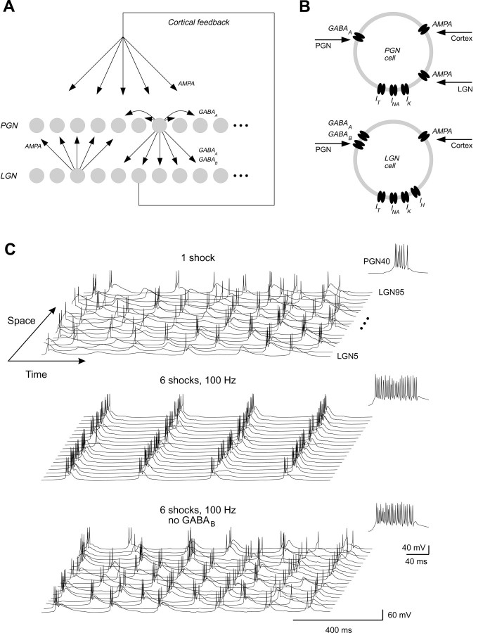
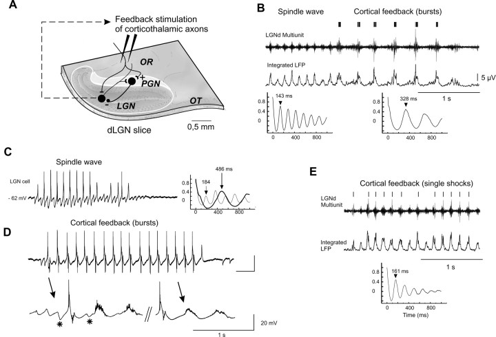
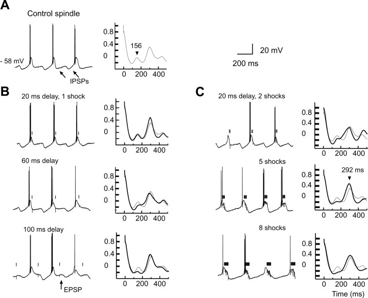
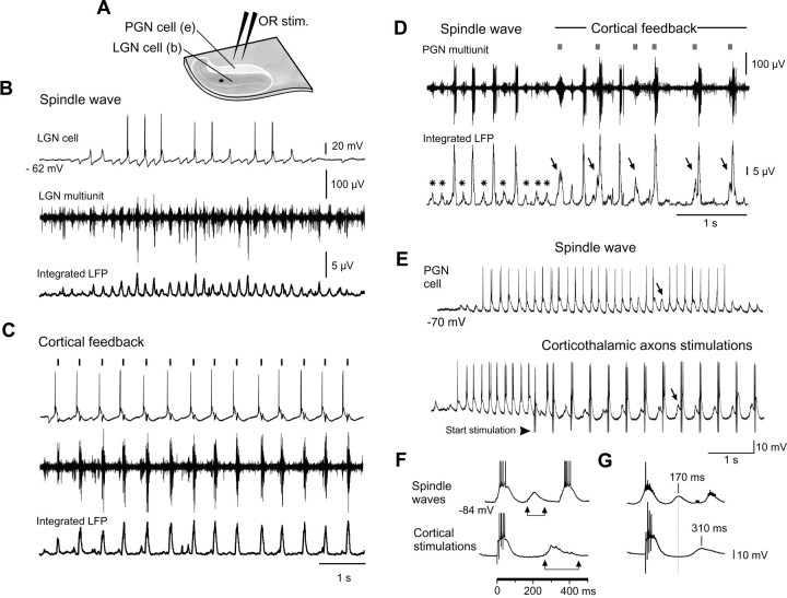
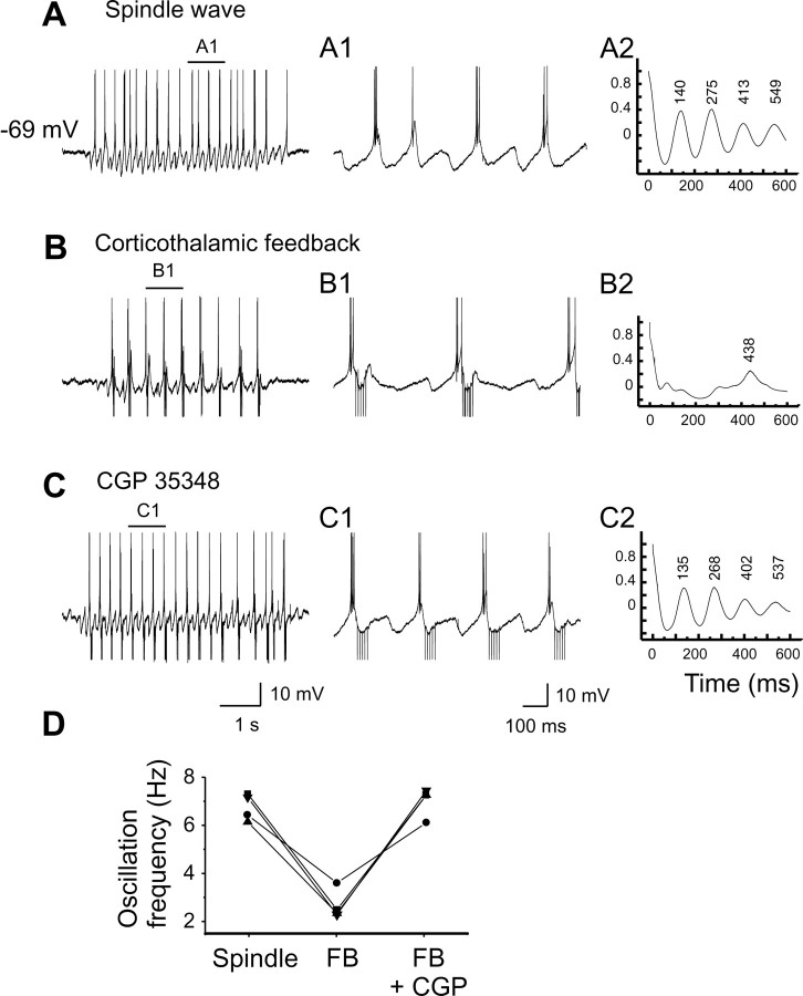
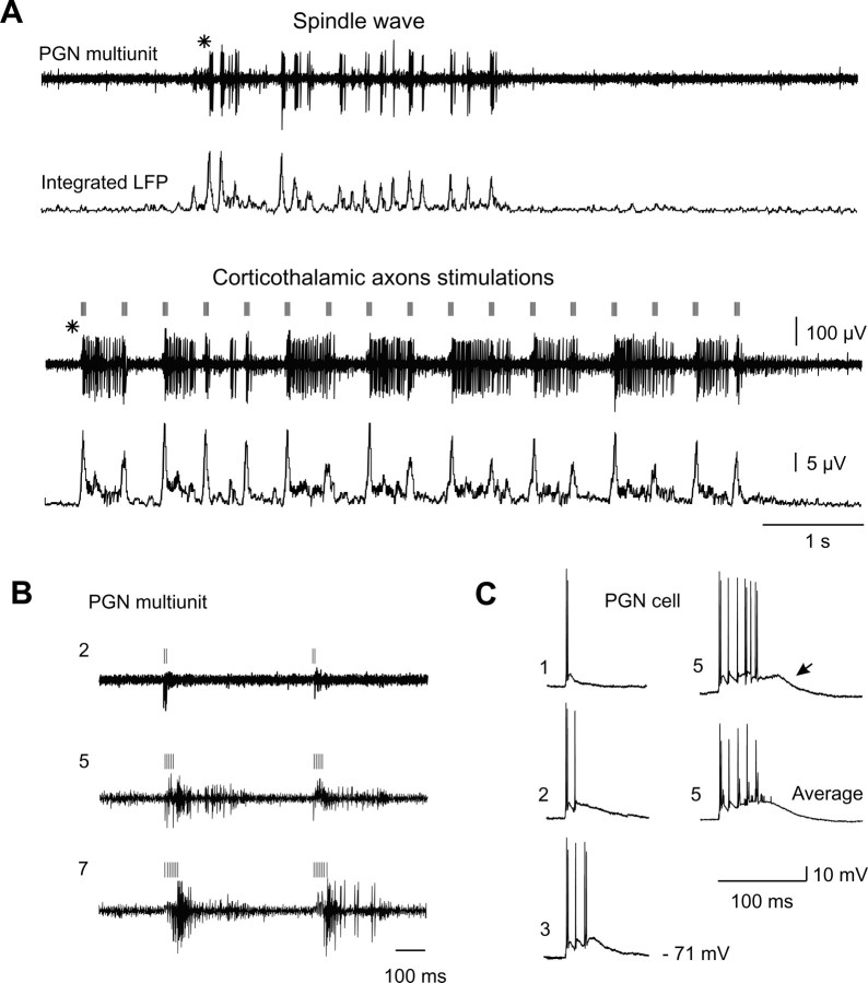
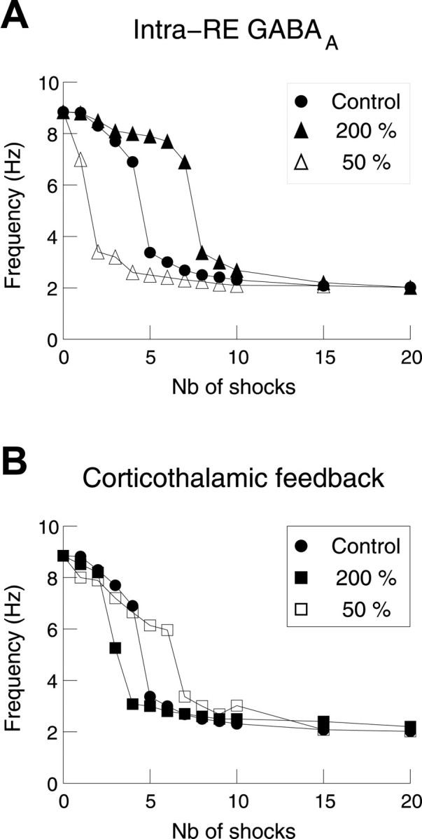
Thalamic circuits have an intrinsic capacity to generate state-dependent oscillations of different frequency and degrees of synchrony, but little is known of how synchronized oscillation is controlled in the intact brain or what function it may serve. The influence of cortical feedback was examined using slice preparations of the visual thalamus and computational models. Cortical feedback was mimicked by stimulating corticothalamic axons, triggered by the activity of relay neurons. This artificially coupled network had the capacity to self-organize and to generate qualitatively different rhythmical activities according to the strength of corticothalamic feedback stimuli. Weak feedback (one to three shocks at 100-150 Hz) phase-locked the spontaneous spindle oscillations (6-10 Hz) in geniculate and perigeniculate nuclei. However, strong feedback (four to eight shocks at 100-150 Hz) led to a more synchronized oscillation, slower in frequency (2-4 Hz) and dependent on GABA(B) receptors. This increase in synchrony was essentially attributable to a redistribution of the timing of action potential generation in lateral geniculate nucleus cells, resulting in an increased output of relay cells toward the cortex. Corticothalamic feedback is thus capable of inducing highly synchronous slow oscillations in physiologically intact thalamic circuits. This modulation may have implications for a better understanding of the descending control of thalamic nuclei by the cortex, and the genesis of pathological rhythmical activity, such as absence seizures.


Figures

Fig. 1.
Fig. 1.
Computational model prediction of the control of thalamic oscillations by corticothalamic feedback. A,Scheme of the thalamic circuit. A network consisting of two one-dimensional layers of LGN and PGN neurons (100 cells each) was simulated with topographic connections mediated by glutamate (AMPA) receptors and GABAergic (GABAA and GABAB) receptors as indicated. One cell (LGN cell 10) was the trigger of the cortical feedback, which was simulated through AMPA conductances in all cell types. The connectivity and conductances used were identical to a previous study (Destexhe et al., 1996) with additional corticothalamic feedback conductances of 1–4 μS in PGN and 0.05–0.5 μS in LGN cells. B, Scheme of the different ionic mechanisms present in each cell type. The voltage-dependent currents IT,Ih,INa, andIK were needed to simulate the intrinsic bursting patterns of thalamic neurons. C, Spatiotemporal network activity raster plots, detailing the results of the simulation of the feedback experiment. Top, Feedback stimuli consisting of a single shock produced bursting patterns typical of spindle oscillations. Middle, Strong feedback (6 shocks at 100 Hz) synchronized the burst discharges of LGN cells and switched the oscillation frequency to 3 Hz in the entire network, although only one cell served as the trigger. Bottom, Suppressing GABAB receptors led to the reverse transformation from 3 to 10 Hz spindle oscillations, and the feedback was ineffective in inducing the 3 Hz rhythm. Each graph represents 19 equally spaced LGN cells in the network, and an example of PGN burst is shown ininset. A delay of 25 msec was used in all simulations.
Fig. 2.
Fig. 2.
Control of thalamic oscillations by corticothalamic feedback in ferret thalamic slices. A,In the self-generating spindling thalamic slice, lateral geniculate (LGN) relay neuron axons projecting to the cortex via the optic radiation (OR) give off a collateral branch to the perigeniculate nucleus (PGN). The GABAergic PGN neurons generate direct inhibitory feedback to the relay neurons of the LGN. Corticothalamic axons run in the OR and synapse on LGN and PGN cells. Bipolar stimulating electrodes were placed in the OR. OT, Optic tract. B, A 7 Hz control spindle is slowed down to a 3 Hz oscillation by the feedback stimulation of OR at a latency of 20 msec after the detection of multiunit bursts activity (5 shocks; 100 Hz). Middle trace, Smooth integration of the multiunit signal (integrated local field potential). Bottom traces, Autocorrelation functions applied on the integrated LFP before and after the transition. C, Intracellular recording of a thalamocortical cell during spontaneous spindle oscillation. The first peak (184 msec) of the autocorrelation function indicates the period of network oscillation (i.e., the inverse of its beating frequency).D, Cortical feedback stimulations (4 shocks; 100 Hz; 50 msec delay) triggered by the burst firing of the cell slows the network oscillation to ∼2 Hz. Downward deflections in the cell are stimulation artifacts. Action potentials were truncated for clarity. The spike-triggering average below was made from before and after the first 2 sec of 12 and 64 oscillatory sequences, respectively; it indicates the persistence of fast compound IPSPs at the beginning of the oscillation (asterisk). An autocorrelation function of this slow oscillation is superimposed in C as thethick trace (486 msec). E, Weak (single shock) feedback stimulation delivered to OR.
Fig. 3.
Fig. 3.
Thalamic oscillation is controlled by the intensity of the cortical feedback. A, Detail of a spindle wave and its control autocorrelogram displayed as thethin trace in B and C.B, Single shocks resulting in monosynaptic EPSPs (arrow) and performed at various delays after the bursts have little effect on the oscillation. C, Increasing the intensity of stimulation to a threshold of four or five shocks leads to the disappearance of fast IPSPs and slows down the network oscillation to 3–4 Hz.
Fig. 4.
Fig. 4.
Synchronization of thalamic cells by the corticothalamic loop. A, Scheme showing the locations of the intracellular recordings that were done successively for the LGN cell and the PGN cell in the same slice, on an anteroposterior axis passing between the branches of the stimulating electrode (the likely orientation for the thalamocortical (TC)–PGN reciprocal connections). Multiunit recordings made at location indicated by thefilled circle. For both cells, stimulation parameters were kept identical (27 μA intensity; 4 shocks at 100 Hz).B, Simultaneous intracellular and multiunit (filled circle) recordings in the LGN during a spindle wave. C, Same recordings during a slow network activity resulting from cortical feedback stimulations (50 msec delay) triggered by the action potentials in the intracellular recording.D, Control multiunit recording in the PGN (spindle wave) and its transformation during cortical feedback stimulation (6 shocks at 100 Hz, 30 msec delay). An extracellular single-unit thalamocortical cell, recorded with another electrode, was the trigger of the feedback (data not shown). The integrated trace shows the amplification of the population bursts (arrows) compared to control (asterisk), indicative of an enhanced synchrony of the activity of PGN cells. E, Intracellular recording of a PGN cell during spontaneous spindle waves and OR stimulation imposed periodically, but this time without feedback (4 shocks at 100 Hz, 400 msec interstimulus interval). Arrows show the EPSPs originated from the rebound firing of thalamocortical cells in adjacent layers. F, Detail of these compound EPSPs and their averages (G) during spindle (top trace; n = 83; triggered on the first spike of the burst) and OR stimulations delivered at 500 msec intervals (bottom trace; n = 56; triggered on the first EPSP). For the average, only EPSPs not leading to the generation of low-threshold calcium spike and bursting were selected.
Fig. 5.
Fig. 5.
Corticothalamic-induced slow oscillation is GABAB-dependent. A, Control 7 Hz spindle oscillation recorded in a thalamocortical cell. B,Corticothalamic feedback stimulation, triggered by the bursts of the same LGN cell, slows down the oscillation at 2–3 Hz. C,The fast 7 Hz rhythm resumes after local application of the GABAB antagonist CGP35348 (1 mm in micropipette) near the recording electrode. D, Graph representing the oscillation frequency of four cells recorded intracellularly versus experimental conditions: control (spindle); corticothalamic feedback (FB); and corticothalamic feedback in presence of CGP35348 (FB + CGP). For all cells, data points correspond to the latencies of the first peak of autocorrelation functions.
Fig. 6.
Fig. 6.
Strong corticothalamic activity enhance the burst discharge of PGN cells. A, Multiunit recording in the perigeniculate nucleus. Short-duration burst discharges of a PGN cell during spindle waves (asterisk; 2 top traces) are transformed in prolonged discharges during the rhythmic stimulations (7 shocks at 140 Hz; 400 msec interval) of corticothalamic axons (2 bottom traces). B, Increasing the number of shocks of stimulation (respectively 2, 5, and 7) increases the intensity of discharges recorded in the PGN. C, Same protocol as inB performed intracellularly for one, two, three, and five shocks. Average for five shocks response consists of 15 traces.
Fig. 7.
Fig. 7.
Conductances that affect the corticothalamic control of oscillations in model LGN–PGN networks. A,Representation of the mean network frequency as a function of the number of shocks given to corticothalamic feedback. The filled circles represent the control transition from 8–9 Hz to 2–4 Hz oscillations (same simulation as in Fig. 1C). The same transition is shown for reinforced (200%; filled triangles) or weakened (50%; open triangles) GABAA conductances within the reticular nucleus. These conductances acted against the slow oscillation. B, Same representation for reinforced (200%; filled squares) or weakened (50%; open squares) AMPAergic conductances underlying cortical EPSPs in thalamic reticular neurons. Reducing these AMPAergic conductances reduced the tendency of the thalamic circuit to switch to slow oscillations.
Fig. 8.
Fig. 8.
Computational evidence that the two oscillations constitute qualitatively distinct rhythmic states. A,Histogram of the number of spikes fired by the LGN population in a simulations of the model shown in Figure 1, A andB. The arrow indicates the onset of the feedback (6 shocks, 100 Hz; other parameters identical to Fig.1C). B, Average output of the LGN population represented against the number of shocks. Left ordinate (circles), Average number of spikes fired by LGN cells per oscillation cycle. Right ordinate(squares), Frequency of the network oscillation.Filled symbols indicate that >25% of GABABconductance was activated in TC cells, in which case the network switched to another type of oscillation with lower frequency and higher synchrony.

Similar articles

Cited by

References

    1. Aghajanian GK, Rasmussen K. Intracellular studies in the facial nucleus illustrating a simple new method for obtaining viable motoneurons in adult rat brain slices. Synapse. 1989;3:331–338. - PubMed
    1. Ahlsen G, Grant K, Lindström S. Monosynaptic excitation of principal cells in the lateral geniculate nucleus by corticofugal fibers. Brain Res. 1982;234:454–458. - PubMed
    1. Andersen P, Andersson SA. Physiological basis of the alpha rhythm. Appelton Century Crofts; New York: 1968.
    1. Bal T, von Krosigk M, McCormick DA. Synaptic and membrane mechanisms underlying synchronized oscillations in the ferret lateral geniculate nucleus in vitro. J Physiol (Lond) 1995a;483:641–663. - PMC - PubMed
    1. Bal T, von Krosigk M, McCormick DA. Role of the ferret perigeniculate nucleus in the generation of synchronized oscillations in vitro. J Physiol (Lond) 1995b;483:665–685. - PMC - PubMed

Publication types

MeSH terms

LinkOut - more resources