

Quantitative flow cytometric evaluation of maximal Cryptosporidium parvum oocyst infectivity in a neonate mouse model
- PMID: 11010875
- PMCID: PMC92301
- DOI: 10.1128/AEM.66.10.4315-4317.2000
Quantitative flow cytometric evaluation of maximal Cryptosporidium parvum oocyst infectivity in a neonate mouse model
Abstract
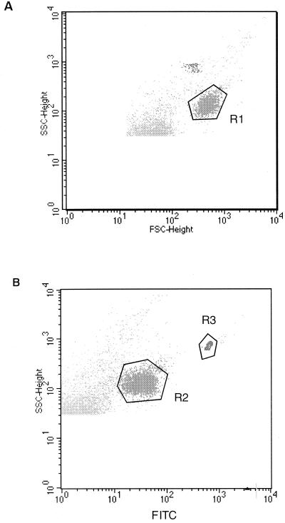
The importance of waterborne transmission of Cryptosporidium parvum to humans has been highlighted by recent outbreaks of cryptosporidiosis. The first step in a survey of contaminated water currently consists of counting C. parvum oocysts. Data suggest that an accurate risk evaluation should include a determination of viability and infectivity of counted oocysts in water. In this study, oocyst infectivity was addressed by using a suckling mouse model. Four-day-old NMRI (Naval Medical Research Institute) mice were inoculated per os with 1 to 1,000 oocysts in saline. Seven days later, the number of oocysts present in the entire small intestine was counted by flow cytometry using a fluorescent, oocyst-specific monoclonal antibody. The number of intestinal oocysts was directly related to the number of inoculated oocysts. For each dose group, infectivity of oocysts, expressed as the percentage of infected animals, was 100% for challenge doses between 25 and 1,000 oocysts and about 70% for doses ranging from 1 to 10 oocysts/animal. Immunofluorescent flow cytometry was useful in enhancing the detection sensitivity in the highly susceptible NMRI suckling mouse model and so was determined to be suitable for the evaluation of maximal infectivity risk.
Figures
References
-
- Arrowood M J, Sterling C R. Isolation of Cryptosporidium oocysts and sporozoites using discontinuous sucrose and isopycnic Percoll gradients. J Parasitol. 1987;73:314–319. - PubMed
-
- Arrowood M J, Hurd M R, Mead J R. A new method for evaluating experimental cryptosporidial parasite loads using immunofluorescent flow cytometry. J Parasitol. 1995;81:404–409. - PubMed
-
- Belosevic M, Guy R A, Taghikilani R, Neumann N F, Gyurek L L, Liyanage L R J, Millard P J, Finch G R. Nucleic acid stains as indicators of Cryptosporidium parvum oocyst viability. Int J Parasitol. 1997;27:787–798. - PubMed
-
- Bennett J W, Gauci M R, Le Moënic S, Schaefer III F W, Lindquist H D A. A comparison of enumeration techniques for Cryptosporidium parvum oocysts. J Parasitol. 1999;85:1165–1168. - PubMed
-
- Brasseur P, Uguen C, Moreno-Sabater A, Favennec L, Ballet J J. Viability of Cryptosporidium parvum oocysts in natural waters. Folia Parasitol. 1998;45:113–116. - PubMed
Publication types
MeSH terms
Substances
LinkOut - more resources
Full Text Sources
Medical
