. 2000 Oct;66(10):4503-9.
doi: 10.1128/AEM.66.10.4503-4509.2000.

Detection and identification of bacterial endosymbionts in arbuscular mycorrhizal fungi belonging to the family Gigasporaceae

Affiliations

Detection and identification of bacterial endosymbionts in arbuscular mycorrhizal fungi belonging to the family Gigasporaceae

V Bianciotto et al. Appl Environ Microbiol. 2000 Oct.

Abstract

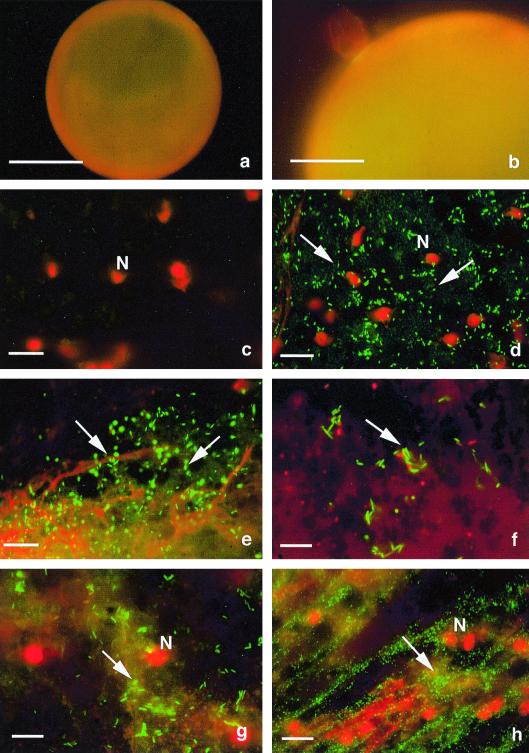
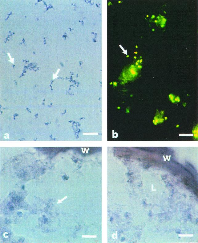
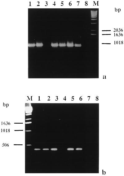
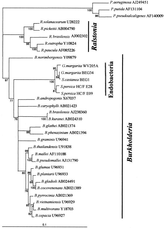
Intracellular bacteria have been found previously in one isolate of the arbuscular mycorrhizal (AM) fungus Gigaspora margarita BEG 34. In this study, we extended our investigation to 11 fungal isolates obtained from different geographic areas and belonging to six different species of the family Gigasporaceae. With the exception of Gigaspora rosea, isolates of all of the AM species harbored bacteria, and their DNA could be PCR amplified with universal bacterial primers. Primers specific for the endosymbiotic bacteria of BEG 34 could also amplify spore DNA from four species. These specific primers were successfully used as probes for in situ hybridization of endobacteria in G. margarita spores. Neighbor-joining analysis of the 16S ribosomal DNA sequences obtained from isolates of Scutellospora persica, Scutellospora castanea, and G. margarita revealed a single, strongly supported branch nested in the genus Burkholderia.


Figures

FIG. 1
FIG. 1
Bacterial endosymbionts in the cytoplasm of manually crushed spores of six fungal isolates (e through h) stained with the Live/Dead Baclight kit and observed by using a Nikon Optiphot-2 microscope with a View Scan DVC-250 confocal system (Bio-Rad, Hemel Hempstead, United Kingdom). Living bacteria fluoresce bright yellow-green under blue light, while dead bacteria fluoresce red under green light. (a and b) No contaminating bacteria were observed on the external surfaces of sterilized and sonicated spores of G. rosea (a) and G. margarita (WV 105A (b). Bars, 100 μm (a) and 50 μm (b). (c) No bacterial endosymbionts were detected in the cytoplasm of G. rosea BEG 9. Only the fungal nuclei (red masses) are visible. Bar, 10 μm. (d) Cytoplasm of a G. margarita WV 205A spore containing many living rod-shaped bacteria that fluoresce green (arrows) and fungal nuclei (red masses). Bar, 10 μm. (e) Cytoplasm of S. persica HC/F E09 containing numerous rod-shaped bacteria (arrows). The nuclei are broken, and red filaments of chromatin are visible. Bar, 10 μm. (f) Appearance of bacteria in S. persica HC/F E28. Bar, 7 μm. (g) S. castanea BEG 1 cytoplasm. Numerous living bacteria are present between the fungal nuclei. Bar, 7 μm. (h) Cytoplasm of G. gigantea containing a very high number of living bacteria that are smaller and rounder than the bacteria in the other isolates. Bar, 7 μm. N, nuclei.
FIG. 2
FIG. 2
In situ hybridization of intracellular symbiotic bacteria in spores of G. margarita BEG 34 and G. rosea BEG 9. BLOr (5′-GTCATCCACTCCGATTATTTA-3′) (6) hybridizes specifically with the 16S rRNA of the G. margarita endosymbiont and was used as probe. (a) In G. margarita a large number of blue rod-shaped spots (diameter, about 1 μm) (arrows) were visible in the fungal cytoplasm. They were especially visible when they grouped together in the cytoplasm. Bar, 10 μm. (b) The shape and position of spots correspond well with those of endobacteria revealed after Baclight kit staining of unfixed spores from the same fungal isolate. Bar, 7 μm. (c) No hybridization signal was obtained when the DIG-labelled probe was omitted. Bar, 10 μm. (d) No hybridization signal was obtained with the cytoplasm of G. rosea when the DIG-labelled BLOr probe was used. Bar, 10 μm. W, spore wall; L, lipid masses.
FIG. 3
FIG. 3
PCR experiments designed to reveal the presence of endobacteria in spores of different AM fungal isolates when two pairs of primers were used. (a) Agarose (1.2%) gel electrophoresis of PCR products amplified with bacterial primers 704f and 1495r when the following templates were used: G. margarita WV 205A (lane 1), G. margarita Brazil isolate (lane 2), G. rosea BEG 9 (lane 3), S. persica HC/F E09 (lane 4), S. castanea BEG 1 (lane 5), G. decipiens (lane 6), G. gigantea (lane 7), and no DNA (lane 8). Lane M contained a 1-kb DNA ladder (Gibco BRL). (b) Agarose (1.2%) gel electrophoresis of PCR products amplified with primers BLOf and BLOr specific for the endobacteria of G. margarita BEG 34 (6) when the following templates were used: G. margarita WV 205A (lane 1), G. margarita Brazil isolate (lane 2), S. persica HC/F E09 (lane 3), G. rosea BEG 9 (lane 4), S. castanea BEG 1 (lane 5), G. decipiens (lane 6), G. gigantea (lane 7), and no DNA (lane 8). Lane M contained a 1-kb DNA ladder (Gibco BRL).
FIG. 4
FIG. 4
Neighbor-joining tree obtained from alignment of the 16S rDNA of the endosymbionts of G. margarita, S. castanea, and S. persica isolates with the closest bacterial sequences retrieved by a BLAST search. Sequences were aligned by using the ClustalX program (31), and the alignment was edited with GeneDoc (22). Neighbor-joining analysis was performed with the ClustalX program using Kimura's distance method. The branch comprising species in the genus Pseudomonas was used as an outgroup. Branches are shown only when the percentage of bootstrap support (1,000 trials) exceeded 70%.

References

    1. Albrecht C, Geurts R, Bisseling T. Legume nodulation and mycorrhizae formation; two extremes in host specificity meet. EMBO J. 1999;18:281–288. - PMC - PubMed
    1. Amann R I, Springer N, Ludwig W, Goertz H D, Schleifer K H. Identification in situ and phylogeny of uncultured bacterial endosymbionts. Nature. 1991;351:161–164. - PubMed
    1. Amann R I, Ludwig W, Schleifer K H. Phylogenetic identification and in situ detection of individual microbial cells without cultivation. Microbiol Rev. 1995;59:143–169. - PMC - PubMed
    1. Andrade G, Mihara K L, Linderman R G, Bethlenfalvay G J. Bacteria from rhizosphere and hyphosphere soils of different arbuscular-mycorrhizal fungi. Plant Soil. 1997;192:71–79.
    1. Bago B, Bentivenga S P, Brenac V, Dodd J C, Piché Y, Simon L. Molecular analysis of Gigaspora (Glomales, Gigasporaceae) New Phytol. 1998;139:581–588.

Publication types

Associated data

LinkOut - more resources