. 2000 Nov;67(5):1095-103.
doi: 10.1086/303105. Epub 2000 Sep 29.

Isolated 2-methylbutyrylglycinuria caused by short/branched-chain acyl-CoA dehydrogenase deficiency: identification of a new enzyme defect, resolution of its molecular basis, and evidence for distinct acyl-CoA dehydrogenases in isoleucine and valine metabolism

Affiliations

Isolated 2-methylbutyrylglycinuria caused by short/branched-chain acyl-CoA dehydrogenase deficiency: identification of a new enzyme defect, resolution of its molecular basis, and evidence for distinct acyl-CoA dehydrogenases in isoleucine and valine metabolism

B S Andresen et al. Am J Hum Genet. 2000 Nov.

Abstract

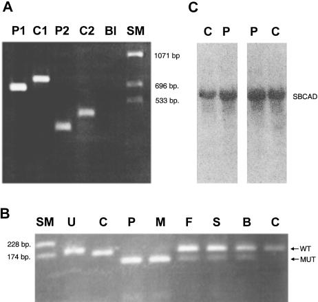
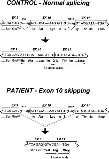
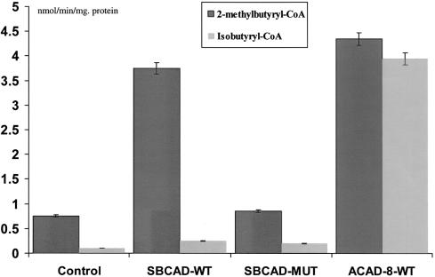
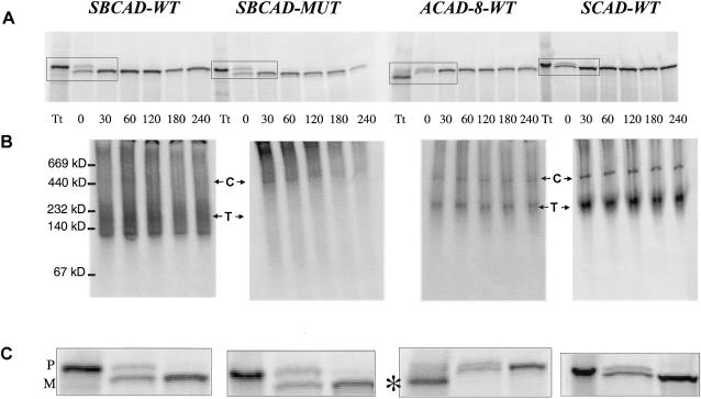
Acyl-CoA dehydrogenase (ACAD) defects in isoleucine and valine catabolism have been proposed in clinically diverse patients with an abnormal pattern of metabolites in their urine, but they have not been proved enzymatically or genetically, and it is unknown whether one or two ACADs are involved. We investigated a patient with isolated 2-methylbutyrylglycinuria, suggestive of a defect in isoleucine catabolism. Enzyme assay of the patient's fibroblasts, using 2-methylbutyryl-CoA as substrate, confirmed the defect. Sequence analysis of candidate ACADs revealed heterozygosity for the common short-chain ACAD A625 variant allele and no mutations in ACAD-8 but a 100-bp deletion in short/branched-chain ACAD (SBCAD) cDNA from the patient. Our identification of the SBCAD gene structure (11 exons; >20 kb) enabled analysis of genomic DNA. This showed that the deletion was caused by skipping of exon 10, because of homozygosity for a 1228G-->A mutation in the patient. This mutation was not present in 118 control chromosomes. In vitro transcription/translation experiments and overexpression in COS cells confirmed the disease-causing nature of the mutant SBCAD protein and showed that ACAD-8 is an isobutyryl-CoA dehydrogenase and that both wild-type proteins are imported into mitochondria and form tetramers. In conclusion, we report the first mutation in the SBCAD gene, show that it results in an isolated defect in isoleucine catabolism, and indicate that ACAD-8 is a mitochondrial enzyme that functions in valine catabolism.


Figures

Figure  1
Figure 1
A, PCR of SBCAD cDNA from the index patient (lane P1) and a normal control (lane C1), using a sense primer located in exon 6 and an antisense primer located in exon 11. “P2” and “C2” result from identical amplifications as P1 and C1, respectively, except that another sense primer, located in exon 8, was used. A blank amplification is marked by “Bl.” The size marker is indicated by “SM.” B, Agarose gel electrophoresis after restriction digestion of PCR products from genomic DNA from the index patient (lane P), his mother (lane M), his father (lane F), his healthy sister (lane S), his healthy brother (lane B), and a normal control (lane C), using a modified sense primer (5′-ATTACCCTGTGGAGAAATACTGAGATGCAAAGACT-3′) located in exon 10 of the SBCAD gene and an antisense primer (5′-AAATCCAGCCACATAACTAGTGCCACCAAACATATACTG-3′) located in intron 10. Undigested PCR products (U) are 187 bp long. After digestion with the SpeI restriction enzyme, PCR products harboring the 1228G→A mutation are 130 bp long, and PCR products with the wild-type sequence are 167 bp. C, Two different northern blot analyses of 15 μg total fibroblast RNA from the patient (lane P) and a control (lane C), using a SBCAD cDNA probe.
Figure  2
Figure 2
Position, in the SBCAD gene, of the 1228G→A mutation, and the resulting aberrant splicing in the patient, compared with the normal splicing pattern in a control person. The position of the 1228G→A mutation observed in the patient is indicated by boldface italic type.
Figure  3
Figure 3
Enzyme activities after expression of wild-type and mutant SBCAD and ACAD-8 in COS-7 cells. The results from measuring ACAD activity in COS-7 cells transfected with pSBCAD-WT, pSBCAD-MUT, or pACAD-8-WT expression vectors are shown. The control is COS-7 cells harboring the expression vector pcDNA3.1+ with no cDNA insert (Control). Activity measurements are given in nmol/min/mg protein. Blackened bars show the enzyme activity measured with 2-methylbutyryl-CoA as substrate, and gray bars show the enzyme activity measured with isobutyryl-CoA as substrate. Error bars indicate the range of the values measured. None of the transfected cells showed activity above background when either butyryl-CoA or isovaleryl-CoA was used as substrate (not shown).
Figure  4
Figure 4
In vitro mitochondrial import and stability of wild-type and mutant SBCAD, wild-type ACAD-8, and wild-type SCAD. Wild-type and mutant SBCAD, wild-type ACAD-8, and SCAD wild-type precursor proteins were synthesized by coupled in vitro transcription/translation and were incubated with isolated rat mitochondria for 30 min. After removal, by washing, of unimported precursor proteins, the fate of the imported proteins was followed for different time intervals. Twenty-microliter samples of the mitochondria were collected at the time points indicated, were washed two times, and were incubated with trypsin, followed by addition of trypsin inhibitor. Then samples were lysed and subjected to centrifugation. Finally, the supernatant fraction was subjected to either SDS-PAGE (A and C) or native PAGE (B). A 1-μl sample of the total translatate (Tt) withdrawn prior to incubation with mitochondria was also subjected to electrophoresis. The positions and molecular masses of coelectrophoresed marker proteins are indicated on the left margin. Positions of the precursor (P) and the mature (M) form of the respective proteins are shown on the right margin, and the position corresponding to the tetrameric proteins (T) and mHsp60 chaperone complexes (C) are indicated. The positions of the chaperone complexes were demonstrated by western blot analysis using an anti-mHsp60 antibody (authors' unpublished results). In the total translatate produced from the pACAD-8-WT plasmid, a lower-molecular-weight band (denoted by the asterisk [*]) was present, but the corresponding polypeptides were not imported into mitochondria. We presume that this band represents ACAD-8 proteins in which an internal in-frame ATG codon has been used as the translation start signal. Such internal ATGs, which are present in a more optimal sequence context for translation initiation (Kozak 1989) than are the correct ATG, are located at positions 115–117 and 178–180 in the ACAD-8 cDNA. Initiation of translation at either of these positions will result in production of proteins without a mitochondrial targeting sequence and with a size corresponding to the lower-molecular-weight band.

Similar articles

Cited by

References

Electronic-Database Information

    1. BLAST, http://www.ncbi.nlm.nih.gov/BLAST/
    1. Genbank Overview, http://www.ncbi.nlm.nih.gov/Genbank/GenbankOverview.html (for human SBCAD gene [accession numbers AF260668–AF260678])

References

    1. Amir N, Elpeleg ON, Shalev RS, Christensen E (1989) Glutaric aciduria type I: enzymatic and neuroradiologic investigations of two kindreds. J Pediatr 114:98–99 - PubMed
    1. Andresen BS, Bross P, Udvari S, Kirk J, Gray RGF, Kmock S, Chamoles N, Knudsen I, Winter V, Wilcken B, Yokota I, Hart K, Packman S, Harpey JP, Saudubray JM, Hale DE, Bolund L, Kolvraa S, Gregersen N (1997) The molecular basis of medium-chain acyl-CoA dehydrogenase (MCAD) deficiency in compound heterozygous patients: is there a correlation between genotype and phenotype? Hum Mol Genet 6:695–708 - PubMed
    1. Andresen BS, Jensen TG, Bross P, Knudsen I, Winter V, Kølvraa S, Bolund L, Ding JH, Chen YT, VanHove JLK, Curtis D, Yokota I, Tanaka K, Kim JJP, Gregersen N (1994) Disease-causing mutations in exon 11 of the medium-chain acyl-CoA dehydrogenase (MCAD) gene. Am J Hum Genet 54: 975–988 - PMC - PubMed
    1. Andresen BS, Olpin S, Poorthuis B, Scholte HR, Vianey-Saban C, Wanders RJA, Ijlst L, Morris A, Pourfarzam M, Bartlett K, Baumgartner ER, deKlerk JB, Schroeder LD, Corydon TJ, Lund H, Winter V, Bross P, Bolund L, Gregersen N (1999) It is possible to correlate genotype with disease phenotype in very-long-chain acyl-CoA dehydrogenase (VLCAD) deficiency. Am J Hum Genet 64:479–494 - PMC - PubMed
    1. Binzak B, Willard J, Vockley J (1998) Identification of the catalytic residue of human short/branched chain acyl-CoA dehydrogenase by in vitro mutagenesis. Biochim Biophys Acta 1382:137–142 - PubMed

Publication types

MeSH terms