Comparative Study
. 2000 Oct 2;19(19):5071-80.
doi: 10.1093/emboj/19.19.5071.

Transfer of palmitate from phospholipids to lipid A in outer membranes of gram-negative bacteria

Affiliations
Comparative Study

Transfer of palmitate from phospholipids to lipid A in outer membranes of gram-negative bacteria

R E Bishop et al. EMBO J. .

Abstract

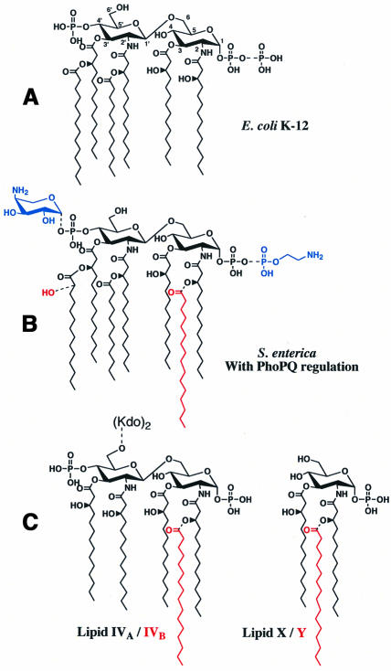
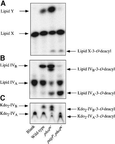
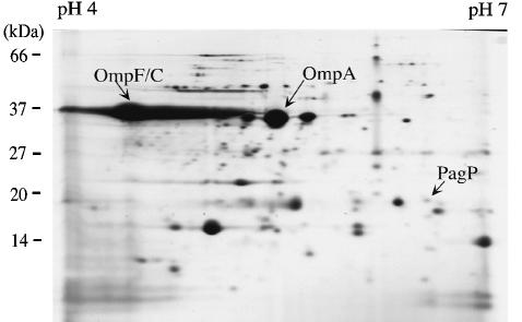
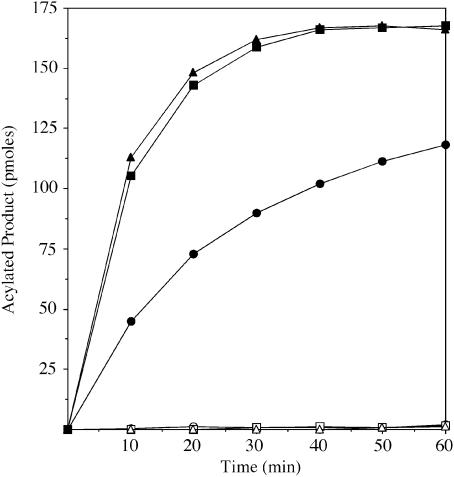
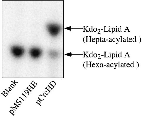
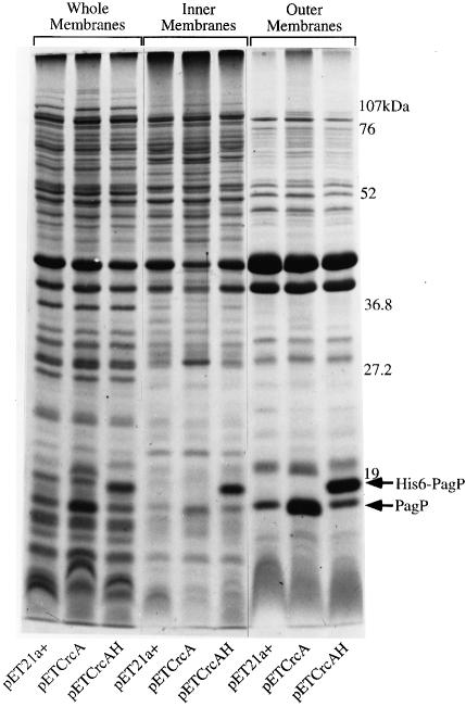
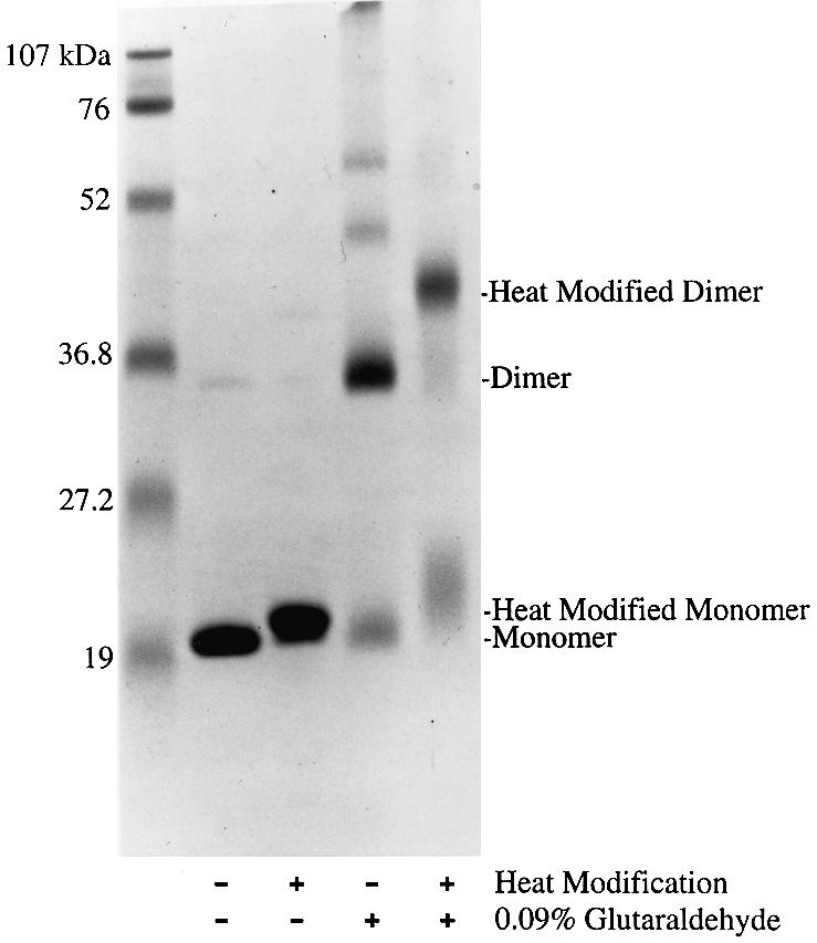
Regulated covalent modifications of lipid A are implicated in virulence of pathogenic Gram-negative bacteria. The Salmonella typhimurium PhoP/PhoQ-activated gene pagP is required both for biosynthesis of hepta-acylated lipid A species containing palmitate and for resistance to cationic anti-microbial peptides. Palmitoylated lipid A can also function as an endotoxin antagonist. We now show that pagP and its Escherichia coli homolog (crcA) encode an unusual enzyme of lipid A biosynthesis localized in the outer membrane. PagP transfers a palmitate residue from the sn-1 position of a phospholipid to the N-linked hydroxymyristate on the proximal unit of lipid A (or its precursors). PagP bearing a C-terminal His(6)-tag accumulated in outer membranes during overproduction, was purified with full activity and was shown by cross-linking to behave as a homodimer. PagP is the first example of an outer membrane enzyme involved in lipid A biosynthesis. Additional pagP homologs are encoded in the genomes of Yersinia and Bordetella species. PagP may provide an adaptive response toward both Mg(2+) limitation and host innate immune defenses.


Figures

None
Fig. 1. Structures of lipid A species and key precursors found in E.coli K-12 and S.typhimurium. (A) About two-thirds of lipid A is normally recovered from E.coli K-12 as a hexa-acylated 1,4′-bis-phosphate with the remaining one-third containing a 1-pyrophosphate group (dashed bond; Zhou et al., 1999). (B) Regulated covalent lipid A modifications found in Salmonella enterica serovars include the l-4-aminoarabinose and phosphoethanolamine substituents (blue), and the S-2-OH and palmitoyl groups (red; Guo et al., 1997). With the exception of the S-2-OH group, latent enzymes for these modifications also are present in E.coli K-12 (Zhou et al., 1999), and are induced by treatment with ammonium metavanadate, or accumulate in polymyxin-resistant mutants. (C) Substrates utilized in this investigation include the disaccharide lipid A precursors lipid IVA and Kdo2–lipid IVA (dashed bond), which can be acylated (red) by PagP to produce lipid IVB and Kdo2–lipid IVB, respectively. The monosaccharide lipid X can also be acylated (red) by PagP to produce lipid Y (Brozek et al., 1987).
None
Fig. 2. Products of PagP reactions using various Salmonella membranes with lipid X, lipid IVA or Kdo2–lipid IVA as acceptors. PagP was assayed at 30°C for 40 min using 0.5 mg/ml membranes from S.enterica serovar typhimurium ATCC 14028 (Wild-type), CS022 (phoPc) and LG069 (pagP, phoPc). Assays contain 100 mM Tris–HCl pH 8, 10 mM EDTA, 0.25% Triton X-100, 1 mM sn-1-(16:0)-2-(18:1cΔ9)-PtdCho as acyl donor and 200 c.p.m./µl of the 32P-labeled lipid X, lipid IVA or Kdo2–lipid IVA acyl acceptors at 10 µM. Metabolites were separated by TLC and visualized by overnight exposure to a PhosphorImager screen. The solvent systems for the indicated acyl acceptors are followed by the Rf values listed in ascending order for each metabolite (identified with arrowheads). (A) Lipid X, chloroform:methanol:water:acetic acid (25:15:4:2 v/v). Rf1 = 0.42, Rf2 = 0.55, Rf3 = 0.71. (B) Lipid IVA, chloroform: pyridine:88% formic acid:water (50:50:16:5 v/v). Rf1 = 0.36, Rf2 = 0.43, Rf3 = 0.52, Rf4 = 0.55. (C) Kdo2–lipid IVA, chloroform:pyridine:88% formic acid/water (30:70:16:10 v/v). Rf1 = 0.38, Rf2 = 0.40, Rf3 = 0.43, Rf4 = 0.45.
None
Fig. 3. Salmonella PagP is an outer membrane protein. Purified outer membrane proteins (350 µg) from the S.typhimurium PhoP-constitutive strain (CS022) were prepared and separated by 15% 2D-PAGE as described previously (Guina et al., 2000). An ∼18-kDa protein species migrating at pI 6.5 was identified as PagP by MALDI-TOF MS tryptic peptide fingerprinting. Corresponding protein was absent from the outer membranes of the pagP mutant strain (LG069). The locations of PagP and the major outer membrane proteins OmpF/C and OmpA are indicated with arrowheads. The positions of molecular weight standards are indicated to the left of the gel and the linear pH gradient generated during the isoelectric focusing step is indicated above the gel.
None
Fig. 4. Outer membrane localization of PagP activity in wild-type E.coli membranes. The inner and outer membranes from E.coli MC1061 were separated by isopycnic sucrose density gradient centrifugation, and ∼0.7 ml fractions were collected. Light scattering material was detected by measuring the A550. The inner and outer membranes were located by measuring NADH oxidase and phospho lipase A activities, respectively. PagP activity was measured as described in the legend to Figure 2, but with 12.5 µl portions from each fraction using 32P-labeled lipid IVA (200 c.p.m./µl) at 100 µM for 220 min. Measurements are expressed as a percentage of the total activity units across the entire gradient. NADH oxidase (squares), phospholipase A (triangles), light scattering (crosses) and PagP (circles).
None
Fig. 5. Relative rates of product formation by E.coli PagP-overproducing outer membranes with lipid X, lipid IVA or Kdo2–lipid IVA as acyl acceptors. PagP was assayed as described in the legend to Figure 2 using outer membranes at 10 µg/ml. Each substrate was present at 10 µM in 25 µl reaction volumes. Induced outer membranes were prepared from E.coli MC1061 transformed with pMS119HE (open symbols) or pCrcHD (solid symbols). Acyl acceptors are lipid X (circles), lipid IVA (squares) or Kdo2–lipid IVA (triangles).
None
Fig. 6. Kdo2–lipid A is a substrate for acylation by PagP. Kdo2–[32P]lipid A (100 c.p.m./µl) at ∼10 µM was assayed for PagP activity for 5 min as described in the legend to Figure 2 using induced outer membranes (10 µg/ml) from E.coli MC1061 transformed with either pMS119HE or pCrcHD. The TLC plate was developed in the solvent system of chloroform:pyridine:88% formic acid:water (30:70:16:10 v/v) and the separated products were visualized by overnight exposure to a PhosphorImager screen. The Rf values for Kdo2–lipid A are 0.52 (hexa-acylated) and 0.60 (hepta-acylated). The positions of the two Kdo2–lipid A derivatives are indicated to the right of the figure.
None
Fig. 7. Outer membrane localization of the PagP and His6-PagP proteins in induced membranes of E.coli BL21(DE3)pLysE transformed with pET21a+, pETCrcA or pETCrcAH. The inner and outer membranes were separated by isopycnic sucrose density gradient centrifugation. Next, 40 µg samples of protein from the whole membranes, the inner membranes, or the outer membranes were solubilized, boiled for 10 min, analyzed by 15% SDS–PAGE and stained with Coomassie Blue dye. The positions of molecular weight standards, PagP and His6-PagP, are indicated to the right of the gel.
None
Fig. 8. Heat modification and chemical crosslinking of purified His6-PagP. Two 17 µg samples of purified His6-PagP were treated with or without prior incubation in 0.09% glutaraldehyde for 100 min at 25°C. Each sample was then split into two 8.5 µg fractions and solubilized, with or without boiling for 10 min, for analysis by 15% SDS–PAGE. The gel was stained with Coomassie Blue dye. The positions of monomeric and dimeric bands, and their heat-modified derivatives, are indicated to the right of the gel. The positions of molecular weight standards are indicated to the left of the gel.

References

    1. Basu S.S., York,J.D. and Raetz,C.R.H. (1999) A phosphotransferase that generates phosphatidylinositol 4-phosphate (PtdIns-4-P) from phosphatidylinositol and lipid A in Rhizobium leguminosarum. A membrane-bound enzyme linking lipid A and PtdIns-4-P biosynthesis. J. Biol. Chem., 274, 11139–11149. - PMC - PubMed
    1. Brabetz W., Muller-Loennies,S., Holst,O. and Brade,H. (1997) Deletion of the heptosyltransferase genes rfaC and rfaF in Escherichia coli K-12 results in an Re-type lipopolysaccharide with a high degree of 2-aminoethanol phosphate substitution. Eur. J. Biochem., 247, 716–724. - PubMed
    1. Brozek K.A. and Raetz,C.R.H (1990) Biosynthesis of lipid A in Escherichia coli. Acyl carrier protein-dependent incorporation of laurate and myristate. J. Biol. Chem., 265, 15410–15417. - PubMed
    1. Brozek K.A., Bulawa,C.E. and Raetz,C.R.H. (1987) Biosynthesis of lipid A precursors in Escherichia coli. A membrane-bound enzyme that transfers a palmitoyl residue from a glycerophospholipid to lipid X. J. Biol. Chem., 262, 5170–5179. - PubMed
    1. Brozek K.A., Hosaka,K., Robertson,A.D. and Raetz,C.R.H. (1989) Biosynthesis of lipopolysaccharide in Escherichia coli. Cytoplasmic enzymes that attach 3-deoxy-d-manno-octulosonic acid to lipid A. J. Biol. Chem., 264, 6956–6966. - PubMed

Publication types

MeSH terms

LinkOut - more resources