. 2000 Oct 2;19(19):5157-66.
doi: 10.1093/emboj/19.19.5157.

H2O2 sensing through oxidation of the Yap1 transcription factor

Affiliations

H2O2 sensing through oxidation of the Yap1 transcription factor

A Delaunay et al. EMBO J. .

Abstract

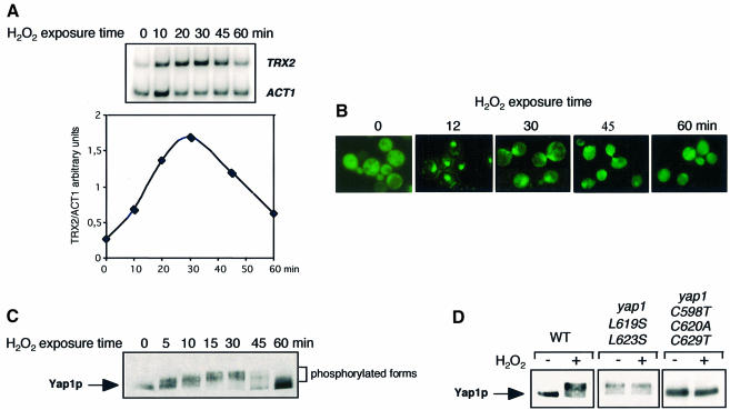
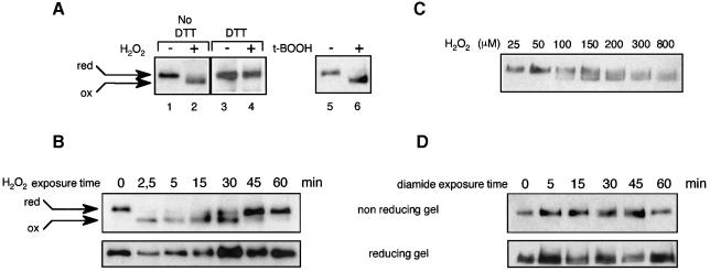
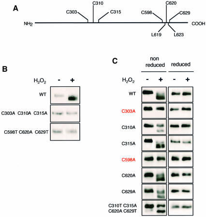
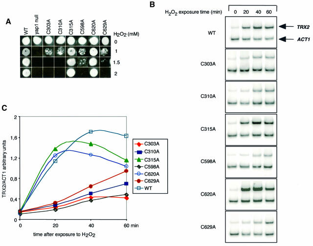
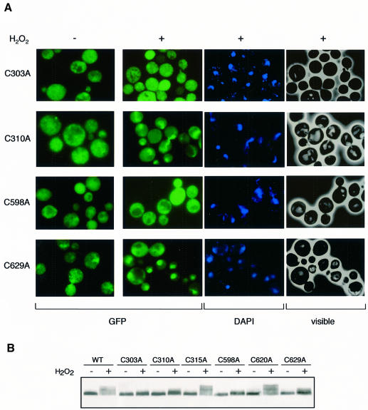
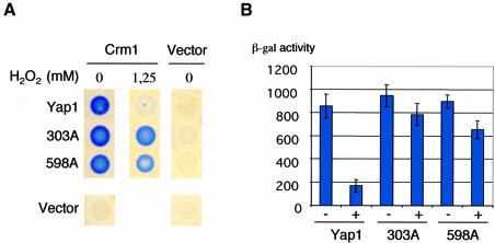
The yeast transcription factor Yap1 activates expression of antioxidant genes in response to oxidative stress. Yap1 regulation involves nuclear accumulation, but the mechanism sensing the oxidative stress signal remains unknown. We provide biochemical and genetic evidence that upon H2O2 treatment, Yap1 is activated by oxidation and deactivated by enzymatic reduction with Yap1-controlled thioredoxins, thus providing a mechanism for autoregulation. Two cysteines essential for Yap1 oxidation are also essential for its activation by H2O2. The data are consistent with a model in which oxidation of Yap1 leads to disulfide bond formation with the resulting change of conformation masking recognition of the nuclear export signal by Crm1/Xpo1, thereby promoting nuclear accumulation of the protein. In sharp contrast to H2O2, diamide does not lead to the same Yap1 oxidized form and still activates mutants lacking cysteines essential for H2O2 activation, providing a molecular basis for differential activation of Yap1 by these oxidants. This is the first example of an H2O2-sensing mechanism in a eukaryote that exploits the oxidation of cysteines in order to respond rapidly to stress conditions.


Figures

None
Fig. 1. Kinetics of Yap1 activation. (A) Expression of TRX2. Total RNA was isolated from a wild-type strain, grown to an OD600 of 0.3 in selective minimal medium, before and 10, 20, 30, 45 and 60 min after treatment with 300 µM H2O2, and TRX2 and ACT1 (control gene) mRNAs were quantified by RT–PCR. An autoradiogram of the separated 32P-labelled PCR products is shown in the upper panel and a plot of the TRX2/ACT1 signal ratio quantified on a PhosphorImager in the lower panel. (B) Nuclear redistribution of Yap1. A Δyap1 strain expressing GFP-Yap1 at an OD600 of 0.3, treated with 300 µM H2O2 for the time indicated and analysed for GFP staining. (C) Yap1 phosphorylation. Extracts of a Δyap1 strain expressing Myc-tagged Yap1 (yMyc-Yap1), before and 5, 10, 15, 30, 45 and 60 min after treatment with 300 µM H2O2 were immunoblotted with the anti-Myc monoclonal antibody after SDS–PAGE. (D) Yap1 phosphorylation correlates with Yap1 nuclear localization. Δyap1 strain cultures expressing Myc-Yap1, Yap1L619S,L623S or Yap1C598T,C620A,C629T, before and 20 min after H2O2 treatment, were processed as in (C).
None
Fig. 2. In vivo oxidation of Yap1. (A) yMyc-Yap1 cultures (grown to an OD600 of 0.4) before and after 2.5 min treatment with 400 µM H2O2 (lanes 1 and 2) or 1 mM t-butyl hydroperoxide (lanes 5 and 6) were lysed using TCA, treated with iodoacetamide and immunoblotted after non-reducing SDS–PAGE. Lanes 3 and 4 are the same as lanes 1 and 2 except that the TCA-precipitated protein pellet was dissolved in the presence of 200 mM DTT, before adding a 3.2 M excess of iodoacetamide. (B) Time course of Yap1 oxidation by H2O2. A yMyc-Yap1 culture before and 2.5, 5, 15, 30, 45 and 60 min after treatment with 400 µM H2O2, processed as in (A) (lanes 1 and 2), except that Yap1 was dephosphorylated and separated under non-reducing or reducing conditions as indicated. (C) The minimum H2O2 concentration required to oxidize Yap1 in vivo. A yMyc-Yap1 culture treated for 5 min with 25, 50, 100, 150, 200, 300 and 800 µM H2O2 and processed as in (A) (lanes 1 and 2). (D) Time-course analysis of Yap1 redox status in response to diamide. A yMyc-Yap1 culture before and 5, 15, 30, 45 and 60 min after treatment with 2 mM diamide, processed as in (A) (lanes 1 and 2) and separated under non-reducing or reducing conditions as indicated.
None
Fig. 3. Identification of Yap1 redox-active cysteines. (A) Schematics of Yap1. (B) Both N- and C-terminal cysteines are required to oxidize Yap1. Δyap1 strain cultures expressing Myc-Yap1, Yap1C303A,C310A,C315A or Yap1C598T,C620A,C629T before and 5 min after treatment with 400 µM H2O2 were processed as in Figure 2A (lanes 1 and 2) and immunoblotted after non-reducing SDS–PAGE. (C) Cysteines C303 and C598 are the redox-active Yap1 cysteines. Δyap1 strain cultures expressing Myc-Yap1, Yap1C303A, Yap1C310A, Yap1C315A, Yap1C598A, Yap1C620A or Yap1C629A, and Yap1C310T,C315A,C620A,C629T before and 5 min after H2O2 treatment, as in Figure 2B and immunoblotted after non-reducing or reducing SDS–PAGE, as indicated.
None
Fig. 4. Activity of Yap1 cysteine substitution mutants. (A) Yap1 cysteine residues important for tolerance to H2O2. The Δyap1 strains not expressing or expressing Myc-Yap1, Yap1C303A, Yap1C310A, Yap1C315A, Yap1C598A, Yap1C620A or Yap1C629A were tested for their tolerance to increasing concentrations of H2O2 by the patch assay on solid medium. (B) The same strains as in (A) were used to measure the amount of TRX2 and ACT1 mRNAs by RT–PCR as in Figure 1A. Cells were treated with 300 µM H2O2 for the time indicated. (C) The TRX2/ACT1 signal ratios quantified from the RT–PCR shown in (B) on a PhosphorImager were calculated and plotted as a function of time of exposure to H2O2.
None
Fig. 5. Essential role of C303 and C598 for H2O2-induced Yap1 nuclear redistribution. (A) Analysis of the cellular distribution of GFP-tagged Yap1C303A, Yap1C310A, Yap1C598A and Yap1C629A. Cells taken before or 12 min after treatment with 300 µM H2O2 were analysed for GFP or DAPI staining or under visible light. (B) Phosphorylation of Yap1 cysteine mutants as an indicator of their subcellular localization. Δyap1 cultures expressing Myc-Yap1, Yap1C303A, Yap1C310A, Yap1C315A, Yap1C598A, Yap1C620A or Yap1C629A were taken before and after treatment with 300 µM H2O2 and processed as in Figure 1C.
None
Fig. 6. The critical role of C303 and C598 is at the level of Crm1–Yap1 interaction. β-galactosidase production from a lacZ reporter gene was assayed in strain EGY48 carrying pKW442 (Crm1lexABD) or pEG202 (empty vector) and one of the following: pLDB439 (YAP1-B42AD or pYAP1C303A-B42AD), pYAP1C598A-B42AD or pJG4-5 (empty vector). (A) Assay performed on solid medium in the absence or presence of H2O2 at the concentration indicated. (B) Assay performed on liquid medium in the absence (–) or presence (+) of 500 µM H2O2. The results are the averages of four independent experiments.
None
Fig. 7. Deactivation (reduction) of Yap1 by the thioredoxin system. (A and B) Expression of TRX2 (A) and TRR1 (B) in wild-type, Δtrr1 (A), Δtrx1Δtrx2 (B), Δgsh1Δglr1pro2-1 and Δtsa1 strains. Cells were treated with 300 µM H2O2 for the indicated time and processed for RT–PCR as in Figure 1A. The ACT1 autoradiograms are not shown. (C) Yap1 is oxidized in mutants of the thioredoxin system. yMyc-Yap1, Δtrx1Δtrx2 and Δtrr1 expressing Myc-Yap1 (grown to an OD600 of 0.3) were processed as in Figure 2A and extracts were analysed by immunoblotting after non-reducing PAGE. (D) Reduction of oxidized Yap1 by the thioredoxin system. yMyc-Yap1 (grown to an OD600 of 0.4), treated with 400 µM H2O2, lysed with glass beads in 100 mM Tris–HCl pH 8, 0.1% SDS, 1 mM EDTA, complete protease inhibitors and PMSF. Twenty-five micrograms of extracts were incubated with 1, 10 or 20 µM thioredoxin (from Spirulina sp.), 1.3 µM E.coli thioredoxin reductase and 1 mM NADPH (Sigma) for 30 min at 37°C. Iodoacetamide (75 mM) was added and the samples were analysed by immunoblotting after non-reducing PAGE.

References

    1. Åslund F. and Beckwith,J. (1999) Bridge over troubled waters: sensing stress by disulfide bond formation. Cell, 96, 751–753. - PubMed
    1. Åslund F., Zheng,M., Beckwith,J. and Storz,G. (1999) Regulation of the OxyR transcription factor by hydrogen peroxide and the cellular thiol-disulfide status. Proc. Natl Acad. Sci. USA, 96, 6161–6165. - PMC - PubMed
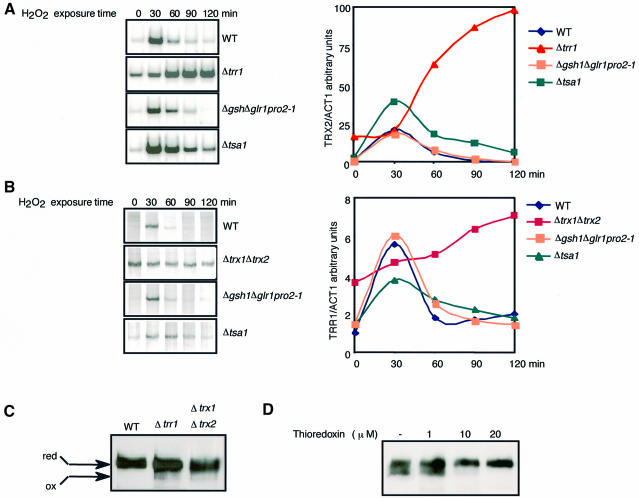
    1. Chae H.Z., Chung,S.J. and Rhee,S.G. (1994) Thioredoxin-dependent peroxide reductase from yeast. J. Biol. Chem., 269, 27670–27678. - PubMed
    1. Coleman S.T., Epping,E.A., Steggerda,S.M. and Moye-Rowley,W.S. (1999) Yap1p activates gene transcription in an oxidant-specific fashion. Mol. Cell. Biol., 19, 8302–8313. - PMC - PubMed
    1. Gaudu P., Moon,N. and Weiss,B. (1997) Regulation of the soxRS oxidative stress regulon. Reversible oxidation of the Fe-S centers of SoxR in vivo. J. Biol. Chem., 272, 5082–5086. - PubMed

Publication types

MeSH terms