. 2000 Oct 2;19(19):5178-93.
doi: 10.1093/emboj/19.19.5178.

Smad2, Smad3 and Smad4 cooperate with Sp1 to induce p15(Ink4B) transcription in response to TGF-beta

Affiliations

Smad2, Smad3 and Smad4 cooperate with Sp1 to induce p15(Ink4B) transcription in response to TGF-beta

X H Feng et al. EMBO J. .

Abstract

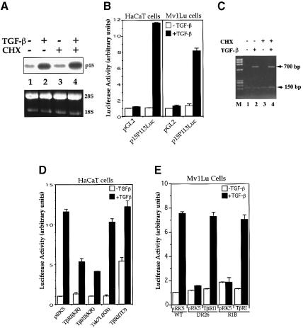
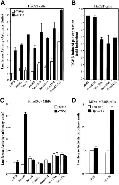
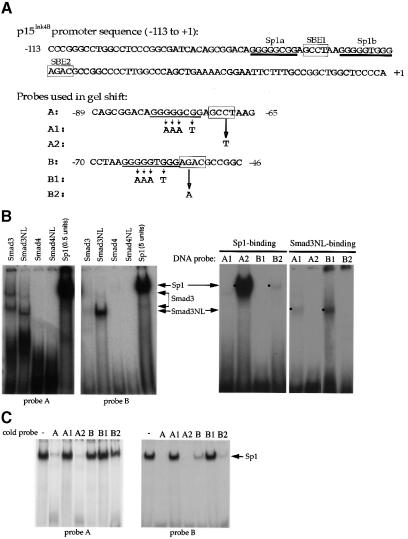
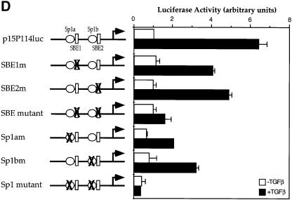
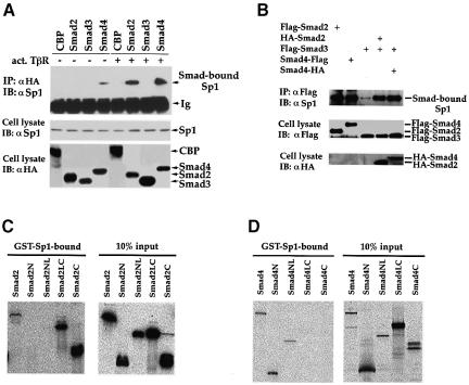
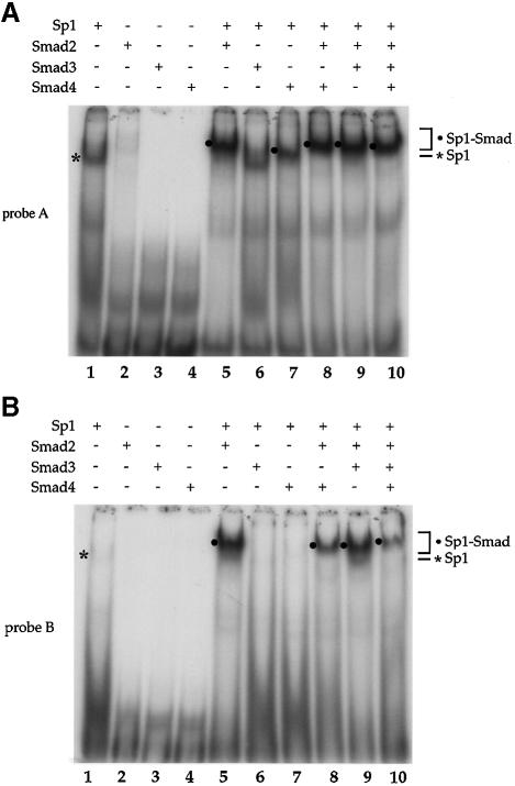
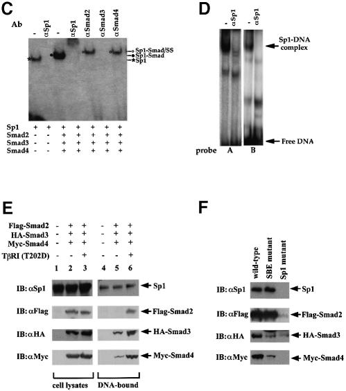
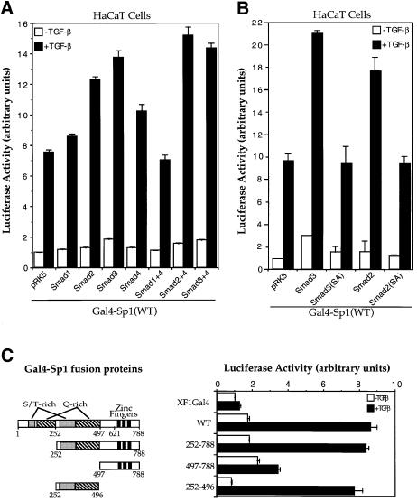
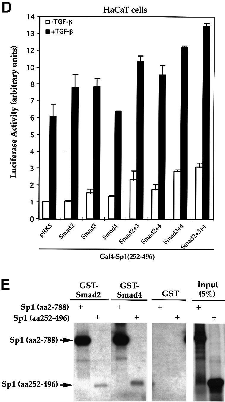
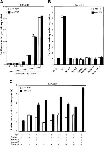
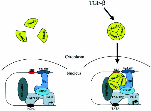
Transforming growth factor-beta (TGF-beta) arrests growth of epithelial cells by inducing the transcription of p15(Ink4B), a cyclin-dependent kinase inhibitor. In this study, we demonstrate that p15(Ink4B) induction was mediated by a TGF-beta-induced complex of Smad2, Smad3, Smad4 and Sp1. Mutations in the Sp1- or Smad-binding sequences decreased or abolished the TGF-beta responsiveness of the p15(Ink4B) promoter. Interference with, or deficiency in, Smad2, Smad3 or Smad4 functions also reduced or abolished the TGF-beta-dependent p15(Ink4B) induction, whereas the absence of Sp1 reduced the basal and TGF-beta-induced p15(Ink4B) transcription. In the nucleoprotein complex, Smad2 interacted through its C-domain with Sp1 and enhanced the DNA binding and transcriptional activity of Sp1. Smad3 interacted indirectly with Sp1 through its association with Smad2 and/or Smad4, and bound directly to the p15(Ink4B) promoter. Finally, Smad4 interacted through its N-domain with Sp1. Our data demonstrate the physical interactions and functional cooperativity of Sp1 with a complex of Smad2, Smad3 and Smad4 in the induction of the p15(Ink4B) gene. These findings explain the tumor suppressor roles of Smad2 and Smad4 in growth arrest signaling by TGF-beta.


Figures

None
Fig. 1. TGF-β-induced p15Ink4B transcription requires functional TβRI and TβRII and is independent of de novo protein synthesis. (A) p15Ink4B expression is an immediate early response gene to TGF-β. Exponentially growing HaCaT cells were treated with or without 400 pM TGF-β or 10 µM cycloheximide (CHX). p15Ink4B mRNA was detected by northern hybridization. Equal levels of RNA were loaded per lane, as illustrated by equal levels of 28S and 18S RNA. (B) Transcriptional activation from the p15Ink4B promoter is induced by TGF-β in HaCaT and Mv1Lu cells. Cells were transfected with the p15P113luc luciferase reporter plasmid and, 40–45 h after transfection, treated with TGF-β for 4 h, and luciferase values were measured. (C) Luciferase mRNA expression from the p15Ink4B promoter in response to TGF-β does not require new protein synthesis. HaCaT cells were transfected with the p15P113luc luciferase reporter plasmid and a control β-galactosidase expression plasmid pSVβgal, incubated with TGF-β and/or cycloheximide for 4 h as shown, and RNA was isolated. The levels of luciferase and β-galactosidase mRNA were assessed by PCR cDNA amplification. While the 150 bp β-galactosidase cDNA band was constant in all lanes, the 700 bp luciferase cDNA band was induced by TGF-β, both in the absence and presence of cycloheximide (CHX). M, DNA fragment length markers (ΦX174 DNA/HaeIII). (D) Dominant-negative inhibition of TGF-β-induced p15Ink4B transcription by kinase-inactive (KR) TβRI and TβRII. HaCaT cells were cotransfected with p15P113luc and the indicated receptor expression plasmids. (E) TGF-β-induced p15Ink4B transcription requires both TβRII and TβRI. The reporter plasmid p15P113luc was transfected into wild-type Mv1Lu cells, which express both receptors, or the derivative DR26 and R1B cells, which lack functional TβRII and TβRI, respectively. Expression plasmids for TβRII or TβRI were cotransfected, as marked. All assays were done in triplicate and all values were normalized for transfection efficiency against the β-galactosidase expression directed from the cotransfected pSV-β-Gal control plasmid.
None
Fig. 2. TGF-β-induced p15Ink4B transcription requires Smad2, Smad3 and Smad4. (A) Transcriptional activation of the p15Ink4B promoter by Smads. HaCaT cells were transfected with the p15P113luc reporter plasmid and indicated combinations of expression plasmids for Smad2, Smad3 and Smad4, and reporter gene expression were measured. (B) C-terminally truncated Smads inhibit TGF-β-induced transcription from the p15Ink4B promoter. HaCaT cells were cotransfected with the p15P113luc reporter plasmid, and indicated expression plasmids for Smad mutants. (C) Smad3 is required for TGF-β-induced transcription from the p15Ink4B promoter. Smad3–/– mouse embryonic fibroblasts (MEFs) were cotransfected with p15P113luc, and indicated expression plasmids for Smad2, Smad3 and Smad3 mutants. Smad3N, Smad3NL, Smad3LC and Smad3C contain the Smad3 regions of amino acids 2–144, 2–231, 144–425 and 232–425, respectively. (D) Smad4 is required for TGF-β-induced p15 transcription. Smad4-defective MDA-MB-468 cells were cotransfected with p15P113luc without or with an expression plasmid for Smad4.
None
Fig. 3. Sp1 binding sites and Smad3-binding elements are required for the activation of the p15Ink4B gene. (A) Nucleotide sequence of the –113 to +1 segment of the p15Ink4B promoter. Predicted Sp1 binding sites (Sp1a and Sp1b) and SBEs 1 and 2 are indicated. The oligonucleotide probes A and B, used in the gel shift experiments, with their inactivating mutations are also shown. (B) Sp1 and Smad3 bind to the p15Ink4B promoter. Purified Sp1 (0.5 or 5 U) or GST–Smad fusion proteins (1 µg) were incubated with the 32P-labeled probe A or B, or the mutant probes A1 or B1, in which the Sp1a or Sp1b sites are mutated, or the mutant A2 or B2 probes, in which the SBE1 or SBE2 sites are inactivated. Gel-shifted DNA–protein complexes are marked. (C) Sp1 binding to the Sp1 binding sites in oligonucleotides A or B can be competed with unlabeled ‘wild-type’ oligonucleotides, but not by oligonucleotides with mutated Sp1 binding sites. Purified Sp1 was incubated with the 32P-labeled probe A or B in the presence of indicated unlabeled DNA. The DNA–Sp1 complex is marked. (D) Mutational analysis of the p15Ink4B promoter. Sp1 sites and SBEs were individually, or in combination, mutated in the p15Ink4B promoter, as shown in Figure 3A. Transcription was measured by luciferase activity. Transfection, TGF-β treatment and luciferase assay in HaCaT cells were performed as in Figure 1.
None
Fig. 4. Physical association of Smad2, Smad3 and Smad4 with Sp1. (A) TGF-β-dependent association of Smads and Sp1 in vivo. HaCaT cells were transfected with HA-tagged Smads or CBP, with (+) or without (–) an expression plasmid for activated TβRI. Cell lysates were immunoprecipitated (IP) with an anti-HA antibody, followed by immunoblotting (IB) with an anti-Sp1 antibody to detect Smad-bound Sp1 (upper panel). Cell lysates were also directly immunoblotted with anti-Sp1 or anti-HA antibodies to demonstrate expression of endogenous Sp1 (middle panel) or transfected Smads (lower panel). (B) Smad3 co-immunoprecipitates with endogenous Sp1 in the presence of Smad2 or Smad4. HaCaT cells were transfected with expression plasmids for Flag-tagged Smad3 and HA-tagged Smad2 or 4. Immunoprecipitation with anti-Flag antibody was followed by anti-Sp1 immunoblotting to detect Smad3-bound Sp1. Expression levels of Flag-tagged Smads (middle panel) or HA-tagged Smads (lower panel) were shown by anti-Flag or anti-HA immunoblotting. (C) Direct interaction of GST–Sp1(1–621) with 35S-labeled Smad2 and its segments. Smad2N, Smad2NL, Smad2LC and Smad2C cover the Smad2 regions of amino acids 2–183, 2–273, 181–467 and 270–467, respectively. (D) Direct interaction of GST–Sp1(1–621) with 35S-labeled Smad4 and its segments. Smad4N, Smad4NL, Smad4LC and Smad4C cover the Smad4 regions of amino acids 2–154, 2–300, 141–552 and 294–552, respectively.
None
Fig. 5. Interaction of Smad2 or Smad4 with Sp1 at the Sp1a and Sp1b binding sites. Purified Sp1 (0.5 U) and GST–Smad fusion proteins (0.2 µg) were incubated with the 32P-labeled probe A, containing the Sp1a binding site (A), or probe B, containing the Sp1b binding site (B) of the p15Ink4B promoter. DNA-bound Sp1 and Sp1–Smad complexes are marked. (C) Gel shift analyses using oligonucleotide A and purified proteins were carried out as in (A). Antibodies, shown above the gel, were added to the gel shift reactions and incubated for 90 min, prior to gel analysis. Sp1–DNA, Sp1–Smad–DNA complex, and the supershifted (SS) complexes are marked. (D) Sp1-binding ability of p15 promoter elements. Nuclear extracts from HaCaT cells were incubated with probe A or B in gel shift reactions, with or without anti-Sp1 antibody. The Sp1–DNA complex, which is displaced in the presence of anti-Sp1 antibody, is indicated. (E) TGF-β-induced formation of Smad–DNA complex in cell lysates. Expression plasmids for Flag–Smad2, HA–Smad3 and Myc–Smad4 were transfected. Forty-eight hours after transfection, cells were lysed and incubated with streptavidin paramagnetic beads (Dynal) with immobilized biotinylated p15 promoter DNA oligonucleotide (nt –84 to –46). After extensive washing, DNA-bound proteins were detected by SDS–PAGE followed by western blotting using the indicated antibodies (lanes 4–6). Immunoblotting of the cell lysates in parallel demonstrates the expression level of endogenous Sp1 and transfected Smads. (F) The TGF-β-induced formation of Smad–DNA complex in cell lysates is largely dependent on intact Sp1 binding sites. Binding of Sp1 and Smad2, Smad3, Smad4 and Sp1 to the wild-type oligonucleotide, as was also done in (E), was compared with their binding to the corresponding mutant oligonucleotides, in which the SBEs and Sp1 binding sequences were mutated, as shown in Figure 3A.
None
Fig. 6. Smads increase the transcriptional activity of Sp1 through its second S/T-rich and Q-rich domains. (A) Smad2 and 3 stimulate the transactivation activity of Gal4–Sp1. HaCaT cells were cotransfected with Gal4–Sp1(WT) and the luciferase reporter plasmid pFR-Luc, and expression plasmids for the indicated Smads. (B) Smad-dependent stimulation of Gal4–Sp1 transactivation activity requires the C-terminal SSXS motif of Smad2 or Smad3. HaCaT cells were cotransfected with Gal4–Sp1(WT) and pFR-Luc, and expression plasmids for the indicated Smads or mutants. (C) Localization of the TGF-β-responsive domain of Gal4–Sp1. HaCaT cells were transfected with expression plasmids for Gal4–Sp1 or its derivatives, as shown, and the pFR-Luc reporter plasmid. pXF1Gal4 is the control plasmid containing only the GAL4 DNA binding domain. (D) Effects of Smads on the activity of Gal4–Sp1(252–496). The Gal4–Sp1(252–496) plasmid, in combination with the indicated expression plasmids for Smads, was transfected into HaCaT cells, together with the pFR-Luc reporter plasmid. (E) Direct interaction of GST–Smads with Sp1 (amino acids 252–496). Equal amounts of GST–Smads or control GST were used to adsorb 35S-labeled full-length Sp1 or its aa 252–496 fragment.
None
Fig. 7. Requirement of Sp1 and its functional cooperation with Smads in the transcription of p15Ink4B in Drosophila S2 cells. (A) Sp1 strongly transactivates the p15 promoter. S2 cells were transfected with the p15P113luc reporter plasmid and increasing amounts of the Sp1 expression plasmid Pac-Sp1, in the presence or absence of an activated TβRI. Luciferase assays were done as in HaCaT cells. (B) Inability of Smads to transactivate the p15 promoter in the absence of Sp1. S2 cells were transfected with the p15P113luc reporter and expression plasmids for Sp1 or for Smads, in the presence or absence of an activated TβRI, as shown. (C) Sp1 and Smads cooperate to activate the p15 promoter in response to TGF-β receptor activation. S2 cells were transfected with the p15P113luc reporter, the Sp1 expression plasmid and expression plasmids for Smads, as marked.
None
Fig. 8. Model for TGF-β-dependent transcriptional activation of the p15Ink4B gene. An oligomeric, likely trimeric, complex consisting of Smad2, Smad3 and Smad4 is translocated into the nucleus upon TGF-β stimulation. Smad3 contacts DNA at the SBE on the p15Ink4B promoter, to which Sp1 was already bound at the Sp1 site (only one Sp1 binding site and one SBE are shown). Additional protein–protein interactions occur between Sp1 and Smad2, Sp1 and Smad4, and Smad2 and/or 3 and CBP/p300. TAF/TBP represents TATA-binding protein (TBP) and its associated factors (TAF), the transcription factors for general transcription associated with RNA polymerase II.

References

    1. Abdollah S., Macías-Silva,M., Tsukazaki,T., Hayashi,H., Attisano,L. and Wrana,J.L. (1997) TβRI phosphorylation of Smad2 on Ser465 and Ser467 is required for Smad2–Smad4 complex formation and signaling. J. Biol. Chem., 272, 27678–27685. - PubMed
    1. Arrick B.A. and Derynck,R. (1996) The biological role of transforming growth factor-β in cancer development. In Waxman,J. (ed.), Molecular Endocrinology of Cancer. Cambridge University Press, Cambridge, UK, pp. 51–78.
    1. Attisano L. and Wrana,J.L. (1998) Mads and Smads in TGFβ signalling. Curr. Opin. Cell Biol., 10, 188–194. - PubMed
    1. Ausubel F.M., Brent,R., Kingston,R.E., Moore,D.D., Seidman,J.G., Smith,J.A. and Struhl,K. (1998) Current Protocols in Molecular Biology. John Wiley and Sons, Inc., New York, NY.
    1. Bae H.W., Geiser,A.G., Kim,D.H., Chung,M.T., Burmester,J.K., Sporn,M.B., Roberts,A.B. and Kim,S.-J. (1995) Characterization of the promoter region of the human transforming growth factor-β type II receptor gene. J. Biol. Chem., 270, 29460–29468. - PubMed

Publication types

MeSH terms

Substances