. 2000 Oct 2;19(19):5212-21.
doi: 10.1093/emboj/19.19.5212.

Quorum-sensing signal binding results in dimerization of TraR and its release from membranes into the cytoplasm

Affiliations

Quorum-sensing signal binding results in dimerization of TraR and its release from membranes into the cytoplasm

Y Qin et al. EMBO J. .

Abstract

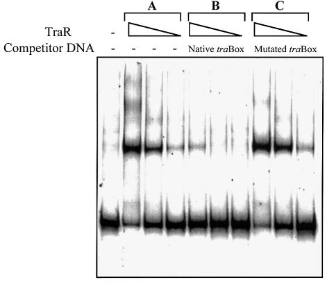
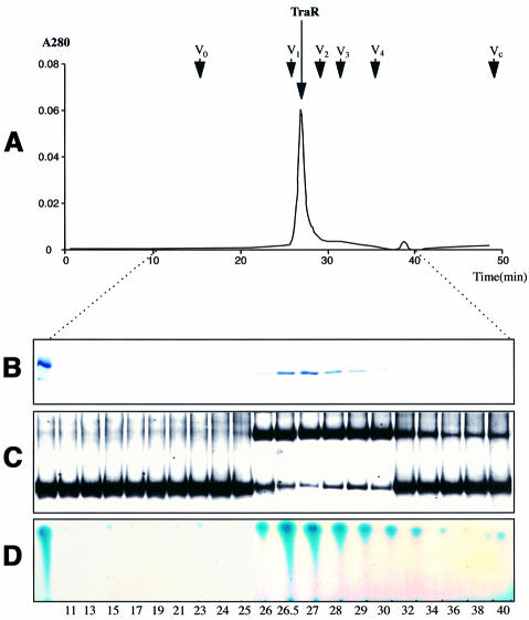
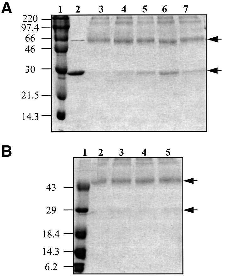
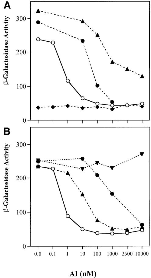
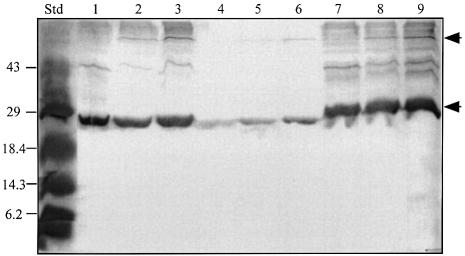
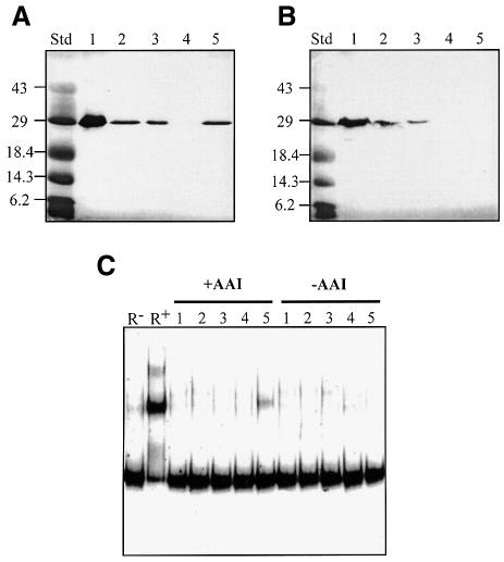
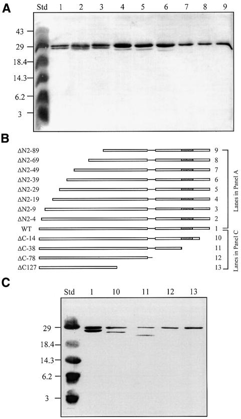
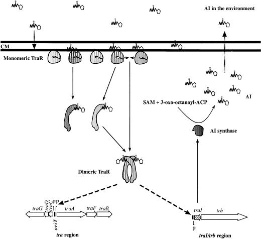
Promoter binding by TraR and LuxR, the activators of two bacterial quorum-sensing systems, requires their cognate acyl-homoserine lactone (acyl-HSL) signals, but the role the signal plays in activating these transcription factors is not known. Soluble active TraR, when purified from cells grown with the acyl-HSL, contained bound signal and was solely in dimer form. However, genetic and cross-linking studies showed that TraR is almost exclusively in monomer form in cells grown without signal. Adding signal resulted in dimerization of the protein in a concentration-dependent manner. In the absence of signal, monomer TraR localized to the inner membrane while growth with the acyl-HSL resulted in the appearance of dimer TraR in the cytoplasmic compartment. Affinity chromatography indicated that the N-terminus of TraR from cells grown without signal is hidden. Analysis of heterodimers formed between TraR and its deletion mutants localized the dimerization domain to a region between residues 49 and 156. We conclude that binding signal drives dimerization of TraR and its release from membranes into the cytoplasm.


Figures

None
Fig. 1. TraR specifically binds DNA containing the tra box. Purified TraR was incubated at 10-fold decreasing protein concentrations (35 to 0.35 ng) with 0.4 ng of a labeled 47 bp DNA fragment containing the tra box. Protein–DNA complexes were detected as described in Materials and methods. Gels contain TraR incubated with: (A) labeled probe only; (B) labeled probe and a 500-fold excess of unlabeled probe; (C) labeled probe and a 500-fold excess of unlabeled probe containing a tra box altered at positions 4 and 15.
None
Fig. 2. TraR isolated from cells grown with 3-oxo-C8-HSL is a dimer. Purified TraR (150 µg) was subjected to size-exclusion chromatography and fractions from the column were assayed for: (A) protein content (A280); (B) monomer protein size by SDS–PAGE; (C) interaction with the tra box by gel retardation; (D) presence of 3-oxo-C8-HSL by thin-layer chromatography, all as described in Materials and methods. Arrows indicate the elution times for a set of standard proteins (Sigma Chemical Co.) including V1, bovine serum albumin (Mr ∼66 000); V2, bovine erythrocyte carbonic anhydrase (Mr ∼29 000); V3, horse heart cytochrome c (Mr ∼12 400); and V4, bovine lung aprotinin (Mr ∼6500). V0 marks the elution time of blue dextran (Mr ∼2 000 000) and Vc denotes the column volume. The peak of absorbance labeled TraR eluted at a time interval corresponding to a protein of Mr ∼52 000.
None
Fig. 3. TraR converts from dimer to monomer during dialysis in the absence of 3-oxo-C8-HSL. Purified dimer TraR (10 µM) was dialyzed against a buffer containing 50 mM HEPES pH 8.5, 150 mM NaCl, 1 mM EDTA, 20% sucrose, 5% glycerol, 0.05% Tween 20 with (B) or without (A) 3-oxo-C8-HSL (10 µM final concentration). At 2-day intervals a volume was removed from each sample, the protein was treated with DSP and subjected to non-reducing SDS–PAGE as described in Materials and methods. (A) Lane 2 contains undialyzed, untreated TraR; lane 3 contains undialyzed, cross-linked TraR; and lanes 4, 5 and 6 contain TraR cross-linked following dialysis in the absence of 3-oxo-C8-HSL for 2, 4 or 6 days, respectively. Lane 7, TraR sampled from day 6 that had been re-incubated with 3-oxo-C8-HSL for 2 h prior to cross-linking. (B) Lane 2 contains undialyzed, cross-linked TraR, and lanes 3, 4 and 5 contain TraR cross-linked following dialysis in the presence of 3-oxo-C8-HSL for 2, 4 and 6 days, respectively. Lane 1 contains size standards (in kDa). The upper and lower arrows mark the locations of the dimer and monomer forms of TraR.
None
Fig. 4. TraR co-purifies with the inner membrane of A.tumefaciens. Membranes prepared from A.tumefaciens NT1(pKKTR2) grown in the absence of signal were fractionated into inner and outer components on a sucrose gradient as described in Materials and methods. Fractions were assayed for NADH oxidase activity (µmol NAD formed/min/ml, filled triangles) and 2-keto 3-deoxyoctanoate (KDO) content (mg/ml, open triangles) (A), and for TraR by western blot analysis (B) as described in Materials and methods. T, M and S represent equal loadings of proteins from the total lysate, the total membrane fraction and the soluble fraction, respectively.
None
Fig. 5. Dimerization of λ cI′ fusions to TraR or LuxR is dependent upon acyl-HSL. Cultures of E.coli JH372, harboring plasmids coding for cI′ fusion proteins were grown for 6 h in L broth with the appropriate acyl-HSL at the indicated concentration. Samples were assayed for β-galactosidase activity as described in Materials and methods. (A) Repression by cI′ fusions to TraR and LuxR but not to EsaR is dependent upon the acyl-HSL concentration. Fusions of cI′ to: TraRpTiC58 (open circles) and TraRpTi15955 (filled circles), both incubated with 3-oxo-C8-HSL, and LuxR (filled triangles) and EsaR (filled diamonds), both incubated with 3-oxo-C6-HSL. (B) Repression by the cI′::TraR fusion is dependent upon the structure of the acyl-HSL. Fusions of cI′ to TraRpTiC58 incubated with: 3-oxo-C8-HSL (open circles), 3-oxo-C6-HSL (filled triangles), C8-HSL (filled circles) and C6-HSL (filled inverted triangles).
None
Fig. 6. 3-oxo-C8-HSL drives dimerization of TraR and its appearance in the cytoplasm. Agrobacterium tumefaciens NT1(pZLQR) was grown in ABM medium to late exponential phase, harvested cells were treated with DSP, broken in the French press, and the lysates (lanes 1–3) were separated into soluble (lanes 4–6) and particulate (lanes 7–9) fractions as described in Materials and methods. Equal amounts of protein of each of the fractions were subjected to SDS–PAGE under non-reducing conditions, and TraR was visualized by far western blot analysis (Luo et al., 2000). Cultures were grown with no ligand (lanes 1, 4 and 7) or with 50 nM (lanes 2, 5 and 8) or 100 nM (lanes 3, 6 and 9) 3-oxo-C8-HSL. The upper and lower arrows mark the locations of the dimer and monomer forms of TraR.
None
Fig. 7. N-terminal His-tagged TraR from cells grown in the absence of 3-oxo-C8-HSL fails to bind to nickel affinity resin. Cultures of A.tumefaciens NT1(pKKHTR9) were grown in ABM with (A) or without (B) 3-oxo-C8-HSL (100 nM). The cells were harvested and lysates were prepared and subjected to nickel affinity chromatography. Equal volumes of samples from each treatment were electrophoresed and TraR was visualized by far western blot analysis (A and B). Samples were also assayed for DNA binding by gel retardation (C), all as described in Materials and methods. Lane 1, total lysate; lane 2, cleared lysate; lane 3, column flow-through; lane 4, 10 mM imidazole wash; lane 5, 100 mM EDTA eluate. R and R+ in (C) contain the labeled probe DNA without or with pure native TraR, respectively.
None
Fig. 8. The dimerization region of TraR is located in the N-terminal half of the protein. Wild-type TraR or its N- or C-terminal deletion mutants were co-expressed with His6-TraR in A.tumefaciens NT1 grown with 100 nM 3-oxo-C8-HSL. Cleared lysates were prepared, subjected to nickel affinity chromatography, and the protein eluted from the resin was analyzed by SDS–PAGE as described in Materials and methods. Heterodimers of His6-TraR and N-terminal deletion mutants (A) were detected by far western blot analysis while heterodimers of His6-TraR and C-terminal deletion mutants (C) were detected by western analysis as described in Materials and methods. The alleles of TraR along with representations of their structures are presented in (B). Std, a mixture of standard markers with sizes in kilodaltons noted to the left of the blots.
None
Fig. 9. Model for the compartmentalization of TraR from intracellular 3-oxo-C8-HSL. Unactivated TraR is associated as monomers with the inner face of the cytoplasmic membrane (CM) where it is shielded from nascent acyl-HSL produced by TraI (Moré et al., 1996) within the cell. The newly synthesized signal passes out through the membrane where it can accumulate in the environment. This extracellular signal diffuses back into the membrane where, when at sufficiently high concentrations, it can interact with its binding site on TraR. Signal binding, at a stoichiometry of 1:1 (Zhu and Winans, 1999), alters some property of TraR, thereby allowing the activator to dimerize and to release from the membrane into the cytoplasm. Alternatively, after binding one molecule of signal, protomers of TraR release from the membrane and dimerize in the cytoplasm. In its soluble dimer form TraR binds to tra box sites located in promoters of the tra regulon and, in conjunction with RNA polymerase, activates transcription.

Similar articles

Cited by

References

    1. Adar Y.Y. and Ulitzur,S. (1993) GroESL proteins facilitate binding of externally added inducer by LuxR protein-containing E.coli cells. J. Biolumin. Chemilumin., 8, 261–266. - PubMed
    1. Beck von Bodman S. and Farrand,S.K. (1995) Capsular polysaccharide biosynthesis and pathogenicity of Erwinia stewartii require induction by an N-acylhomoserine lactone autoinducer. J. Bacteriol., 177, 5000–5008. - PMC - PubMed
    1. Beck von Bodman S., Majerczak,D.R. and Coplin,D.L. (1998) A negative regulator mediates quorum-sensing control of exopolysaccharide production in Pantoea stewartii subsp. stewartii. Proc. Natl Acad. Sci. USA, 95, 7687–7692. - PMC - PubMed
    1. Cangelosi G.A., Best,E.A., Martinetti,C. and Nester,E.W. (1991) Genetic analysis of Agrobacterium tumefaciens. Methods Enzymol., 145, 177–181. - PubMed
    1. Cha C., Gao,P., Chen,Y.-C., Shaw,P.D. and Farrand,S.K. (1998) Production of acyl-homoserine lactone quorum-sensing signals by Gram-negative plant-associated bacteria. Mol. Plant Microbe Interact., 11, 1119–1129. - PubMed

Publication types

MeSH terms