

Inhibition of acetylcholine muscarinic M(1) receptor function by the M(1)-selective ligand muscarinic toxin 7 (MT-7)
- PMID: 11015294
- PMCID: PMC1572361
- DOI: 10.1038/sj.bjp.0703606
Inhibition of acetylcholine muscarinic M(1) receptor function by the M(1)-selective ligand muscarinic toxin 7 (MT-7)
Abstract
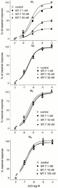
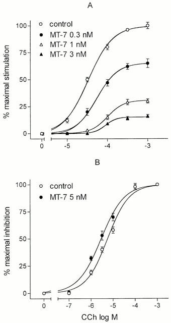
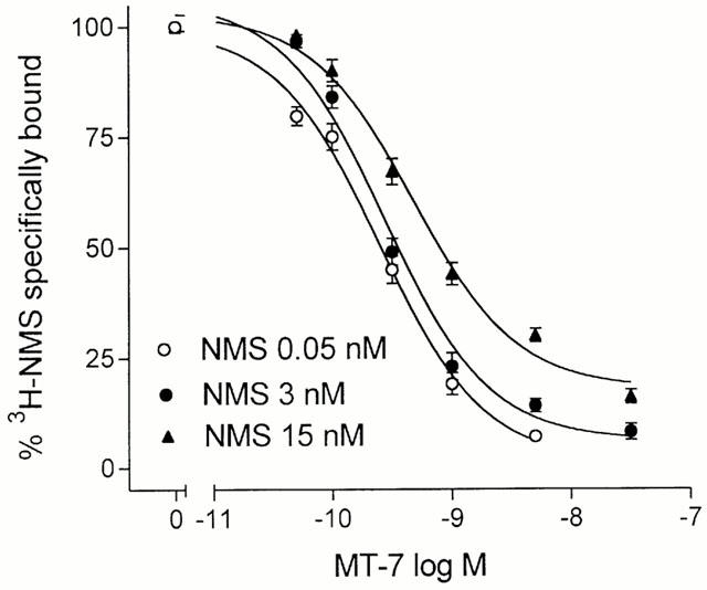
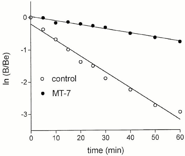
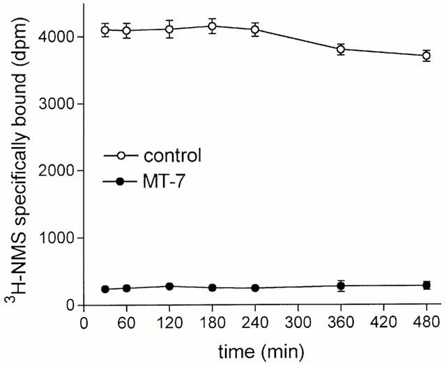
MT-7 (1 - 30 nM), a peptide toxin isolated from the venom of the green mamba Dendroaspis angusticeps and previously found to bind selectively to the muscarinic M(1) receptor, inhibited the acetylcholine (ACh)-stimulated [(35)S]-guanosine-5'-O-(3-thio)triphosphate ([(35)S]-GTPgammaS) binding to membranes of Chinese hamster ovary (CHO) cells stably expressing the cloned human muscarinic M(1) receptor subtype. MT-7 failed to affect the ACh-stimulated [(35)S]-GTPgammaS binding in membranes of CHO cells expressing either the M(2), M(3) or M(4) receptor subtype. In N1E-115 neuroblastoma cells endogenously expressing the M(1) and M(4) receptor subtypes, MT-7 (0.3 - 3.0 nM) inhibited the carbachol (CCh)-stimulated inositol phosphates accumulation, but failed to affect the CCh-induced inhibition of pituitary adenylate cyclase activating polypeptide (PACAP) 38-stimulated cyclic AMP accumulation. In both CHO/M(1) and N1E-115 cells the MT-7 inhibition consisted in a decrease of the maximal agonist effect with minimal changes in the agonist EC(50) value. In CHO/M(1) cell membranes, MT-7 (0.05 - 25 nM) reduced the specific binding of 0.05, 1.0 and 15 nM [(3)H]-N-methylscopolamine ([(3)H]-NMS) in a concentration-dependent manner, but failed to cause a complete displacement of the radioligand. Moreover, MT-7 (3 nM) decreased the dissociation rate of [(3)H]-NMS by about 5 fold. CHO/M(1) cell membranes preincubated with MT-7 (10 nM) and washed by centrifugation and resuspension did not recover control [(3)H]-NMS binding for at least 8 h at 30 degrees C. It is concluded that MT-7 acts as a selective noncompetitive antagonist of the muscarinic M(1) receptors by binding stably to an allosteric site.
Figures
References
-
- ADEM A., KARLSSON E. Muscarinic receptor subtype selective toxins. Life Sci. 1997;60:1069–1076. - PubMed
-
- ADEM A., ASBLOM A., JOHANSSON G., MBUGUA P.M., KARLSSON E. Toxins from the venom of the green mamba Dendroaspis angusticeps that inhibit the binding of quinuclidinyl benzilate to muscarinic acetylcholine receptors. Biochim. Biophys. Acta. 1988;968:340–345. - PubMed
-
- BRADFORD M.M. A rapid and sensitive method for the quantitation of microgram quantities of protein utilizing the principle of protein-dye binding. Anal. Biochem. 1976;72:248–254. - PubMed
-
- CARSI J.M., VALENTINE H.H., POTTER L.T. m2-Toxin: A selective ligand for M2 muscarinic receptors. Mol. Pharmacol. 1999;56:933–937. - PubMed
Publication types
MeSH terms
Substances
LinkOut - more resources
Full Text Sources
Molecular Biology Databases
