

Use of a repetitive DNA probe to type clinical and environmental isolates of Aspergillus flavus from a cluster of cutaneous infections in a neonatal intensive care unit
- PMID: 11015372
- PMCID: PMC87445
- DOI: 10.1128/JCM.38.10.3612-3618.2000
Use of a repetitive DNA probe to type clinical and environmental isolates of Aspergillus flavus from a cluster of cutaneous infections in a neonatal intensive care unit
Abstract
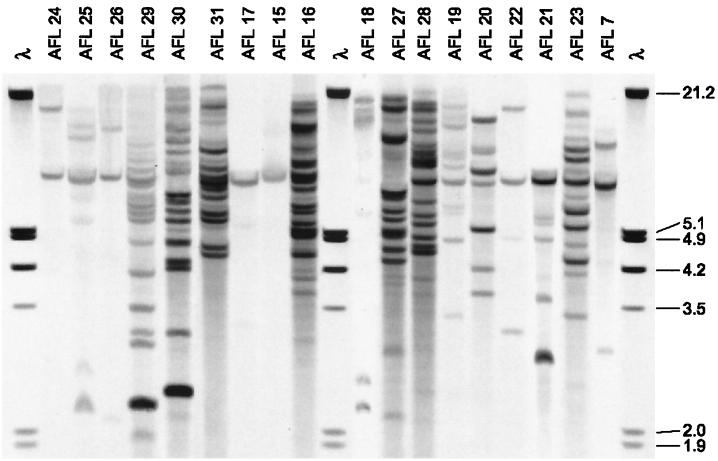
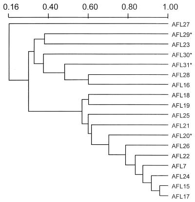
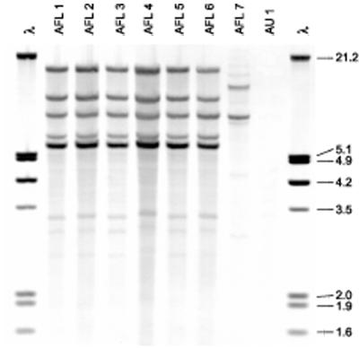
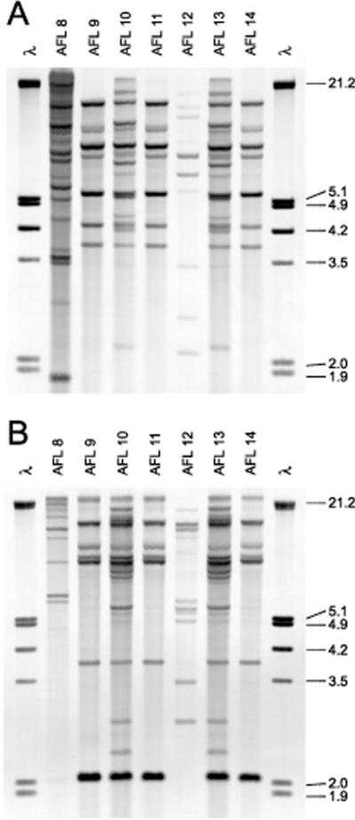
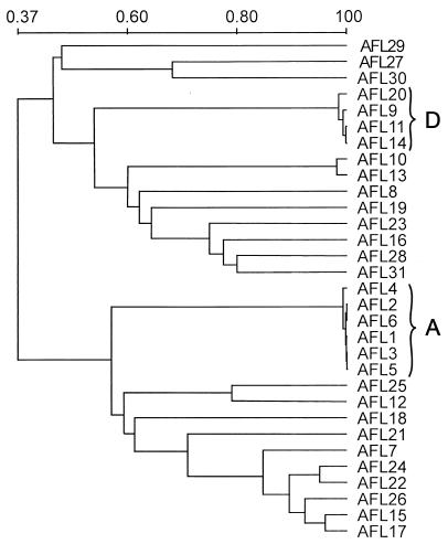
Aspergillus flavus is second to A. fumigatus as a cause of invasive aspergillosis, but no standard method exists for molecular typing of strains from human sources. A repetitive DNA sequence cloned from A. flavus and subcloned into a pUC19 vector, pAF28, was used to type 18 isolates from diverse clinical, environmental, and geographic sources. The restriction fragment length polymorphisms generated with EcoRI- or PstI-digested genomic DNA and probed with digoxigenin-labeled pAF28 revealed complete concordance between patterns. Eighteen distinct fingerprints were observed. The probe was used to investigate two cases of cutaneous A. flavus infection in low-birth-weight infants in a neonatal intensive care unit (NICU). Both infants were transported by the same ambulance and crew to the NICU on the same day. A. flavus strains of the same genotype were isolated from both infants, from a roll of tape used to fasten their umbilical catheters, from a canvas bag used to store the tape in the ambulance, and from the tape tray in the ambulance isolette. These cases highlight the need to consider exposures in critically ill neonates that might occur during their transport to the NICU and for stringent infection control practices. The hybridization profiles of strains from a second cluster of invasive A. flavus infections in two pediatric hematology-oncology patients revealed a genotype common to strains from a definite case patient and a health care worker. A probable case patient was infected with a strain with a genotype different from that of the strain from the definite case patient but highly related to that of an environmental isolate. The high degree of discrimination and reproducibility obtained with the pAF28 probe underscores its utility for typing clinical and environmental isolates of A. flavus.
Figures
References
-
- Bryce E A, Walker M, Scharf S, Lim A T, Walsh A, Sharp N, Smith J A. An outbreak of cutaneous aspergillosis in a tertiary-care hospital. Infect Control Hosp Epidemiol. 1996;17:70–72. - PubMed
-
- Buffington J, Reporter R, Lasker B A, McNeil M M, Lanson J M, Ross L A, Mascola L, Jarvis W R. Investigation of an epidemic of invasive aspergillosis: utility of molecular typing with the use of random amplified polymorphic DNA probes. Pediatr Infect Dis J. 1994;13:386–393. - PubMed
-
- Challen M P, Moore A J, Martínez-Carrera D. Facile extraction and purification of filamentous fungal DNA. BioTechniques. 1995;18:975–976. - PubMed
Publication types
MeSH terms
Substances
LinkOut - more resources
Full Text Sources
Medical
